

 PDF
PDF ePub
ePub Citation
 Print
Print


INTRODUCTION
Myolipoma is a rare benign tumor, first described by Meis and Enzinger in 1991 (1), which occurs most frequently in adults. These tumors are composed of irregularly admixed mature adipose tissue and smooth muscle fibers (2). Myolipoma has been described in the retroperitoneum (1-3), the orbital region (4), subcutaneous locations (1), the pericardium (5), the rectus sheath of the anterior abdominal wall, and the abdominal cavity with attachment to the abdominal wall (1). However, there is a paucity of literature describing the radiologic appearance of myolipoma, particularly that of soft tissue. No magnetic resonance imaging (MRI) findings in subcutaneous myolipoma of an extremity have been reported. Here, we present MRI findings on a subcutaneous myolipoma of the ankle. This is the first reported case of this tumor in such a location.
CASE REPORT
A 34-year-old woman was admitted to our hospital with a 3-year history of a palpable mass in the left ankle. The mass had been slowly increasing in size. Physical examination revealed a non-tender, non-fluctuant, and non-pulsatile mass in the left ankle region. On plain ankle radiography, mild soft tissue bulging was observed around the medial malleolus.
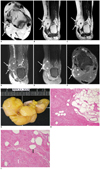
In order to better characterize the lesion, we performed MRI of the left ankle (Fig. 1A-F). The lesion was located in the subcutaneous region at the level of the posteromedial ankle joint. The mass was lobulated in contour, with some septations, and showed a hyperintense T1, T2 signal intensity (SI). On fat-suppressed T2-weighted imaging (WI), the lesion showed significant signal loss. However, focal, non fat-suppressed, intermediate T1 and T2 SI were noted at the medial peripheral aspect of the mass. Following administration of gadolinium diethylenetriamene pentaacetate (DTPA), the main lesion showed no remarkable enhancement; however, the peripheral portion of the mass showed heterogeneous enhancement.
Our radiological differential diagnoses included lipoma variant, atypical lipoma, or liposarcoma. Surgical excision was performed. Subcutaneous dissection revealed a yellowish well-encapsulated mass. The mass was well-defined, multi-lobulated, and oval-shaped in character, and was located in the medial subcutaneous tissue of the ankle. The mass measured 4.5 × 3.2 × 2.4 cm and was made up of yellowish soft tissue. Upon gross pathologic examination (Fig. 1G), the tumor was found to be completely encapsulated and was yellow-to-light pink in color. Histologic analysis showed a variable admixture of smooth muscle and mature adipose tissue (Fig. 1H, I). Our pathologic diagnosis was myolipoma.
DISCUSSION
Myolipoma of soft tissue is an extremely rare benign lipomatous lesion. Such lesions are usually located in the retroperitoneum or abdomen, including the inguinal region and the abdominal wall (1-3, 6). Less common locations include subcutaneous tissue, the orbit, the mediastinum, and the erector spinae (1, 4, 5, 7, 8). In the uterus, a similar tumor is known as lipoleiomyoma (3). Patients may present with evidence of a soft tissue mass; however, large retroperitoneal lesions may be identified as incidental findings (1). Because they can be detected earlier, subcutaneous lesions are much smaller (9). The condition is most prevalent in the fifth and sixth decades of life, with a slight predilection for woman (8). Neither local recurrence nor metastatic disease has been reported (7). The tumor is benign, and is composed of variable amounts of smooth muscle fibers and mature adipose tissue, but with no lipoblasts, floret-like giant cells, or zones of atypia (2). Histopathologically, the smooth muscle component of the tumor is usually regularly interspersed with adipose tissue, resulting in a sieve like appearance (1). Only a few reports on the radiological appearance of myolipoma have appeared in the literature (2, 6, 8, 9).
Imaging reflects the histologic composition of the lesion, in which the proportions of adipose and non-adipose tissue can vary. Due to hyperechogenicity of the lipomatous regions of the tumor, ultrasound findings are suggestive, but not diagnostic. Lipomatous components show CT and MRI features characteristic of fat tissue. However, the non-lipomatous component exhibits nonspecific solid intrinsic features with soft tissue attenuation on CT scans and an intermediate SI on T1-weighted MR images and an intermediate to high SI on T2-weighted images (9). In our patient, most of the mass yielded an SI characteristic of fat, whereas the peripheral region showed an intermediate T1 SI value, but with a T2 SI value isointense to muscle, with heterogeneous enhancement, indicative of the presence of both fat and muscle components. Tumors consisting of a mix of mature adipose and smooth muscle tissue, including those designated as lipoleiomyomas, fibrolipoleiomyomas, and myolipomas are exceedingly rare. Usually, the muscular component is predominant (2, 6, 9). However, on histological examination, the mass of the patient in our case consisted mainly of a fatty component with only a small amount of muscle component, which differed from the known findings reported in the literature of myolipomas involving other regions of the body. In order to define the typical radiologic findings of myolipoma affecting the extremities, we think that analysis of additional cases of myolipoma will be necessary.
Soft tissue myolipoma is a benign lesion, which must be distinguished from lesions with malignant or uncertain biologic behavior. The pathogenesis of myolipoma remains unclear. There are two main theories, namely adipose metaplasia and a multipotential Mullerian cell origin (2).
The main differential diagnoses of myolipoma include well-differentiated liposarcoma, spindle-cell lipoma, angiomyolipoma, leiomyoma with fatty degeneration, and lipoleiomyosarcoma (2, 3). Liposarcomas contain lipoblasts or floret-like giant cells (enlarged eosinophilic cells with atypical nuclei) or zones of atypia. Due to similar fat content, a well-differentiated liposarcoma can mimic myolipoma. Internal thick, irregular fibrous septations of well-differentiated liposarcomas show CT attenuation and MR signal intensity similar to those of muscle, and may become enhanced after gadolinium administration (10). However, a well-differentiated liposarcoma shows a fatty mass with poorly defined internal areas of non-adipose tissue. Non-adipose components are most often seen as prominent thick septa (> 2 mm), which may show nodularity. Focal nodular or globular non-adipose areas may show significant enhancement (6, 10). Histopathologically, myolipoma differs from liposarcoma; the former tumor type is encapsulated, and myolipoma is non-invasive. In addition, when examined microscopically, a myolipoma does not contain lipoblasts, atypical cells, or mitotic figures (3). Spindle cell lipomas in the typical locations of myolipoma are rare, and are composed of bland spindle-shaped cells, lacking smooth muscle differentiation (8). On MRI, non-adipose components have an SI value similar to that of muscle on T1WI and equal to or greater than that of fat on T2WI, and marked enhancement is evident (9). Angiomyolipoma differs from myolipoma in that the former tumor shows a regular arrangement of medium-sized arteries with thick muscular walls and narrow lumina, contains epithelioid smooth muscle cells showing occasional cellular atypia, displays melanoma marker positivity, and is frequently associated with tuberous sclerosis (3). CT permits confident, noninvasive diagnosis of angiomyolipoma. Heterogeneous soft tissue attenuation (due to hemorrhage or fibrosis or representing muscular and vascular components) may be evident. Contrast enhancement is variable and is dependent on the amount of soft tissue and vascularity (10). A leiomyoma with fatty degeneration lacks the regular distribution of fat characteristic of a myolipoma (2-4, 8). Myolipoma also differs significantly from lipoleiomyosarcoma in that the former does not show the histologic features of a well-differentiated liposarcoma and shows entirely banal smooth muscle tissue. This is unlike the mild-to-moderate extent of muscle atypia seen in lipoleiomyosarcoma (3).
Surgical excision is the treatment of choice for myolipoma (8). No reports of local recurrence, metastatic disease, or malignant transformation have appeared in the literature.
In conclusion, myolipoma of soft tissue is a rare benign lipomatous tumor. We report our MRI findings of a tumor occurring in an extremity, a location where myolipomas have not been previously documented. Despite the benign nature of these tumors, radiological investigations and correct diagnosis are of significant importance, because such masses need to be considered in the differential diagnosis of fat-containing lesions of soft tissue.
 XML Download