

 PDF
PDF ePub
ePub Citation
 Print
Print


INTRODUCTION
Primary amyloidosis (AL-type) is a rare disease that is characterized by deposition of proteinaceous material in various organs (1), with the liver and spleen being the major sites of involvement. However, the deterioration of liver function is usually mild. The clinical manifestations include hepatomegaly, jaundice and cholestasis (2). To date, there has only been a few case reports that have described primary amyloidosis associated with multiple myeloma and this resulted in subacute or acute, fulminant hepatic failure (3-5). Although several case studies have reported positive 18F-fluorodeosxy glucose (18F-FDG) uptake in pulmonary amyloidosis (6, 7), to the best of our knowledge, there have not been any reports of the 18F-FDG positron emission tomography (PET)/computed tomography (CT) findings in hepatic amyloidosis or primary hepatic amyloidosis associated with multiple myeloma. Herein, we report on a case of primary AL hepatic amyloidosis associated with multiple myeloma and this resulted in hepatic failure and we describe the 18F-FDG PET/CT image findings of the patient.
CASE REPORT
A 64-year-old female patient was admitted at our hospital with progressive jaundice and right upper quadrant pain for two days. The patient had a history of laparoscopic cholecystectomy due to gall stones two months previously. One month ago, a percutaneous drainage catheter had been inserted due to ascites. The laboratory studies performed at admission showed elevated total bilirubin (13.2 mg/dL; normal range, 0.2-1.0 mg/dL) and direct bilirubin (10.4 mg/dL; normal range, 0-0.3 mg/dL). The hepatic functional enzymes were within the normal limits.
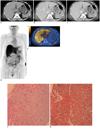
Abdominal CT had been performed on the 11th day before admission at an outside hospital. These abdominal CT images demonstrated mild hepatomegaly with decreased attenuation on the precontrast phase images (Fig. 1A). The arterial phase images revealed heterogeneous enhancement of the liver with low-density lesions in the periportal areas (Fig. 1B). Ascites was also observed. The portal phase scan images showed homogeneous enhancement of the liver with focal hypoattenuating areas (Fig. 1C). These findings may be consistent with amyloidosis of the liver. Diffuse decreased splenic attenuation with contrast enhancement could be seen on the CT scans, but the size and shape of the spleen were within normal limits. The magnetic resonance cholangiopancreatographic images taken outside our hospital showed no evidence of biliary obstruction.
PET/CT using a Discovery STe scanner (GE Healthcare, Milwaukee, WI) was performed to rule out the possibility of a hidden malignancy. The patient fasted for six hours before scanning. Before injection, the patient's glucose levels were within the normal range. One hour after injection of 18F-FDG (5.5 MBq/kg), a non-contrast CT scan and subsequent emission scan (2.5 min/bed, 3D mode) were obtained from the mid femur to the base of the skull. The attenuation-corrected PET images were reconstructed with a 3D iterative ordered subsets expectation maximization algorithm (2 iterations, 20 subsets). In the final step, a 3D isotropic Gauss filter was applied for a final image resolution of 4.29 mm full width at half maximum.
The maximum intensity projection and transverse fusion PET/CT images revealed marked diffusely increased 18F-FDG uptake in the enlarged liver with a maximum standardized uptake value (SUVmax) of 7.2 (Fig. 1D, E). The average hepatic SUV (SUVave) was measured on three contiguous transverse slices at the hepatic hilum using a cut-off SUV of 1.8 for boundary determination (derived from the blood pool activity of the aortic arch), which resulted in a value of 4.1. Other findings on the PET/CT images included high 18F-FDG uptake (SUVmax, right:left = 10.9:14.6) in the bilateral adrenal glands without a definite mass seen on the non-contrast CT images that would suggest benign lesion. However, the spleen and bone marrow did not show increased 18F-FDG uptake, and the size of spleen was within the normal limit.
On the 2nd admission day, a percutaneous ultrasound-guided biopsy targeting the right hepatic lobe was performed. The obtained liver specimen stained positive with Congo red. There were irregularly expanded portal triads infiltrated with an amorphous eosinophilic material, and diffuse amyloid (AL type) deposition had replaced most of the normal liver tissue, which is all consistent with amyloidosis (Fig. 1F, G). On the 7th admission day, the patient's blood creatinine level increased (1.34 mg/dL) and she developed anuria. On the 8th day, bone marrow biopsy was performed due to detecting serum monoclonal gammopathy. This led to the definitive diagnosis of multiple myeloma through the demonstration of a 35% plasma cell population and the presence of kappa light chain-restricted monoclonal plasma cells. The patient then suffered gradual progression of the hepatic coma, and she subsequently underwent liver transplantation, followed by hepatic artery revision and a 2nd liver transplantation due to hepatic artery occlusion. The patient finally died of hepatic failure with sepsis on the 29th admission day.
DISCUSSION
Amyloidosis is an uncommon disease that is characterized by abnormal extracellular deposition and accumulation of protein and protein derivatives. Primary amyloidosis (AL-amyloidosis), in which the amyloid deposition is derived from the light chain fragment of immunoglobulin, constitutes a wide spectrum of monoclonal gammopathies that includes multiple myeloma, Waldenstrom's macroglobulinemia, heavy chain disease and monoclonal gammopathies of unknown significance (8). Approximately 12-15% of the patients with multiple myeloma develop systemic amyloidosis during the course of disease, and up to 30% of patients have subclinical amyloid deposits (9).
Hepatic involvement is common in systemic amyloidosis and in patients with B-cell and plasma cell dyscrasia. However, hepatic amyloidosis generally presents as hepatomegaly and minimally abnormal liver function test findings, while it rarely causes symptoms from deteriorated liver function (4). Jaundice, for instance, occurs in only 5% of the patients with hepatic amyloidosis (3). Furthermore, serious symptoms such as fatal hepatic failure are extremely rare in patients with multiple myeloma-associated amyloidosis (3-5).
The findings of hepatic amyloidosis on ultrasound (US), CT or magnetic resonance imaging (MRI) are generally non-specific. The ultrasound exams of patients with hepatic amyloidosis display such non-specific findings as heterogeneous echogenicity and diffuse decreased parenchymal echogenicity (10). The findings on contrast-enhanced CT are also non-specific, including diffuse or focal hypoattenuating areas in the liver with or without extensive calcification (11). Similar diffuse hypoattenuated liver parenchyma was reported in a case with multiple myeloma-associated amyloidosis in a patient with hepatic failure (3). The presence of asymmetric and triangular hepatomegaly with the apex at the falciform ligament is a characteristic feature of hepatic amyloidosis, which may help its differentiation from other infiltrative diseases (10). In our patient, the portal phase CT demonstrated mild hepatomegaly with heterogeneous enhancement and low-density lesions in the periportal areas. MRI of hepatic amyloidosis has been reported to show increased signal intensity of the hepatic parenchyma on the T1-weighted images without a signal change on the T2-weighted images (12).
There have been several case studies reporting the accumulation of pentavalent or trivalent technetium-99m-labeled dimercaptosuccinic acid (99mTc-DMSA) in amyloidosis associated with multiple myeloma, which is unique in that the increased splenic uptake significantly contributed to reaching the final diagnosis (13, 14). However, there have been no reports regarding 99mTc-DMSA scanning in a patient with hepatic amyloidosis. In patients with pulmonary amyloidosis, increased 18F-FDG uptake has been shown in the multiple pulmonary nodules (6). In our patient, PET/CT revealed markedly diffusely increased 18F-FDG uptake in the enlarged liver. The laboratory results were not consistent with hepatitis at the time of PET/CT, whereas staining of hepatic tissue confirmed the presence of amyloidosis. Considering those results, markedly diffusely increased 18F-FDG uptake in the liver may be a PET/CT finding of hepatic amyloidosis.
 XML Download