

 PDF
PDF ePub
ePub Citation
 Print
Print


INTRODUCTION
Angiogenesis is the process by which new blood vessels develop from existing microvasculature in a variety of physiologic states. Angiogenesis is essential for solid tumor growth, invasion and metastatic pathway. Without angiogenesis, tumor growth cannot proceed beyond 1-2 mm2 because tumor growth is severely limited by oxygen, the nutrient supply, and removal of waste products into the surrounding medium (1-3). For many tumors, vascular density provides a prognostic indicator of metastatic potential, with highly vascular primary tumors having a higher incidence of metastasis than poorly vascular tumors. Tumor angiogenesis is a promising targeting for development of novel anti-cancer therapies because endothelial cells are thought to be genetically stable compared with tumor cells.
Tumor angiogenesis is regulated by the production of angiogenic stimulators and inhibitors. In recent years, it has been well-established that tyrosine kinase receptors of vascular endothelial growth factor (VEGF), fibroblast growth factor (FGF), and platelet-derived growth factor (PDGF) are strongly implicated in angiogenesis associated with solid tumors (4-8). One of the most specific and critical regulators of angiogenesis is VEGF, which regulates endothelial proliferation, permeability, and survival. VEGF is a mitogen for vascular endothelial cells derived from arteries, veins, and lymphatics (6, 9). VEGF has been shown to play a coordinated role in endothelial cell proliferation and assembly of the vessel wall in a variety of normal and abnormal circumstances by activating the mitogen-activated protein kinase/extracellular signal-regulated kinase and phosphatidylinositol 3'-kinase/AKT pathways, and VEGF is also a survival factor for normal and tumor endothelium (5, 10). The VEGF signaling pathway is activated by ligand-induced phosphorylation of the VEGF receptors (VEGFRs). The blocking of VEGFR phosphorylation by a kinase inhibitor is expected to disrupt VEGF signaling pathways resulting in changes in tumor vascular characteristics and growth (10-13).
(2R,3R,4S)-6-amino-4-[n-(4-chlorophenyl)-N-(1H-imidazon-2ylmethyl)amino]-3-hydroxyl-2-methyl-2dimenhoxymethyl-3,4-dihydro-2H-1-benzopran (KR-31831) is a newly developed small molecular weight drug used as an anti-angiogenic inhibitor in our co-worker group. KR-31831 has been reported to suppress endothelial cell proliferation, tube formation, invasion, and migration in vitro (14, 15, 17). Also, KR-31831 inhibits vessel formation in the mouse Matrigel plug assay in vivo (15-17). Although the specific mechanisms underlying the anti-angiogenic effects of this new synthetic agent are not fully understood, the inhibitory mechanism of KR-31831 on tumor angiogenesis, especially on the VEGF-signaling pathway in human umbilical vein endothelial cells (HUVECs), has been thoroughly studied. KR-31831 down-regulates VEGF-induced tumor formation and proliferation of HUVECs by inhibiting intracellular Ca2+ release and phosphorylation of extracellular regulated kinase 1/2 (Erk1/2). Moreover, the expression of VEGF receptor 2 (VEGFR2 [also known as Flk-1 or KDR]) was also reduced by the treatment of KR-31831 (16, 17).
Technical advances in magnetic resonance imaging (MRI) have improved the non-invasive measurement of functional information about tumors for the diagnosis and assessment of treatment response (18, 19). Dynamic contrast-enhanced (DCE) MRI has the ability to non-invasively detect morphologic and functional characteristics of tumor vasculature, including the anti-angiogenic response of tumors by the differential distribution of contrast agent in tissues (20). By quantification of tumor perfusion and capillary permeability, this technique allows for assessment of anti-angiogenic treatment response more readily than indirect and delayed assessment of tumor size.
In the current study we tested the in vivo anti-angiogenic efficacy of the newly developed angiogenesis inhibitor, KR-31831, using quantification of DCE-MRI in mice bearing SKOV3 tumor.
MATERIALS AND METHODS
Cell Culture
The human ovarian carcinoma cell line, SKOV3, was obtained from the American Type Culture Collection (Manassas, VA) and cultured in RPMI-1640 medium supplemented with 10% fetal bovine serum (FBS) and 1% ampicillin and streptomycin. Cells were cultured at 37℃ in a humidified incubator containing 5% CO2 and routinely passaged twice a week at a split ratio of 1:3. All experiments were performed with 70-80% confluent cultures.
Subcutaneous Ectopic Xenograft Tumor Model and Treatment
The anti-angiogenic efficacy of KR-31831 was tested in mice bearing ectopic SKOV3 tumors. Female BALB/c nu/nu mice (7 weeks old) were purchased from Orient Bio (Seoul, South Korea) and housed in specific pathogen-free conditions. The mice were cared for in accordance with guidelines set forth by the American Association for the Accreditation of Laboratory Animal Care (AAALAC), and all studies with mice were reviewed and approved by the Institutional Animal Care and Use Committee (IACUC) of Samsung Biomedical Research Institute (SBRI). SBRI is an Association for Assessment and Accreditation of Laboratory Animal Care International-accredited facility and abides by the Institute of Laboratory Animal Resources (ILAR) guide. Ectopic xenograft tumors were established by subcutaneous injection of 2 × 10⁶ SKOV3 cells in a total volume of 0.1 mL of a serum-free medium containing 50% Matrigel (BD Bioscience, Erembodegem, Belgium) into the right thigh under isoflurane anesthesia. As the tumors became palpable, the mice were randomly assigned into treatment and control groups (six mice per group). KR-31831 was obtained from the Korea Research Institute of Chemical Technology and prepared as a suspension in vehicle (10% cremophor [vol/vol] and 10% absolute ethyl alcohol [vol/vol] in normal saline) for intraperitoneal injection into xenograft-bearing athymic nude mice. KR-31831 therapy was initiated 14 days after THE cell line injection. In the treated group (n = 6), mice were treated daily with KR-31831 (50 mg/kg). In the control group (n = 6), mice were also treated daily with vehicle alone intraperitoneally for 21 days. Immediately after DCE-MRI data acquisition, all of the mice were maintained under anesthesia using 1.5% isoflurane, 70% N2O, and 30% O2, and transcardially perfused with 0.9% normal saline followed by 10% neutral buffered formalin (NBF) solution. For the subsequent histologic analysis, the tumor tissue was removed, fixed further in 10% NBF for 24 hrs, then sliced in accordance with MR images.
MRI Data Acquisition
All data were obtained using a 7.0 Tesla (T) micro-MRI System (20 cm gradient set, and 72 mm i.d. birdcage coil; Bruker-Biospin, Fallanden, Switzerland). Mice were anesthetized with 1.5% isoflurane in 70% N2O and 30% O2 using an MR-compatible mobile inhalation anesthesia system. DCE-MR imaging was performed using a coronal T1-weighted 3D gradient echo sequence, as follows: FLASH sequence; TR = 67 ms; TE = 3 ms; flip angle = 70°; FOV = 30 × 30 mm; imaging matrix = 128 × 128; slice thickness = 2.5 mm; temporal resolution = 6 s; and 120 dynamics. For T1 mapping, 5 pre-contrast scans were acquired with the same post-contrast parameters, but different flip angles (5°, 15°, 35°, 60°, and 70°). Baseline images were acquired for approximately 60 s, followed by an automatic injection over 4-5 s of 0.1 mmol/kg of Dotarem via the tail vein, followed by further acquisitions, up to a total time of 12 min (120 images).
MRI Data Analysis
The concentration of contrast agent was estimated by determination of the difference in longitudinal relaxation rates, as follows: C(t) = (1/T1[t] - 1/T1[0])/r1, where T1 (t) and T1 (0) are the post- and pre-contrast T1 values, respectively, and r1 denotes the longitudinal relaxivity (2.92 s-1mM-1) (20). DCE-MRI data were converted into the concentration of the contrast agent using a different flip angle method (21). The general equation for SI values at a given flip angle is as follows:
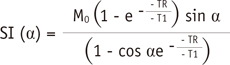
where TR is the repetition time, α is the flip angle, and M0 is the equilibrium longitudinal magnetization. The M0 and T1 parameter were estimated by a linear least-squares method using 5 MR images (5°, 15°, 35°, 60°, and 70°). Following the estimation of M0 and T1 (0) values for the pre-contrast image, post-contrast T1 value (T1 [t]) can be estimated as a function of time from the SI (t) for the post-contrast image with an α = 70°.
We used the extended Kety two-compartment model for quantification of tissue concentration (22), as follows:
(18)
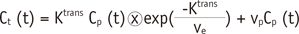
where Ct is the concentration of the contrast agent in the observed tissue, CP is the concentration in blood plasma, vp is the fractional blood plasma volume per unit volume of tissue, Ktrans is the volume transfer constant, and ve is the fractional extravascular extracellular space (EES) per unit volume of tissue. The homemade software was implemented for performance of non-linear fitting of tissue concentration curves using the MATLAB lsqnonlin function.
The arterial input function (AIF) was manually defined at baseline for each mouse using Analysis of Functional NeuroImages (AFNI [http://afni.nimh.nih.gov/afni/)] (23), and used for quantification of DCE-MRI on days 0, 3, and 21. With AFNI software, users move the cursor to the voxel in a concentration image window; the corresponding concentration time curve is then displayed in a time profile window. The AIF voxels were manually selected in a voxel-by-voxel manner by visual inspection of voxel concentration profiles in AFNI software.
Immunohistochemical Analysis for Quantification of Microvessel Density
Formalin-fixed, paraffin-embedded tumor tissues were serially sliced into 5 µm sections corresponding to the magnetic resonance images for quantification of microvessel density (MVD) with CD31 staining by the immunohistochemical method. After heat-induced antigen retrieval for 20 min in citrate buffer (pH 6.0), endogenous peroxidase was blocked with 3% hydrogen peroxide in phosphate-buffered saline (PBS) for 10 min. Non-specific epitopes were blocked with blocking solution (2.5% normal horse serum) for 20 min at room temperature followed by incubation with anti-mouse CD31 (1:150; BD-Pharmingen, Erembodegem, Belgium) antibody diluted in PBS containing 1% bovine serum albumin at room temperature for 30 min. After washing with PBS, the tissue sections were incubated with biotin-conjugated IgG for 30 min, then washed again followed by incubation with horseradish peroxidase (HRP)-conjugated streptavidin for 30 min. The color reaction was developed using the chromogen 3,3-diaminobenzidine (DAB) for 5 min. After washing, the tissue sections were lightly counterstained with Mayer's hematoxylin before dehydration and mounting. MVD was determined by examining the viable tumor area without necrosis in 12 mice (control [n = 6] and treated [n = 6]), as described previously (24). Individual microvessels were counted in 5 random fields per individual tumor section at 200X magnification. Brown-staining endothelial cells that were clearly separate in brown appearance were counted as individual vessels. The results were expressed as an average of the total number of microvessels observed in each individual tumor.
Statistical Analysis
The pharmacokinetic parameters (Ktrans, ve, and vp) were estimated from DCE-MRI data using the pharmacokinetic modeling method before and after treatment with KR-31831 (3 and 21 days). The median and range of pharmacokinetic parameters were examined. The non-parametric statistical analysis (Wilcoxon signed-rank test) was performed to test significant differences in pharmacokinetic parameters before and after treatment with KR-31831 at a p < 0.05.
RESULTS
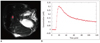
The AIF was manually defined by visual inspection of concentration profiles before treatment (day 0). Figure 1A shows representative images of the AIF near the femoral artery, and Figure 1B demonstrates the concentration profile of the AIF, which was averaged across the manually-defined AIF voxels in Figure 1A.
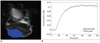
For quantification of DCE-MRI parameters, the regions of interest (ROI) were manually-defined on anatomic T2-weighted images, as shown in Figure 2A. A non-linear fitting method was used to fit the observed concentration profiles of tumor tissues (Ct) and arterial vessels (Cp) into the pharmacokinetic model (the extended Kety two-compartment model). The fitted concentration of tumor tissues is shown in Figure 2B; the results showed that the estimated Ktrans parameter was 0.145, the ve was 0.868, and the vp was 0.0441.
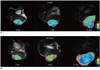
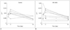
Figure 3 shows the representative pharmacokinetic parametric mapping obtained from the control mice (Fig. 3A) and the KR-31831-treated mice (Fig. 3B) on days 0, 3, and 21 after treatment. To quantify changes in parameters between before and after treatment, the medians of the pharmacokinetic parameters (Ktrans, ve, and vp) were extracted from the tumor region for each subject, and these parameters were compared on days 0, 3, and 21 after treatment for each group (Table 1). Figure 4 demonstrated that there were significant decreasing Ktrans values on day 21 compared to days 0 and 3 in the KR-31831-treated group (p < 0.05), whereas there was no significant difference in the control group (p = 0.84) using the Wilcoxon signed-rank test. There was no significant difference between the control and KR-31831 groups with respect to Ktrans values at baseline (day 0); other parameters (ve, and vp) were not significantly different in the KR-31831-treated and control groups.
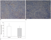
Immunohistochemical analysis for MVD performed on control and KR-31831-treated tumor tissues 21 days after treatment. As shown in Figure 5, tumor microvessels immunohistochemically analyzed with CD31 antibody reactivity in vascular tumor areas without necrosis in corresponding MRI sections were also significantly reduced in KR-31831-treated tumors compared to controls. Quantitative analysis of MVD was performed by examining individual microvessels in five random fields per tumor section under high-field magnification. The MVD was 18.6 ± 5.2 in control mice, while the MVD was 14.9 ± 4.0 and significantly reduced by approximately 20% in KR-31831-treated mice (p < 0.05) (Fig. 5C).
DISCUSSION
Angiogenesis, the recruitment of new blood vessels, is the process by which new blood vessels grow in a variety of physiologic and pathophysiologic states. However, it is still a matter of debate whether or not MVD is a good indicator of the therapeutic efficacy of anti-angiogenic drugs.
Targeted therapy for angiogenesis represents a promising strategy for the development of anti-cancer drugs as several pre-clinical studies have shown that angiogenesis is a key pathway for tumor growth, invasion, and metastasis (3, 6, 10, 25). Furthermore, clinical trials in cancer patients are ongoing with several VEGF inhibitors, including a humanized monoclonal antibody (26) and various small molecules inhibiting signal transduction (9, 27, 28).
A number of endogenous proteins that act as positive regulators or activators of tumor angiogenesis have been identified, including VEGF, basic fibroblast growth factor (bFGF), tumor necrosis factor alpha (TNF-α), angiopoietin-1 and -2, interleukin (IL)-8, and platelet-derived growth factor beta (PDGF-β) (10, 27). One of the more potent endothelial mitogens is VEGF, also known as vascular permeability factor (VPF) because it was initially recognized for its ability to increase microvascular permeability. VEGF is widely distributed and has been shown to play a coordinated role on the vascular endothelium, including endothelial mitogenesis, permeability, vascular tone, the production of vasoactive molecules, and the stimulation of monocyte chemotaxis (6, 13, 26). There are five members of the VEGF family in addition to four members of the angiopoietin family and at least one member of the ephrin family of regulators; the regulators must all work in a complementary and coordinated manner to form functional vessels (5, 25). A large number of anti-angiogenic drugs have been targeted against the angiogenic cytokine, VEGF. This cytokine is a principal mediator of vascular permeability, which has been measured by DCE-MRI and used as a pharmacodynamic end point for the development of these new compounds. Therefore, we designed this study to evaluate the anti-angiogenic efficacy of KR-31831 using DCE-MRI.
KR-31831 is a 4-(N-imidazol-2-ylmethyl) aminobenzopyran analogue originally designed for the treatment of ischemic diseases, such as myocardial infarction and stroke, by a group of our co-workers (16, 17). Benzopran is one of the most frequently used backbones of synthetic drugs, including anti-oxidants, anti-hypertensives, and therapeutic agents for ischemia-related diseases. A variety of amines were introduced at the 4-position of benzopyran for the identification of adenosine triphosphate (ATP)-sensitive potassium channel (KATP) openers targeting ischemic disease, such as myocardial infarction and stroke. Previously, the group of co-workers demonstrated the ability of KR-31831 to inhibit VEGF-induced vascular permeability in in vitro and in vivo experiments (14, 15). Specifically, KR-31831 was shown to exert anti-angiogenic activities as measured by the inhibitory effect on HUVEC tube formation. Additionally, KR-31831 also showed inhibitory effects on VEGF-activated cell proliferation, migration, and invasion.
Dynamic contrast-enhanced MRI involves the acquisition of sequential images during the passage of a contrast agent through a particular tissue of interest. Analysis of DCE-MR images allows the generation of signal intensity versus time graphs. There have been two kinds of methods for quantification of DCE-MRI. Semi-quantitative analysis is based on the characterization of shape of time-to-signal curves which enable measurement of maximum enhancement, peak enhancement, before arrival time, and wash-in and -out slopes (29-31). However, these parameters are not associated with physiologic parameters, and are also not comparable among different MRI scanners. Alternative methods have been developed based on the pharmacokinetic model to estimate physiologic parameters, which have been proposed by Tofts and Kermode (32), Brix et al. (33), and Larsson et al. (34). These different models have been standardized in terms of quantities and symbols based on the assumption that tissue is composed of the extravascular extracellular space (EES) and the plasma space (vp), and the transport between EES and vp (volume transfer constant, Ktrans) (22). In the current study, we used the generalized Tofts model (extended Kety model) to quantify pharmacokinetic parameters form murine data, and our results showed the feasibility of DCE-MRI tools for evaluation of anti-angiogenic efficacy of the newly developed angiogenesis inhibitor, KR-31831.
In conclusion, the effect of KR-31831 on xenografted human ovarian tumor vasculature has been studied using DEC-MRI and a CD31 immunohistochemical method. As expected, a significant reduction in MVD was in agreement with the DCE-MRI finding of a decrease in fractional plasma volume and transendothelial permeability, thus providing a very robust demonstration of KR-31831 efficacy. Our results indicate that KR-31831 may offer opportunities for novel therapeutic strategies in ovarian carcinoma and DCE-MRI may be a useful tool to evaluate the effect of newly developed candidate drugs (such as KR-31831) on angiogenesis.
 XML Download