

 PDF
PDF ePub
ePub Citation
 Print
Print


INTRODUCTION
Spontaneous intracranial hypotension (SIH) is rare, with an estimated annual incidence of five per 100,000. The female : male ratio is about 2:1 and the peak incidence is reached at the fourth decade (1). The pathogenesis remains unclear but is thought to be the result of spinal cerebrospinal fluid (CSF) leaks (1). Although a definite diagnosis is made when CSF opening pressure is less than 60 mm H2O at the lumbar puncture (1, 2), some authors have reported that the CSF leaks can be depicted on a MR myelography with the use of a heavily T2-weighted pulse sequence (3, 4). Clinical manifestations in SIH are widely variable with the most characteristic manifestation being a postural headache, which is defined by the revised International Classification of Headache Disorders criteria as a headache occurring or worsening within 15 minutes while assuming the upright position and gradually improving within 15 to 30 minutes after lying down (1, 5). Other commonly related symptoms include a stiff neck, nausea, vomiting, diplopia, cranial neuropathies, and marked exacerbation of the headache by Valsalva maneuver (1, 5). There are multiple available treatment options which include bed rest, analgesics, sedatives, oral caffeine, intravenous hydration, epidural blood patch (EBP), epidural saline infusion, and direct surgical repair of spinal dura tear (1, 6). Among them, the mainstay of treatment is EBP, a procedure to secure a CSF leak that in turn restores brain buoyancy (1). Surgical repair of a dura tear is reserved for a patient with an identifiable site for a CSF leak that is refractory to EBP (1, 4, 7). The majority of patients with SIH have favorable outcomes and the resolution of symptoms may be achieved in a period of weeks and months to years after treatments (1, 8-11). The advent of MRI has revolutionized the understanding of SIH. Typical MR imaging features include subdural collections, dural thickening and pachymeningeal enhancement, engorged venous structures, pituitary hyperemia, and sagging of the brain (1, 12). The superior ophthalmic vein (SOV) drains into the cavernous sinus and is theoretically influenced by the alteration in intracranial pressure. However, the relationship between the SIH and the SOV diameter, before and after EBP, was rarely investigated. Our case demonstrates a striking regression of the SOV diameter three days after an EBP. The "reversal of the SOV" sign is introduced, which implies the reconstruction of intracranial pressure and is also highly suggestive of a successful EBP procedure.
CASE REPORT
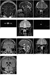
A 65-year-old man began to suffer from an intermittent right fronto-parietal headache and right neck soreness seven months prior to admission. The headache, as he described, was not related to his posture. He sought medical help because of the development of an orthostatic headache a few days prior to admission. Brain and spinal MRI studies were performed. The brain MRI revealed dural thickening with pachymeningeal enhancement, subdural effusion, pituitary hyperemia, and a slight brain sag (Fig. 1A, B), which are typical findings of SIH. In addition, bilateral ophthalmic veins were significantly engorged (Fig. 1C). MR myelography, using the heavily T2-weighted pulse sequence, revealed CSF leaks in the thoracic epidural space from the T1 to T6 levels (Fig. 1D, T4 level illustrated). An EBP was performed the next day by injection of 30 c.c. of autologous blood into the T6 epidural space. The patient was kept in the Trendelenburg position for two hours after the procedure to allow for the ascension of blood over several segments towards the sites of CSF leaks. However, he did not experience instantaneous relief of the postural headache. The first follow-up MRI was performed on the third day after the EBP, which showed the complete seal of spinal CSF leaks and a striking reversal of the bilateral SOV diameters (Fig. 1E, F). The second follow-up MRI on the eleventh day after EBP failed to demonstrate any resorption of bilateral subdural collections or regression of dural venous engorgement (Fig. 1G, H). The diameters of the bilateral SOVs were stable (Fig. 1I). The postural headache persisted, but subsided dramatically about one and a half months later. Serial follow-up brain and spinal MRI studies at one-month intervals in the following four months showed gradual resolution of the subdural collections (Fig. 1J) without recurrent spinal CSF leak. Of note, the diameters of the bilateral SOVs were stable without further engorgement.
DISCUSSION
The clinical manifestation, typical MRI features, and treatment options in SIH have been well described in the literature (1-12). In this article, we introduce the "reversal of the SOV" sign following the EBP procedure, which we believe may imply the reconstruction of intracranial pressure.
According to the Monroe-Kellie hypothesis, the sum of the volumes of intracranial blood, CSF, and cerebral tissue should be constant in an intact cranium (13). Both the clinical symptoms and MRI features in SIH can be the result of a spinal CSF leak. The postural headache can be explained by CSF volume depletion with loss of CSF buoyancy, which in turns leads to brain sagging and traction of various intracranial pain-sensitive structures (1). For pachymeningeal enhancement, engorgement of the venous structures and pituitary hyperemia could be the results of compensatory effects for increasing the vascular component (1). As for the subdural collection, it is intended for increasing the intracranial CSF component (1).
The majority of CSF is produced by the choroid plexuses in the brain. Circulating over the brain and spinal cord, the CSF provides the supporting buoyancy for the central nervous system. The estimated CSF space is about 160 to 170 mL in a young adult and CSF is produced at a rate of 0.4 mL/min (14). As a result, the turnover rate is about four times a day. Based on this inference, the CSF volume should become normal within one day if the CSF leak has been secured by a successful EBP.
Dural venous engorgement is one of the imaging features in SIH. In one series, the authors observed that 14 of 15 (93%) patients with intracranial hypotension were associated with engorgement of the transverse sinus (15). Since the cavernous sinuses are not completely isolated from the dura matter, they are easily influenced by alteration in intracranial hydrodynamics (16). The SOV, which serves as one of the tributaries of the cavernous sinus and brings the venous flow from the orbit back to the cavernous sinus, is theoretically also influenced by alteration in intracranial hydrodynamics. As can been seen in our case, the diameters of bilateral SOVs had already returned to a normal status before the evolution of other imaging features were perceptible. Therefore, the diameter of SOV may indirectly reflect the status of intracranial pressure.
However, the relationship between the diameter of SOV and the intracranial pressure has been rarely investigated. In one series, the authors measured the average diameter of SOV on MRI and found that it was smaller in SIH where the term, "collapsed" SOV was used to describe this feature (17). In another study, the authors, by color Doppler imaging, found both the mean diameter and the flow of the SOV were substantially greater in SIH (18).
We cannot explain the discrepancy in the aforementioned studies, but our case supports that an engorged SOV might be one of the features of SIH, a mechanism such as an engorged dural vein in SIH. Nevertheless, it remains unclear whether the "reversal of the SOV" in our case definitely implies a complete seal of the CSF leak and restoration of intracranial pressure or whether the finding was incidental. Therefore, further case reports or studies are encouraged to further delve into this phenomenon. It is recommended that Color Doppler imaging be performed to access the diameter of the SOV, because it can be done at bed side without the need for sending the patient to an MRI room, and thus helps to prevent recurrent CSF leaks.
In summary, a high index of clinical suspicion and familiarity with MRI features are essential for a diagnosis of SIH to be made with confidence. Heavily T2-weighted MR myelography can benefit from its non-invasiveness. It is thus worth recommendation, not only for localization of CSF leak, but also for evaluation of post-treatment efficacy. The "reversal of the SOV" sign may provide adjunctive information for knowledge of post-treatment intracranial hemodynamic change. However, the application of "reversal of the SOV" sign to patients with SIH, in whom the SOVs are collapsed, is uncertain and needs substantially greater investigation.
 XML Download