

 PDF
PDF ePub
ePub Citation
 Print
Print


Dear Editor:
We read with great interest the article by Algin et al. (1) "Role of duplex power Doppler ultrasound in differentiation between malignant and benign thyroid nodules", in the December 2010 issue of the Korean Journal of Radiology. The authors concluded that the vascularity pattern was not useful to distinguish malignant from benign thyroid nodules.
In this article, it is worthy to note that the cytology examinations were dichotomized between the benign and malignant samples. May we suggest that the use of the cytologic Bethesda system 2008 classification (2) would probably have shown some correlations between one or more of each of the Bethesda classification categories and the pattern of thyroid nodule vascularity. As the management of thyroid nodules is currently based upon the Bethesda classification, highlighting such a correlation would probably have been useful and more up-to-date for nodule selection.
Furthermore, as the decision to perform fine-needle aspiration biopsy (FNAB) or surgery for thyroid nodules is made based on the presence of at least one malignant criteria on B-mode ultrasonography (US) (e.g., microcalcifications, irregular margins, hypoechogenicity, etc.), a Doppler US pattern analysis would also have been useful for the nodules that showed only benign features. We do emphasize that some Doppler US features in this subgroup may have correlation either with malignancy or a category of the 2008 Bethesda classification.
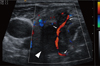
Another point to consider is that the authors' analysis was restricted to the distribution of vascularization within the thyroid nodule. We stress that quantitative analysis of the nodule vascularization could be helpful to differentiate benign from malignant nodules (3). Furthermore, two other true qualitative Doppler US criteria that reflect angiogenesis may strongly suggest malignancy. The first is anarchic intralesional vascularization with winding vessels, and the second is the presence of a peripheral large afferent (penetrating) vessel (4) (Fig. 1). As a matter of fact, penetrating "sword-like" vessels seen on Doppler US may be present in poor differentiated papillary or anaplasic thyroid nodules at an early stage, and this can lead to a diagnosis of early-stage malignancy and the subsequent optimizing of these patients' therapeutic management.
 XML Download