

 PDF
PDF ePub
ePub Citation
 Print
Print


INTRODUCTION
Although there is debate about superior vena cava (SVC) filtration in patients with upper extremity deep venous thrombosis (DVT), a percutaneous SVC filter insertion is now regarded as safe, feasible, and effective in preventing symptomatic pulmonary embolisms due to upper extremity DVT in patients in whom anticoagulation or thrombolysis has failed or is contraindicated (1-3). Additionally, a retrievable SVC filter can be used as a temporary and effective barrier against a symptomatic pulmonary embolism in settings where the risk is transient (e.g., after trauma or during pregnancy). Such a device is also useful during aggressive, catheter-directed thrombolysis or mechanical thrombectomy, which can induce thrombus migration to the pulmonary artery (4).
The Tempofilter II (B. Braun, Melsungen, Germany) is a kind of temporary caval filter. The cone-shaped filter unit is deployed and retrieved using a tethered catheter. The filter unit has eight smooth legs and no hooks. The filter cone is held in place in the vena cava by a tethering catheter with a subcutaneous anchoring device. Most often, the Tempofilter II is used in the inferior vena cava (IVC) for protection against a pulmonary embolism due to lower extremity DVT (5). To our knowledge, there are no printed reports concerning the placement of the Tempofilter II in the SVC to prevent a pulmonary embolism in patients with upper extremity DVT. In this article, we present a case of SVC filtering using the Tempofilter II in patients with upper extremity DVT.
CASE REPORT
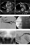
A 67-year-old woman was brought to the emergency unit after a traffic accident. She suffered multiple traumatic injuries including liver laceration, left femoral shaft fracture, pelvic bone fractures, and multiple rib fractures. Roughly three weeks later, she developed dyspnea and swelling of the left upper extremity. A contrast-enhanced chest computed tomography (CT) revealed a pulmonary embolism in the right pulmonary artery and thrombosis in the left brachiocephalic vein (Fig. 1A, B). On a lower extremity CT angiography and Doppler ultrasound taken after the diagnosis of pulmonary thromboembolism, a lower extremity DVT was not detected. A left upper extremity venogram revealed complete obstruction of the brachiocephalic vein with lack of collaterals, implying the development of acute upper extremity DVT (Fig. 1C).
To prevent the recurrence of a pulmonary embolism from the left upper extremity venous thrombi, and to reduce her left arm swelling, removal of venous thrombi was necessary. Because anticoagulation and thrombolysis were contraindicated due to her recent major trauma history, we decided to perform a thrombus aspiration. To prevent a further possible pulmonary embolism during the procedure, we planned temporary filtration in SVC.
One day following the diagnosis of upper extremity DVT and pulmonary thromboembolism, the Tempofilter II was placed in the SVC percutaneously, via the right femoral vein. After a right femoral vein puncture, a guide wire was threaded through the IVC, right atrium, and up into the SVC. The subcutaneous puncture site was enlarged by about 10 mm with an incision and an approximately 20-mm-sized subcutaneous pocket was created by tissue dissection. Next, the introducer system (dilator and sheath) was inserted along the guide wire into the SVC under fluoroscopic guidance. After the dilator and guide wire were removed, iodinate contrast agent was injected to obtain a superior vena cavogram to clarify the exact location of the thrombus and to confirm the optimal landing zone for a filter in the SVC. Next, a filter unit was introduced into the SVC via the sheath. By pushing and pulling the tethered catheter, the filter was deployed correctly in the supra-azygos SVC. After checking the position of the filter, the anchoring device was attached to the tethering catheter just around right femoral vein, and the excess length of the catheter was sectioned immediately above the olive shaped button. After burying the anchoring device into the subcutaneous pocket, the incision was sutured (Fig. 1D, E).
After placement of the Tempofilter II in the SVC, thrombus aspiration was performed through left basilic vein with a 100 cm length 8-Fr guiding catheter (Guider SofTip; Boston Scientific, Natick, MA). Thrombus aspiration via a catheter was performed with a 20 cc syringe. During the thrombus aspiration, anticoagulation or thrombolysis was not performed. A follow-up venography performed after thrombus aspiration showed recanalization of the left brachiocephalic vein, regression of collateral veins (Fig. 1F), and didn't show any thrombus capture within the unit. A CT angiography obtained two weeks after Tempofilter II placement demonstrated a patent SVC with no thrombus around the filter, as well as resolution of the pulmonary embolism in the right pulmonary artery. Moreover, no additional anticoagulation or thrombolysis was noted. Just one day after a follow up CT angiography, we successfully removed the filter without complication. Retrieval of the filter was achieved by making a skin incision around the palpable anchoring device under local anesthesia. The tethered catheter and filter were removed by simply pulling the tethered catheter outward.
DISCUSSION
The unique design of the Tempofilter II consisting of a long tethering catheter and filter device with no hook is not seen with other filters and because of this, has many advantages. First, the Tempofilter II is a retrievable, temporary filter and this characteristic could make it an effective SVC filter. Other advantages of the Tempofilter II include a long tethering catheter, which makes filter retrieval easy and rapid due to the simple traction on the tethering catheter.
Yet another advantage is that the Tempofilter II is less likely to tilt. Its unique design, which has been mentioned in the above paragraph, makes it possible that the filter possesses a more vertical orientation within the SVC (the tethered catheter, which is fixed around inguinal area, acts like a pendulum). The vertical axis and the lack of tilting of the filter within the SVC may increase the ability of the filter to capture clots (6).
Additionally, the Tempofilter II has a decreased incidence of vena cava injury compared to other filter types, because it does not have any hooks on the filter strut. There have been several reports about complications related to hooked temporary SVC filters, such as SVC perforation, cardiac tamponade, and aortic injuries (7-9).
Finally, the Tempofilter II allows for easier optimal filter placement. The tethered catheter enables the fine correction of filter position by allowing for the movement of the filter to-and-fro within the SVC. Usually, placement of a filter at the optimal level in the SVC is more technically difficult than placement in the IVC, due to the shorter length of the optimal landing zone in the SVC (2).
The use of the Tempofilter II in the SVC also has several potential limitations or drawbacks. First, the Tempofilter II cannot be used permanently; it has been validated for indwelling times of up to six weeks, although the manufacturer states that retrieval is feasible and should require no additional equipment, up to three months after placement (5). Therefore, if patients need a permanent SVC filter, the Tempofilter II must be replaced with another type of filter device. Second, the Tempofilter II should be used in only bed-ridden patients. In mobile patients, the tethering catheter can bend or buckle due to the effect of gravity and the patient's movement. Furthermore, while the lack of hooks on the struts has benefits, this also makes the Tempofilter II filter more prone to migration within the relatively short SVC. In cases where the Tempofilter II was used in the IVC, there have been reports of intracardiac migration inducing fatal outcomes (5, 10).
A third disadvantage is that the use of the Tempofilter II is also limited by the length of the introducer sheath and the tethered catheter, making it impossible to use the Tempofilter II in tall patients. When the Tempofilter II is inserted into the SVC, the tethered catheter is introduced through the common femoral vein and travels through the iliac vein, IVC, and SVC. As a result, the length of tethered catheter remaining outside the body may be insufficient to withdraw the introducer sheath up to length of 70 cm. However, if the manufacturer were to provide a longer tethered catheter, this limit could be easily overcome.
In conclusion, temporary filters should meet two important requirements. First, they must be effective for several weeks to several months. Second, they must be removable from any amount of organized and adherent thrombus after anticoagulation therapy has been completed and the indication for caval filtration has ceased (5). Based on these requirements and despite its drawbacks and limitations, the Tempofilter II may be one of the good options for the temporary filtration of the SVC to prevent pulmonary embolisms in bed-ridden patients with upper extremity DVTs.
 XML Download