

 PDF
PDF ePub
ePub Citation
 Print
Print


INTRODUCTION
Primary cardiac tumors have a reported incidence of 0.002-0.3% in autopsy series. Cardiac osteosarcomas are very rare, accounting for less than 10% of primary malignant cardiac tumors (1, 2). An osteosarcoma is a malignant osteoid or bone producing tumor. Most reports in the radiology literature describe the characteristic CT imaging findings of osteosarcomas as mineralized lesions (3). However, its MRI findings have not been described in detail. Herein, we present a case of a 47-year-old woman with primary cardiac osteosarcoma which metastasized to the femur, 18 months after tumor resection and we describe MRI findings of the primary cardiac tumor and its bone metastasis with histopathologic correlation.
CASE REPORT
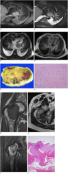
A 47-year-old woman was admitted to the hospital with gradually worsening dyspnea, orthopnea, and nocturnal paroxysmal dyspnea. A non-contrast chest CT (Mx 8000 CT scanner, Philips) revealed bilateral pulmonary congestion and pleural effusion with left atrial enlargement and a low-attenuated mass in the left atrium (LA). Neither distinct calcification within the mass nor mediastinal lymph node enlargement was noted. MR images (Intera 1.5T, Philips) demonstrated a 46 × 41 mm, well defined mass with a broad base at the posterolateral wall of the LA. The mass showed mildly heterogeneous and high signal intensity (SI) on double inversion-recovery T1-weighted images (T1WI), with fat saturation (Fig. 1A). In addition, triple inversion recovery T2-weighted images (T2WI) with fat saturation and cine-cardiac images showed heterogeneously high SI (Fig. 1B, C). On delayed contrast-enhancement MR images, the mass was heterogeneously enhanced (Fig. 1D).
The patient underwent surgical removal of the mass, which was found to be smooth, well-encapsulated with focal necrosis and hemorrhage (Fig. 1E). Histologically, the tumor was mainly composed of atypical spindle cells with high nuclear density and frequent mitosis in the background of abundant collagen and had foci of hemorrhage and coagulative necrosis. In addition, the presence of minimal osteoid deposition foci confirmed the tumor as a fibroblastic osteosarcoma (Fig. 1F). At that time, a bone scan was performed to identify any primary bone lesion or bone metastasis. The result was negative and, therefore, osteosarcoma of bone-origin and the presence of bone metastasis were ruled out. The patient was further treated with adjuvant chemotherapy and radiotherapy. Periodic check-ups with chest CT revealed no evidence of regional recurrence.
Eighteen months after surgery, the patient complained of right hip pain and consequently underwent a hip MRI. The examination revealed a mass at the right femoral neck. The lesion was isointense to muscle on T1WI (Fig. 1G) and showed a heterogeneously high SI on T2WI (Fig. 1H). The mass was well-demarcated, with a peripheral low SI rim on both T1WI and T2WI. Gadolinium-enhanced T1WI showed heterogeneous enhancement with central non-enhancing portions (Fig. 1I). A bone scan using technetium-99m (Vertex™ V60, Philips, Best, The Netherlands) was performed to evaluate the presence of other bone lesions, and showed an increased uptake at the known lesion in the right femur and no other pathologic uptake. A biopsy and following histological analysis confirmed the lesions as metastases from the previously resected cardiac tumor on the basis of histologic similarity between them (Fig. 1J).
DISCUSSION
Most cardiac tumors are metastatic tumors which are 20-40 times more common than primary tumors. About 75% of all primary cardiac tumors are benign and the remaining 25% being primary malignant tumors. The vast majority (95%) of primary malignant tumors are sarcomas and osteosarcomas, which are relatively quite rare, accounting for less than 10% primary cardiac malignant tumors (1, 2). Primary cardiac osteosarcomas exhibit a predilection for the LA, whereas the majority of malignant cardiac tumors, such as metastatic tumors, angiosarcomas or lymphomas, commonly arise in the right atrium (4). Due to its left atrial location, patients with cardiac osteosarcomas generally present as congestive heart failure with respiratory symptoms as in our case (5). Moreover, they tend to have a broad base of attachment, at a location away from the fossa ovalis and invading the surrounding structures, such as the mitral valve or pulmonary veins (2).
Osteosarcomas are a heterogeneous group of tumors containing malignant, bone-producing cells. Histologically, the tumor contains variable amounts of spindle-cells, osteoid, bone, or cartilage. Depending on the predominant component, osteosarcomas can be subgrouped as osteoblastic, chondroblastic, or fibroblastic (3). Macroscopically, the tumor may be well circumscribed and pseudoencapsulated, or infiltrate into the surrounding tissues and calcification, necrosis, or hemorrhage within the tumor can be seen (5, 6). The characteristic CT findings of cardiac osteosarcomas have been reported as a low attenuation mass with dense mineralization (3, 7). Moreover, osteosarcomas often have dense calcification to form hardy stone masses or may also have minimal calcification in the early stage. However, a tumor lacking the identifiable calcification on CT presents as a nonspecific soft tissue mass as it did in our case, which made us miss the osteosarcoma as part of our differential diagnosis. As the microscopic examination showed, the tumor had too minimal a portion of osteoid deposition to reveal calcification on CT.
The first report of MRI findings of cardiac osteosarcomas was presented in 2001 by Yamagishi et al. (8) in which the tumor appeared as a huge mass of heterogeneous SI in the LA. The report focused on the role of MRI in differentiating malignant from benign tumors based on a broad-based attachment and invasive features. Since then, a few cases have been reported with MR images, however, with no detailed description of MRI findings and no contrast-enhancement study included. According to previous reports, cardiac osteosarcomas appear to be irregularly lobulated and may have variable heterogeneous SI on T1WI and heterogeneously high SI on T2WI (2, 4, 6, 7, 9). In our case, the tumor showed mildly heterogeneous and high SI on both T1WI and T2WI. In addition, the heterogeneity of the signal on T2WI was more definite on cine images with long echo time (Fig. 1C). This heterogeneity is attributed to the heterogeneous histologic composition since some portion of the tumor was composed of spindle cells with collagen material, while the other portion consisted of necrosis and hemorrhage. It is known that hypercellular areas are well-enhanced after gadolinium administration and show high SI on T2WI, whereas low SI on T2WI corresponds to hypocellular areas (10). Our case showed heterogeneous enhancement and the areas of enhancement are well-matched to the areas of high SI on T2WI, suggesting hypercellular areas. As discussed earlier, the tumor showed no calcification which would have shown low SI on all pulse sequences, due to minimal osteoid formation discernable only on histologic examination.
Primary cardiac osteosarcomas are very aggressive with a high frequency of recurrence and metastasis. Metastasis was observed at various sites including skin, lung, liver, bones, brain and adrenal glands (5). Our case also revealed metastasis at the femur, although the patient underwent chemotherapy and radiotherapy after surgical removal of the tumor. A comparison of the metastatic bone lesion with the cardiac lesion indicated less heterogeneity and denser enhancement with the lesser portion of non-enhancement which meant lesser extent of central necrosis in the metastatic bone lesion. The peripheral rim of low SI on both T1WI and T2WI showed enhancement on gadolinium-enhanced images. This finding is considered to correspond to a fibrous pseudocapsule, which has been reported in some extraosseous osteosarcomas (10).
We present a case of primary cardiac osteosarcoma with a detailed description of MRI findings and histopathologic correlation of cardiac osteosarcoma and its bone metastasis.
 XML Download