

 PDF
PDF ePub
ePub Citation
 Print
Print


INTRODUCTION
Self-expandable metallic stent placement is now widely used to treat acute malignant colorectal obstruction. It can serve as a bridge to surgery, and for allowing preoperative decompression and cleansing of the colon. Further, it can be used for palliation in patients who are not candidates for surgery (1-4).
Self-expandable stent placement has been performed under fluoroscopic or endoscopic guidance or both. Interventional radiologists usually perform stent placement under fluoroscopy-guidance and they are much more experienced in using angiographic catheters, passing guide wires through obstructive lesions and deploying self-expandable metallic stents (5-7). However, lesions located at or more proximal to the descending colon are more technically challenging because of the tortuous, curved angulations of the sigmoid and descending colon. Furthermore, the angiographic catheter is short (8). Therefore, even if a guide wire can reach the obstructive colonic lesion proximal to the descending colon, the tortuous angulations of the descending colon and the short catheter length may make it difficult for an angiographic catheter to reach a given lesion over a guide wire. Such a catheter may only prolapse into the greater curvature of the tortuous left colon. In fact, some reports have suggested that stent placement is difficult to perform in the descending colon and splenic flexure compared to that of the rectum and sigmoid colon (5, 9, 10).
Therefore, to overcome the limitations of placing angiographic catheters proximal to the descending colon, we inserted a 6-Fr stiff, long introducer sheath into the sigmoid and descending colon to prevent prolapse of the catheter and guide wire into the greater curvature of the tortuous sigmoid colon. We then advanced the catheter over the guide wire to the lesion proximal to the descending colon. The purpose of this study was to evaluate the efficacy of the coaxial technique using a stiff, long sheath to obtain easy advancement of the angiographic catheter over malignant obstructive colonic lesions.
MATERIALS AND METHODS
Patients
Between August 2005 and September 2010, we attempted to place fluoroscopy-guided self-expandable metallic stents in 77 consecutive patients who had symptoms of subtotal obstruction (bowel distension, difficulty passing solid stool, the presence of narrow caliber stools or ability to pass only small amounts of liquid stool or gas) or total obstruction (nausea, vomiting, abdominal distension, decreased or absent bowel sounds or the inability to pass any stool or gas) (1).
All the patients underwent plain abdominal radiography and contrast-enhanced CT scanning, which showed acute colorectal obstruction. The obstruction sites included the rectum in eight patients, the rectosigmoid colon in 31 patients, the sigmoid colon in 22 patients, the descending colon in five patients, the splenic flexure in five patients and the transverse colon in six patients. We excluded those patients whose obstruction sites were located proximal to the hepatic flexure. The cause of obstruction was primary colorectal carcinoma in 75 patients, advanced gastric cancer in one patient and pancreatic cancer in one patient. This retrospective study was approved by our institutional review board.
Procedure
After written informed consent was obtained, each patient was brought to the fluoroscopy suite and placed in the lateral decubitus position. Analgesia (pethidine 25 mg; Je Il Pharm, Daegu, Korea) was administered just before the procedure. There was no routine antibiotic or sedative administration.
An enema tube was introduced into the rectum. A small amount of nonionic contrast media (Pamiray; Dongkook Pharm, Jincheon, Korea) was injected through the tube to outline the stricture. Under fluoroscopy guidance, a 0.035-inch hydrophilic guide wire (Terumo, Tokyo, Japan) and a 135 cm, 5-Fr angiographic catheter (HN1; Jung Sung Medical, Gyeonggi-do, Korea) were then inserted coaxially into the anus. After the angiographic catheter was then advanced over the guide wire beyond the obstructive lesion, the guide wire was used to negotiate over the obstructive lesion. The angiographic catheter was then passed over the obstruction and the guide wire was removed. The length and position of the obstruction were determined by injecting contrast media prior to stent placement. A 0.035-inch stiff guide wire (Amplatz Superstiff; Lunderquist Superstiff) was then introduced so the stent introducer could be easily advanced into the obstructive lesion.
Stent (Taewoong Medical Co., Gimpo, Korea) placement was performed through the stiff guide wire under fluoroscopy guidance. The length of the metallic stent was determined so at least four additional centimeters (2 cm extra-long on each side of the proximal and distal portions of the actual stricture) would be provided in addition to the actual stricture length for covering the entire stricture.
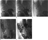
However, if the angiographic catheter could not be advanced over the guide wire into the obstructive lesion because the lesion was too far from the anus or the angiographic catheter had prolapsed into the greater curvature of the tortuous sigmoid colon (Fig. 1), then the angiographic catheter was removed, and a 6-Fr shuttle introducer sheath (Cook, Bloomington, IN) was inserted over the guide wire in place of the angiographic catheter. This prevented prolapse of the catheter into the greater curvature of the tortuous sigmoid colon during advancement of the catheter over the guide wire. The angiographic catheter was then reinserted into the shuttle sheath over the guide wire coaxially, in attempt to negotiate over the obstructive lesions (Fig. 1).
If fluoroscopic-guided negotiation of the angiographic catheter or guide wire failed regardless of the use of the shuttle introducer sheath, then combined endoscopic and fluoroscopic guidance was attempted. If this combined guidance also failed, then the patient was referred for surgery.
Assessment
Technical success was defined as the ability to pass the angiographic catheter over the guide wire and past the obstructive lesion with or without the support of the shuttle sheath, as well as successful stent coverage of the obstructive site. Clinical success was defined as improvement of the obstructive symptoms, and this was indicated by restored gas and stool passage. The technical success rate, the clinical success rate and complications were analyzed.
RESULTS
Successful stent placement was achieved in 75 of 77 patients (97%). The stent used in all the patients was 22 mm and 24 mm in diameter and 6-12 cm in length. The median follow-up was eight months (range: 3-13 months). The procedure could not be completed in two patients. Both of these patients had lesions in the sigmoid colon. One patient had failed with negotiation of a guide wire over the obstruction in the sigmoid colon even with fluoroscopy and endoscopy guidance. Because the obstructive lesion was close to the anus, the shuttle sheath was not used in this case. An emergency laparotomy was scheduled for the patient, but he refused the surgery. The other patient had a perforation of the sigmoid colon caused by the guide wire and catheter during the negotiation, and the perforation was diagnosed based on peritoneal spillage after the injection of contrast media through the catheter. Thereafter, the patient died of sepsis caused by the colon perforation and generalized weakness.
Eleven patients (M:F = 7:4; mean age, 65.5 years; range, 37-83 years) who had malignant colonic obstruction at the level of the splenic flexure or transverse colon failed advancement of an angiographic catheter to the obstructive lesion because of the tortuosity of the sigmoid and descending colon and the short length of the catheter. Hence, the coaxial technique using a 6-Fr shuttle introducer sheath and an angiographic catheter was implemented in all of these 11 patients. The 6-Fr shuttle introducer sheath was successfully located around the obstructive lesions in all of these patients. No patients required additional pain control during the manipulation of these sheaths. The angiographic catheter inserted through the sheath was able to successfully reach the obstructive lesion in all the patients. Stent placement was successfully performed in all the patients after the guide wire and the angiographic catheter passed over the obstructive lesion (Fig. 2).
Relief of clinical bowel obstructive symptoms (indicating clinical success) was obtained within 24 hours in all 75 patients, including the patients in whom the shuttle sheath was used.
There were no the shuttle sheath-related complications such as perforation or bleeding. Stent migration occurred in two patients (3%). One patient had transverse colon invasion by gastric cancer and the patient was treated using a shuttle sheath. The other patient had primary rectosigmoid colon cancer. Each of these patients underwent 22 mm diameter stent placement. Larger replacement stents (24 × 100 mm) were successfully implanted using same method, and clinical success was achieved.
One patient had stent reocclusion secondary to tumor ingrowth during the follow-up. This occurred in the 10 months following stenting, and the obstruction was relieved by placement of a second stent.
DISCUSSION
Self-expandable metallic stent placement for colorectal obstruction has been performed in the past using radiological or endoscopic guidance, or a combination of both (5). de Gregorio et al. (11) have reported that the guide wire cannot be advanced under fluoroscopic guidance, but it can reach the neoplastic stricture under endoscopic guidance. However, interventional radiologists are much more experienced in passing the guide wires through obstructive lesions and deploying metallic stents (6, 7). Therefore, Fan et al. (5) have reported that fluoroscopy in combination with endoscopy improves the rate of successful self-expandable metallic stent deployment. However, even in this study, which used endoscopy for additional guidance, stent implantation was still rather difficult in the descending colon and splenic flexure compared to that in the rectum and sigmoid colon. Even though fluoroscopy-guided self-expandable metallic stent placement proximal to the descending colon is much more technically difficult than is the endoscopy-guided or combination-guided self-expandable metallic stent placement, many interventional radiologists still implant stents at the obstruction site using fluoroscopic guidance because additional endoscopy guidance can be painful and it is not always feasible. Kim et al. (8) introduced a multifunctional gastrointestinal coil catheter to obtain the easy access to the colonic obstruction proximal to the descending colon and to measure the stricture length without removal of the guide wire. Technically successful stent placement was obtained in 93 of 98 patients (95%). However, in patients with near complete or complete obstruction of the colorectal tract, the clinician may experience difficulty introducing the tip of the multifunctional gastrointestinal coil catheter through the stricture, and this is largely due to the tip's flatness. In fact, they encountered technical failure in five patients. Two of the five patients with technical failure had obstructive lesions proximal to the descending colon. We used the shuttle sheath only to uphold the angiographic catheter in the sigmoid and descending colon. Negotiation over the obstruction was performed using the angiographic catheter and guide wire instead of the multifunctional coil catheter. Hence, we had a 100% technical success rate of stent placement in the transverse colon and splenic flexure.
Kim et al. (12) placed an 8-Fr guiding sheath (Balkin sheath; Cook, Bloomington, IN) in cases of obstruction proximal to the splenic flexure in order to facilitate easier selection of the obstructing lesion. This guiding sheath kept the sigmoid colon straight and it helped to select the obstructing core by enhancing the torque manipulation of the 5-Fr catheter and the 0.035-inch guide wire; the usefulness of this method was very similar to that of the shuttle sheath used in our study. However, the Balkin sheath is larger than the shuttle sheath (8-Fr versus 6-Fr) is and it may make the patient uncomfortable during the procedure. It is also too short to overcome the long, tortuous, curved angulation of the sigmoid and descending colon when compared with the shuttle sheath, which is longer (45 cm versus 80 cm, respectively). Shuttle introducer sheaths are intended to introduce balloon catheters, closed or non-tapered end catheters, and other catheters. The sheath system is manufactured with a stainless steel wire embedded in the sheath material and a high flex polyurethane dilator. The stiff shaft material provides torque control and curve support for the angiographic catheter inserted into the sheath. This sheath has a radiopaque band incorporated into the tip, as well as a soft, a traumatic tip. Therefore, it is usually implemented as a long introducer sheath during neurointerventional procedures such as carotid arterial stenting. To the best of our knowledge, no published reports have discussed the efficacy of the shuttle sheaths in facilitating stent placement in patients with malignant colonic obstruction.
Our study has some limitations. First, the patients with lesions proximal to the transverse colon were excluded due to poor site accessibility. However, the incidence of successful stent placement for colonic obstruction proximal to the transverse colon is low. A review of a colonic stenting series covering a 10-year period showed that only five of 310 patients had stents placed proximal to the descending colon (13). Another study of 1349 patients showed that only five stents were sited in the right colon, with the failure rates increasing according to the more proximal the stenting attempts (6% rectosigmoid versus 15% descending colon versus 15% proximal to the descending colon) (14). This is due to the fact that right colonic obstruction is typically managed differently than left colonic obstruction is. Right-sided lesions can be managed through a one-stage operation with resection and ileocolonic anastomosis, without the need for formal bowel preparation. Therefore, stents may be used as a bridge to surgery in cases where emergency surgery is considered too risky or when the disease is very advanced and palliation is needed. In fact, the majority of the reported cases of colonic stenting have involved the distal colon (1). Our study was also limited in that the number of cases was not large, although symptomatic colonic obstruction proximal to the descending colon is not common. Furthermore, the shuttle sheath was not invented for colonic stent placement and it is a more expensive alternative means of therapy.
Despite these limitations, fluoroscopy-guided self-expandable metallic stent placement was effective and useful for relieving the obstructive symptoms, and even in patients with colonic obstruction proximal to the descending colon. In particular, the coaxial technique using a shuttle sheath and angiographic catheter can increase the technical success rate of fluoroscopy-guided self-expandable metallic stent placement in patient with colonic obstruction proximal to the descending colon.

 XML Download