

 PDF
PDF ePub
ePub Citation
 Print
Print


Mammary hamartomas are localized overgrowths of fibrous, epithelial, and fatty elements which occur primarily in adults during the reproductive years (1). Mammary hamartomas are widely known to have no special propensity to undergo malignant transformation, and are not a marker for increased relative risk for breast cancer development (1); however, some investigators have suggested that malignant transformation of mammary hamartomas is possible, given the presence of glandular tissue in this type of lesion (2-4). In fact, 14 cases of carcinomas associated with mammary hamartomas have been previously documented in the literature (2-12). In this report, we review the literature and describe a new case of invasive ductal carcinoma (IDC) arising in a mammary hamartoma that was detected while the patient was being followed for a known hamartoma.
CASE REPORT
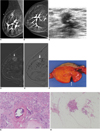
A 72-year-old woman presented for further examination of a right breast lump. According to the patient, the lump had been present for at least 10 years. She complained of discomfort of recent onset in the right breast. On physical examination, a soft, mobile, 10 cm mass, which occupied nearly the entire right breast, was palpated. On mammography, a large circumscribed mass surrounded by a water-density capsule was shown in the right breast (Fig. 1A, B). The mass was a mixture of isodense and fat densities and had dystrophic calcifications in a branching pattern. The mammograms were initially interpreted as a benign hamartomatous lesion (Fig. 1A, B). On ultrasonography (US), the mass was very heterogeneous and completely encompassed in a thin echogenic pseuduocapsule of compressed breast tissue, which was compatible with hamartoma. However, a careful US examination revealed an irregular hypoechoic mass of 1.4 cm with a non-parallel orientation within the hamartoma (Fig. 1C). A retrospective review of the mammograms revealed focal asymmetry, which correlated with the suspicious mass on US (Fig. 1A, B). The lesion was early enhanced and a washout on enhanced MRI scans (Fig. 1D, E), thus it was thought to be a malignancy. Surgical excision for entire mass was done and the diagnosis from the frozen specimen of the suspicious area was IDC.
The patient therefore underwent a modified radical mastectomy. The microscopic examination showed an IDC arising in a hamartoma (Fig. 1F-H). There was no IDC which was outside the hamartoma. There was no intraductal component and there was no axillary lymph node metastasis. The remaining palpable mass was hamartoma with dystrophic calcifications. The patient subsequently underwent adjuvant chemotherapy. At 24 months post surgery, there was no evidence of local recurrence of the IDC or any distant metastasis.
DISCUSSION
Mammary hamartomas, a term applied to breast tumors in 1971 by Arrigoni et al. (13), have also been referred to as lipofibroadenomas, adenolipofibromas, and fibroadenolipomas (1). Hamartomas may present as tender or non-tender palpable lumps, but are often discovered incidentally during a screening mammography (1, 14). The typical mammographic feature of hamartomas is a circumscribed fibrofatty mass (15). On US, most mammary hamartomas have circumscribed margins, an oval shape, and heterogeneous internal echogenicity (16, 17).
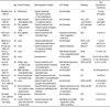
Malignancies associated with hamartomas are rare. The clinical, radiologic, and histologic findings of the previously described 14 cases of malignant hamartomas and the current case are summarized in Table 1. The mean patient age was 56.3 years (range, 25-78 years). The lesions are frequently recognized as palpable masses. The size of the hamartomas range from 1.5-12.0 cm in diameter and the size of the associated carcinomas range from 0.3-3.5 cm in diameter.
Of the described 15 cases, mammography was obtained in 12 cases, of which 10 showed the typical appearance of hamartomas with suspicious features, such as clusters of microcalcifications, pleomorphic micocalcifications, and spiculated masses. The remaining two cases had the typical appearance of a hamartoma with no suspicious features, thus co-existing malignancies were unexpected findings at the time of tumorectomy. US findings were available in only six cases, of which four had suspicious masses with irregular margins, hypoechogenicity, or a non parallel orientation within the hamartomas; and two cases were diagnosed pre-operatively as carcinomas by US-guided fine needle aspiration or core needle biopsies and underwent one-step curative surgery (6, 7). As stated above, the majority of cases had suspicious findings within the hamartoma on mammography or US. Radiologists therefore need to pay careful attention in order to detect subtle suspicious findings, even though mammography or US may show typical hamartomas.
Among the 15 cases described here, 12 had carcinomas that were confined to the hamartomas and the remaining three cases had carcinomas that involved both the hamartomas and adjacent normal breast tissue. If carcinomas involve both the hamartoma and normal breast tissue, it is difficult to determine whether the carcinoma arises within the hamartoma or an isolated carcinoma which initiates growth nearby later extends into the hamartoma. However, in the majority of cases described here, including the current case, the carcinomas were located within the hamartomas, (2-5, 7, 8, 10-12) thus we believe that the malignancies arose within the hamartomas.
In conclusion, breast hamartomas have generally been classified as rare, benign tumors, and carcinomas occur only rarely. However, hamartomas may be diagnosed with greater frequency due to widespread screening mammography. Radiologists should recognize that malignancy may co-exist or develop in hamartomas and be alert to the presence of suspicious features within a hamartoma.
 XML Download