

 PDF
PDF ePub
ePub Citation
 Print
Print


Yolk sac tumor (YST) is also known as endodermal sinus tumor and Teilum tumor, and is one type of germ cell neoplasm. YST is considered to be a highly malignant tumor that primarily occurs in the testis or ovary. Ten to fifteen percent of the cases may arise in a variety of midline extragonadal sites that mostly display an axial distribution pattern, such as the mediastinum, the pineal region and sacrococcygeal region and the female reproductive tract. The possible etiopathogenesis of these extragonadal YSTs is still controversial. One hypothesis suggests these tumors may arise from misplaced or arrested germ cells during embryogenesis (1), while others have proposed these tumors arise from aberrant differentiation of somatic cells (2).
We report here on a rare case of primary YST arising in the pancreas with hepatic metastasis in an adult woman, and we focus on the CT findings and making the differential diagnosis.
CASE REPORT
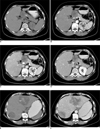
A 57-year-old woman was admitted to our hospital for further evaluation of a mass located in the upper abdomen, and this mass was incidentally detected by an abdominal ultrasound exam during a medical checkup. The patient presented with recurrent abdominal distension without any other complaints. Her medical history was unremarkable except for cholecystolithiasis and a fatty liver that were diagnosed two years previously. On admission, the physical examination revealed mild hepatic percussion pain, but no mass was palpated. The laboratory tests showed an extremely high level of α-fetoprotein (AFP) > 35,350 ng/ml (reference: < 20 ng/ml). The carbohydrate antigen (CA) 19-9, CA125, blood amylase, aspartate transferase (AST), alanine transferase (ALT) and bilirubin levels were within the normal ranges. Pre-contrast and contrast-enhanced spiral CT of the abdomen was performed, including the arterial phase, portal venous phase and equilibrium phase following bolus injection of intravenous contrast material. The pre-contrast CT scan revealed a 4.5 cm in diameter, smooth-marginated, solitary, heterogeneous mass occupying the pancreatic neck and body (Fig. 1A). On the post-contrast CT images, the mass displayed moderate inhomogeneous enhancement with central hypoattenuation (Fig. 1B-D). Two hypodense lesions were discovered in the liver and they measured 4.7 × 4.3 cm and 7.0 × 6.8 cm, respectively, displaying minimal diffuse heterogeneous enhancement (Fig. 1E, F). The high density within the large mass in the left hepatic lobe indicated central hemorrhage.
Since pancreatic cancer is sometimes associated with an elevated AFP level (although not so high as in this patient) and more often the hepatic metastases, an initial diagnosis of conventional pancreatic adenocarcinoma was made. The patient underwent neoadjuvant chemotherapy that consisted of fluorouracil, tetrahydrofolate and cisplatin, yet this had little effect and the serum AFP level did not decrease. Exploratory laparotomy was performed. An encapsulated mass measuring about 4 cm in diameter was discovered in the pancreatic neck and body. In addition, two metastatic lesions 5.0 × 4.0 cm and 4.0 × 3.0 cm in size, were found in the III, IV and VII hepatic segments. Tumor resection that included the primary pancreatic neoplasm and the right hepatic metastatic mass was carried out. For the left hepatic mass, hepatic segmentectomy (segments II and III and part of segment IV) was performed. Central hemorrhage was verified in the large metastatic lesion. The pathologic diagnosis revealed malignant yolk sac tumor of the pancreas. Microscopically, the tumor cells were arranged in a typical reticular or glandular pattern. Eight of the 13 dissected lymph nodes were involved with tumor. The immunohistochemical examination demonstrated that the tumor cells were diffusely positive for AFP, placental alkaline phosphatase (PLAP) and carcino-embryonic antigen (CEA), but they were negative for CA19-9.
The AFP level was notably decreased after surgery (2,097, > 1,200, > 500 ng/ml on the postoperative 3rd, 9th and 25th day, respectively). To exclude the existence of primary gonadal germ cell tumor, further clinical and ultrasound examinations were conducted on the genital system of this patient, but no abnormality was found. A month after the operation, the patient received systematic chemotherapy with the BEP regimen (bleomycin, etoposide and cisplatin). A total of three cycles of therapy were performed with an interval of three weeks between cycles. During chemotherapy, the CT examination was repeated and it revealed no tumor recurrence in the pancreas.
DISCUSSION
Of all the genital tumors, yolk sac tumors are relatively uncommon and they are mostly discovered in infants and adolescents (median age, 19 years) (3). Although they typically arise from the gonads, YSTs have already been reported in many extragonadal sites. Generally, the mediastinum, the sacrococcygeal region, the brain, the retroperitoneal space and the female reproductive tract are relatively common locations. Exceedingly rare sites such as liver, kidney, omentum, stomach, spinal cord, etc have also been previously reported (4-6). To the best of our knowledge, there has been no report about primary YST arising in the pancreas, and especially occurring in a 57-year-old woman.
Because of their rarity, there has been no systematic study on the CT features of extragonadal YSTs with a large number of cases. In a previous case report, this tumor was usually described as a large, multi-lobulated, solid heterogeneous, hypodense mass that showed moderate and heterogeneous enhancement (7). Central necrotic and/or cystic changes were observed on the CT images by Wong et al. (8) who reviewed seven cases of primary YSTs in the liver. Choi et al. (9) evaluated the CT findings of YSTs of the ovary in ten patients, and they characterized these tumors as large, smooth-marginated, well enhancing, solid masses with a cystic, hemorrhagic or necrotic portion. In the present case, the non-contrast CT scan showed an oval-shaped, heterogeneous soft-tissue mass with a central hypodense area, and the mass displayed moderate inhomogeneous enhancement. The imaging findings were verified by the morphological observations of an encapsulated tumor with focal necrosis. Central hemorrhage was detected in the hepatic metastatic lesion, which might be attributed to the notable proliferative and vascularization activities of the tumor cells. In some studies, YSTs have shown infiltrative growth into the adjacent soft tissues, and higher magnification revealed a microcystic growth pattern of tumor cells that formed reticular or glandular structures, which contributed to the cystic or necrotic changes seen on the CT images.
Since YSTs usually show high malignancy, the duration from the onset of symptoms to the admission is always short and metastasis may already exist at the time of the patient's admission, like the case presented here. An extremely high AFP level may be a clue for making the diagnosis of YSTs. Because of the extreme rarity of primary YST of the pancreas, metastatic germ cell tumor from the gonads or other primary or metastatic pancreatic neoplasms should be excluded. Needle aspiration biopsy should be performed for the confirmative diagnosis.
Chemotherapy has greatly improved the survival of patients with YST. Surgical excision with combined adjuvant chemotherapy is the treatment of choice. However, the prognosis is poor if there is metastasis.
In conclusion, we present here the first case of primary YST arising in the pancreas with hepatic metastasis in an adult woman. This unique case is of value for reminding radiologists and oncologists to be aware of the diagnosis of YST when a patient presents with an extremely high level of AFP and an oval-shaped or multilobulated, heterogeneous, soft tissue mass is discovered in the pancreas displaying moderate and inhomogeneous enhancement on CT images. Making a careful differential diagnosis should be done so that the optimal therapy can be administered.
 XML Download