

 PDF
PDF ePub
ePub Citation
 Print
Print


Intracranial foreign body granulomas are very rare, with only a few cases reported in the literature. Such granulomas are usually caused by hemostatic agents that are deliberately left behind during craniotomies (1, 2). Other causative synthetic materials include silicone-coated sheets used as dura mater substitutes, chemotherapy wafers, and agents employed for embolization of highly vascular tumors or arteriovenous malformations such as polyvinyl alcohol or N-butyl-2-cyanoacrylate (1, 3).
Reports on MR imaging of foreign body granulomas are even rarer. MR imaging findings included a low signal intensity on T1-weighted images, low or high signal intensity on T2-weighted images, and nodular or ring enhancement after contrast infusion in a few cases (1, 4-6). Conventional MR imaging findings are not consistent, and foreign body granulomas may be indistinguishable from recurrent tumors, radiation necrosis, abscess, resolving infarction, hematoma, or even unrelated primary or metastatic neoplasms. Recently, advanced MRI techniques such as perfusion MRI and MR spectroscopy have increasingly been used for tumor grading or differentiation between tumors and non-neoplastic conditions. We present here a case of postoperative intracranial granuloma of the foreign body type, examined by conventional MR imaging, perfusion MRI, and MR spectroscopy.
Case Report
A 35-year-old woman was admitted complaining of a headache over the last two months. She had undergone a right temporal lobectomy for generalized tonic-clonic seizures 10 years earlier. The pathology at that time revealed hippocampal sclerosis.
Laboratory tests were normal except for an increased erythrocyte sedimentation rate of 45 mm/hr.
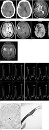
A pre-contrast CT showed a linear calcification surrounded by a lobular-shaped mass of increasing attenuation in the right frontal lobe (Fig. 1A, B). A postcontrast CT scan revealed that the mass showed strong enhancement. The mass was accompanied by extensive edema surrounding it, with a midline shifting to the left side.
On MR imaging, the mass showed a heterogeneous dark signal intensity on T2-weighted images (Fig. 1C), and a slightly high signal intensity on T1-weighted images (Fig. 1D). The mass showed intense contrast enhancement (Fig. 1E), and was located in the cortical and subcortical region. Moreover the mass had an irregular margin and was classified as an intra-axial mass with cortical involvement.
Perfusion MRI was performed by a gradient-echo echo planar imaging technique. Three regions of interest (ROI) were determined based on the gradient-echo echo planar imaging source images and the matching T2 and gadolinium-enhanced T1-weighted images. The ROI with the highest regional cerebral blood volume (rCBV) was selected and was found to be almost the same as that of the contralateral side (rCBV ratio = 1.04) (Fig. 1F). Localized proton MR spectroscopy was performed using a multivoxel point-resolved spectroscopy sequence with an echo time of 144 msec. The spectroscopic data showed a slightly elevated choline (Cho) and slightly decreased creatine (Cr) and N-acetyl aspartate (NAA) peaks with a small increase in the choline/creatine (Cho/Cr) ratio in the enhancing lesion (Fig. 1G-I). Our preoperative diagnosis was a high grade glioma accompanied with calcification and hemorrhage. This diagnosis was based on the strong enhancement of the mass accompanied with severe peritumoral edema and a mass effect. However, the perfusion data and MR spectroscopy findings suggested that the mass was benign.
The mass was surgically removed and the pathology revealed a foreign body-associated granulomatous inflammation with marked lymphocytic and histiocytic infiltration, microcalcification, and fibrous scar tissue formation (Fig. 1J, K). Following surgery, the patient's headache was relieved and her postoperative course was uneventful.
DISCUSSION
The chemical agents used in neurosurgery to achieve intra-operative hemostasis can cause foreign body reactions. Historically, several terms such as textiloma, gossypiboma, gauzoma, and muslinoma have been employed, reflecting the hemostatic materials involved. In our case, gelfoam is thought to be the foreign body that caused the granuloma formation.
The signal intensities of the foreign body granulomas are variable on the T1- and T2-weighted images. However, such granulomas invariably show nodular or ring-enhancing mass lesions. Variable signal intensity on T2-weighted images may reflect a complex pathology including acute or chronic inflammation, granuloma formation, fibrosis, collagen deposition, and degeneration of foreign materials. In our patient, the mass showed heterogeneous high and low signal intensities on T2-weighted images. The low signal intensity proportion of the T2-weighted images may be explained by the fibrosis seen in the histologic specimen. In the case of ring-enhancing mass lesions, the central portions may show low signal intensities on T2-weighted images. These represent degenerated foreign material and may be helpful in distinguishing a muslinoma from brain abscess (7).
Foreign body reactions to hemostatic materials may mimic brain tumors on MR imaging. This was true of our patient based on the MR imaging findings showing intense mass enhancement with severe surrounding edema. However, on perfusion MRI, the rCBV did not increase and, on multi-voxel MR spectroscopy, the mass Cho/Cr ratio increased only slightly, which was not consistent with a high-grade tumor.
Previous reports on perfusion and MR spectroscopic findings in patients with non-neoplastic tumor-mimicking masses showed mildly decreased-to-increased perfusion, and small rises in Cho/Cr ratios, with decreased NAA. However, there were some unusual exceptions. Perfusion MRI and MR spectroscopic findings from our patient were very similar to those of patients from previous reports with non-neoplastic mass lesions (8, 9). Retrospectively, we think we could have excluded the possibility of malignancy in our patient based on the perfusion MRI and MR spectroscopy, although benign tumorous conditions may not be totally excluded by such exploratory modalities.
Although the perfusion MRI and MR spectroscopic findings are not specific for foreign body granulomas, these techniques may be helpful in excluding a high-grade tumor diagnosis, which may be the most important differential diagnosis. By combining clinical history of any previous operation, conventional MR imaging findings, and perfusion MRI and MR spectroscopy, it may be possible to correctly diagnose a foreign body granuloma.
In summary, conventional MRI findings in foreign body granuloma patients may sometimes mimic those of brain tumors. To differentiate the diagnoses, the performing a perfusion MRI and MR spectroscopy may be helpful.
 XML Download