

 PDF
PDF ePub
ePub Citation
 Print
Print


Abbreviations
CSF
cerebrospinal fluid
DTI
diffusion tensor imaging
FA
fractional anisotropy
FDR
false discovery rate
FLAIR
fluid attenuated inversion recovery
FOV
field of view
FWE
family-wise error
FWHM
full-width at half maximum
GM
gray matter
GMC
gray matter concentration
MNI
Montreal Neurological Institute
mTLE
mesial temporal lobe epilepsy
NEX
number of excitations
rCBF
regional cerebral blood flow
SPGR
spoiled gradient recalled
SPM
statistical parametric mapping
VBM
voxel-based morphometry
WCAT
white matter changes of the anterior temporal lobe
WM
white matter
WMAT
white matter of anterior temporal lobe
WMC
white matter concentration
WMV
white matter volume
Mesial temporal lobe epilepsy (mTLE) is the most common type of intractable partial epilepsy, and hippocampal sclerosis is the most frequent pathological finding in mTLE (3). Brain MRI has been successfully used for detecting epileptogenic lesion (4). Hippocampal sclerosis is easily visible on MR images, but it is difficult to detect mild structural abnormalities in other brain structures of mTLE patients. A voxel-based morphometry (VBH) provides a quantitative means of examining brain MR images in terms of structural differences or for performing statistical analysis, and has been widely used to detect subtle changes of the brain structures in the cerebral cortex, limbic structures, subcortical nuclei (2, 3), and cerebral white matter (1, 5) of mTLE patients. A previous study was conducted to identify correlations between gray matter concentrations (GMCs) and hippocampal volumes (6).
In VBM, the GMCs generally reflect the amount of neuronal concentration, whereas white matter concentrations (WMCs) indicate the amount of nerve fibers or myelinated axons. Past reports have described the presence of heterotopic neurons in the anterior temporal stem of mTLE patients (7, 8). In addition, the relationship between heterotopic neurons of the anterior temporal stem and the ictal hyperperfusion was previously suggested (9). From these observations, we speculated that a correlation exists between the GMCs and the heterotopic neuron counts in the white matter of the anterior temporal lobe.
The aim of the present study was to investigate structural changes in pathologically proven unilateral mTLE patients using optimized VBM, as well as determine whether a correlation exists between heterotopic neuron counts for the gray and WMCs.
MATERIALS AND METHODS
Patients
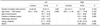
We included 31 unilateral mTLE patients (16 left [5 men, 11 women] and 15 right [7 men, 8 women]) who achieved excellent surgical outcomes (Engel class I) after an anterior temporal lobectomy with amygdalohippocampectomy (Table 1). To include only patients whose seizures arise from the mesial temporal lobe, we excluded patients who did not have a postoperative seizure-free outcome because they may have had a seizure focus in another region such as the lateral temporal cortex or extratemporal regions. Hippocampal sclerosis was pathologically confirmed in all cases. The clinical characteristics collated for each subject were at the age of seizure onset, duration of epilepsy, white matter changes of the anterior temporal lobe (WCAT), as well as though the number of heterotopic neurons by MRI and by the pathology. The data of all patients were collected at the Samsung Medical Center from 2004 to 2006.
For comparative purposes twenty four healthy volunteers (8 men, 16 women) made up the left mTLE group, while 23 healthy volunteers (12 men, 11 women) made up the right mTLE group (Table 1). Because the age and sex distributions of the left and right mTLE groups were different, two cohorts of normal controls were prepared to match the respective age and sex distribution. These healthy subjects had no history of head trauma or of a neurological or psychiatric disorder and were not on a medication. Moreover, the healthy subjects all had a normal spoiled gradient recalled in the steady state (SPGR) MRI findings, and showed no signal changes in the fluid attenuated inversion recovery (FLAIR) or T2-weighted images.
Magnetic Resonance Imaging
MRI scanning was performed using a GE Signa 1.5 Tesla scanner (GE Medical Systems, Milwaukee, WI). All subjects underwent SPGR, T2-weighted, and FLAIR imaging protocols. Coronal SPGR MR images were obtained using the following scanning variables; 1.6 mm thickness, no gap, 124 slices, repetition time/echo time (TR/TE) = 30/7 milliseconds (msec), flip angle (FA) = 45°, number of excitations (NEX) = 1, matrix = 256×192, and field of view (FOV) = 22×22 centimeters (cm). The voxel dimension of the SPGR MR images was 0.86×0.86×1.6 mm. An oblique coronal FLAIR MRI was performed using a 4.0 mm slice thickness, 1.0 mm gap, 32 slices, TR/TE = 10002/127.5 msec, 1 NEX, matrix = 256×192, and a FOV = 20×20 cm. The oblique coronal T2-weighted MR images were obtained with a 3.0 mm slice thickness, 0.3 mm gap, 56 slices, TR/TE = 5300/99 msec, FA = 90°, 3 NEX, matrix = 256×192, and FOV = 20×20 cm.
Voxel-Based Morphometry
Using SPM2 (Wellcome Department of Cognitive Neurology, Institute of Neurology, University College London) and MATLAB 7.0 (The MathWorks, Natick, MA), an optimized VBM protocol (9) was performed to determine GMCs and WMCs and regional volume changes. To create customized templates and prior images of gray and white matter, all MR images from the mTLE patients and normal controls were spatially normalized to a standard T1 template. Spatial normalizations were applied using the following parameters: voxel size = 1×1×1 mm, cutoff spatial normalization of 25 mm, nonlinear regularization = medium, and 16 nonlinear iterations. The normalized images were segmented into gray matter, white matter, cerebrospinal fluid (CSF), and sub-sampled into a voxel size of 2×2×2 mm. The spatially normalized raw images, segmented gray matter, white matter, and CSF images were averaged and saved into the customized T1 template, gray matter, white matter, or CSF prior images, respectively. Finally, three previously obtained images and the customized T1 template were smoothed using an 8 mm full-width at half-maximum (FWHM) isotropic Gaussian kernel. The raw T1 images of all subjects were automatically segmented into gray matter, white matter, and CSF partitions in native space, and volumes of gray and white matter images were then calculated. The spatial normalization parameters were estimated by matching each individual's own gray matter with SPM to the gray matter template of this study, to create spatially normalized versions of the original images. Spatially normalized images were segmented using each patients' own prior images (gray matter, white matter, and CSF partitions), and then spatially normalized. Moreover, the segmented gray and white matter images were modulated for regional volume change analyses. Modulated images were smoothed using an 8 mm FWHM isotropic Gaussian kernel, whereas unmodulated images were smoothed using a 12 mm FWHM isotropic Gaussian kernel. An unmodulated VBM is indicative of concentration differences, whereas modulated VBM is indicative of volume differences.
Neuronal Counting and Criteria for White Matter Changes of Anterior Temporal Lobe
Temporal lobe specimens were fixed in buffered formalin overnight and sectioned in a coronal plane at 5 mm intervals and at a thickness of 4-µm after routine processing for histology. The sections were stained with Hematoxylin and Eosin, followed by Luxol fast blue, only for cases with an ill-defined gray-white junction. All glass slides were reviewed by a neuropathologist. The histologic features were assessed in white matter at 1 or 2 cm from the temporal lobe pole. In each case, three different sections of the temporal lobe specimens containing a considerable amount of white matter were selected to facilitate the counts of isolated heterotopic neurons (7). Under an optical microscope (Olympus BX50, Olympus Optical Co., Japan), isolated heterotopic neurons counts were obtained in the white matter at greater than 3 mm deep from the gray-white junction and at an objective magnification of 200X. The counts were taken by a neuropathologist, who counted the total number of heterotopic neurons in three different fields (field size of 1 mm2) per section. Heterotopic neuron numbers are presented as percentages of the total numbers found in nine different fields per case.
The criteria used to determine WCAT included a decreased demarcation of the gray and white matter boundaries, or an increased signal intensity of white matter at the anterior temporal lobe on oblique coronal T2-weighted MR images (7).
Statistics
For VBM analyses, a one way ANOVA (analysis of variance) was used for the concentration analyses of unmodulated gray and white matter images. In addition, an ANCOVA (analysis of covariance) with gray matter volume (GMV) as a covariate was used for the regional volume change analyses using modulated images. To correct for multiple comparisons, the results were corrected using a false discovery rate correction at a significance level of p < 0.05. The voxel clusters were corrected with an extent threshold of kE > 200 voxels.
Correlation analyses were performed using SPM2 using a whole brain mask between the heterotopic neuron counts and the GMCs and WMCs. The mean values of significant clusters were extracted from gray and white matter images at an uncorrected significance level of p < 0.001, followed by a simple and partial correlation with the control of age at disease onset and disease duration between GMCs or WMCs and the heterotopic neuron counts. Gender distributions were tested using the Pearson Chi-square test. Age, disease duration, and age at disease onset were tested using the two-tailed t-test. Statistical analyses were performed using SPSS version 11.5 (SPSS Inc, Chicago, IL).
Coordinates were defined in accordance with the Montreal Neurological Institute (MNI) coordinate system, and cluster regions were named as described in Duvernoy's atlas (11).
RESULTS
Clinical Data
Age and sex were not different between patients with left mTLE and normal controls. The same is true for right mTLE patients and normal controls. Age at seizure onset and disease duration were not different in the left and right mTLE patient groups, but the frequency of WCATs was significantly higher in the right mTLE group compared to the left mTLE group, and the number of heterotopic neurons of the right mTLE group was greater than that of the left mTLE group (Table 1).
Gray Matter Concentration Analysis
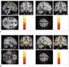
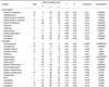
In left mTLE patients, GMCs were reduced in the left hippocampus, bilateral thalami, precentral gyri, superior frontal gyri, cingulate gyri, left supramarginal gyrus, and cerebellum (Fig. 1A) (Table 2). Regional GMVs in left mTLE were reduced in the bilateral frontopolar gyri, cingulate gyri, caudate nuclei, left thalamus, both hippocampi, left insular gyrus, pons, and cerebellum (Fig. 2A).
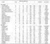
In right mTLE patients, GMCs were reduced in the right hippocampus, bilateral thalami, precentral gyri, frontopolar gyri, left inferior frontal gyrus, bilateral superior frontal gyri, superior temporal gyri, middle temporal gyri, and cerebellum (Fig. 1B) (Table 3). Regional GMVs in right mTLE were reduced in the bilateral frontopolar gyri, caudate nuclei, thalami, hippocampi, left orbital gyrus, left superior and middle temporal gyri, and cerebellum (Fig. 2B). There was no increased GMC or volume in both mTLEs.
White Matter Concentration Analysis
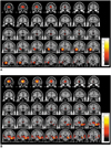
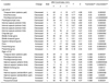
In the left mTLE group, WMCs were reduced in the left temporal stem (anterior part), left entorhinal area, both parahippocampal areas, both internal capsules (anterior limb), and in both cingulate gyri (Fig. 3A), whereas the WMCs of the pons and both precentral gyri were increased (Fig. 3B) (Table 4).
In right mTLE group, WMCs were reduced in the right temporal stem (anterior part), right internal capsule (anterior limb), and both the parahippocampal areas, whereas the WMCs increased in the pons, both internal capsules (retrolentiform part), right precentral gyrus, and the left paracentral lobule (Fig. 3C, D).
Correlation between Heterotopic Neuron Numbers and Gray, White Matter Concentrations
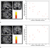
The WMCs (maximum: 1, minimum: 0) of the anterior temporal stems of the left mTLE patients were found to be negatively correlated with heterotopic neuron counts (x, y, z = -44, -5, -24, voxel level: uncorrected p = 0.000025, cluster level: family-wise error [FWE] corrected p = 0.041, z = 4.05, kE = 912 voxels). A simple correlation analysis indicated that WMCs and heterotopic neuron counts were negatively correlated (r = -0.839, p = 0.00005), and a partial correlation analysis (controlling for age at onset and disease duration) confirmed this (r = -0.861, p < 0.001). Whole brain masks, uncorrected p < 0.001, and an extent threshold kE > 200 voxels, were utilized for the correlation analyses (Fig. 4A).
Heterotopic neuron counts were positively correlated with GMCs (maximum: 1, minimum: 0) of the anterior temporal stems of left mTLE patients (x, y, z = -43, -4, -26, voxel level: uncorrected p = 0.000018, cluster level: FWE corrected p = 0.170, z = 4.13, kE = 956 voxels). A simple correlation analysis between the GMCs and the heterotopic neuron counts showed a strong positive correlation (r = 0.819, p = 0.00005). Similarly, a partial correlation analysis with a control of age at seizure onset and disease duration also showed a positive correlation (r = 0.810, p < 0.001) (Fig. 4B). No correlations were found between heterotopic neuron counts and GMCs or WMCs in right mTLE patients.
DISCUSSION
Clinical Considerations
In this study, we uncovered four significant findings; first, the abnormality of the cortico (precentral gyrus)-thalamo-hippocampal network; second, a large GMV reduction in the anterior frontal lobe; third, a structural abnormality in the pontine area; and fourth, a significant correlation between heterotopic neuron counts and GMC or WMC in the left mTLE patients.
Gray matter concentrations were reduced in the hippocampus, thalamus, precentral gyrus, cerebellum, as well as the wide frontal and temporal areas. Previous studies have reported decreased GMCs (1-3, 12, 13), GMVs (4), GMCs and GMVs (14) in the hippocampus, thalamus, and cerebellum. Our finding of decreased GMC in the precentral gyrus supports a previous study (15). We previously reported that ictal hyperperfusion and interictal hypoperfusion of the cortico-thalamo-hippocampal network in patients with mTLE (9), which may be related to the GMC reduction observed in the precentral gyrus in the present study.
In the GMV analysis with modulated gray matter images, GMVs were also decreased in the caudate nucleus, anterior temporal lobe, and frontopolar gyrus. Moreover, hilar cell densities of the resected hippocampi were found to positively correlate with the regional cerebral glucose metabolic rate in the bilateral thalamus, putamen and globus pallidus, and ipsilateral caudate (16). This relationship suggests that hippocampal cell loss causes efferent synaptic activity reductions to the thalamus and basal ganglia, hence decreasing neuronal activity and consequently decreased cerebral glucose metabolism in these structures. Similar to a previous study (17), volume reductions via manual volumetry have been reported in the entorhinal, perirhinal, and temporopolar cortices of drug-refractory mTLE in the present study. The large volume reductions of the anterior frontal lobe in the present study appear to be a new finding. A positive correlation has been reported between hippocampal volume and regional GMVs in the hippocampus, thalamus, and cingulated gyrus, whereas normal controls showed no such correlation (6). Previously, we reported ictal hypoperfusions of the anterior frontal lobe (9, 18). Three mechanisms of ictal hypoperfusion have been suggested, which include a steal phenomenon (19), ictal surround inhibition (20), and regional postictal hypoperfusion during the ictal state due to rapid seizure propagation to other brain regions (9, 21).
The white matter abnormalities in mTLE have been previously reported by VBM and diffusion tensor imaging studies (1, 5, 14, 22). Decreased WMC and white matter volume (WMV) has been reported in the ipsilateral parahippocampal gyrus as well as in the contra-lateral internal capsule (14), and decreased WMC in the ipsilateral temporopolar, temporal stem, entorhinal, and perirhinal areas (1). In addition, reduced WMV were also observed in ipsilateral temporopolar, temporal stem, corpus callosum, and anterior frontal areas (5). Decreased fractional anisotropy has been reported in the entorhinal cortex, corpus callosum, and in the anterior limb of the internal capsule, indicating diffuse white matter loss in the limbic system (22). The results of the present study support these findings.
A previous study reported WMC reduction (1) and WMV reduction (5) in the ipsilateral temporopolar region or temporal stem in mTLE patients. The authors presumed that a cause of WMC reduction may be the secondary damage from repetitive seizure propagations or the deafferentation from GMV reduction of ipsilateral temporal lobes. However, we showed that the WMC in the ipsilateral temporopolar region is negatively correlated with the number of heterotopic neurons in the white matter of the anterior temporal lobe in the left mTLE group (Fig. 4), but not in the right mTLE group. We also previously reported heterotopic neurons in the ipsilateral anterior temporal stem from histologic and ictal rCBF hyperperfusion in the temporal stem of mTLE patients (9). It is unclear why there was no significant correlation between GMC and the heterotopic neuron number of the anterior temporal stem in the right mTLE. The significantly higher frequency of white matter signal changes on T2-weighted MRI and the large number of heterotopic neurons in right mTLE than in left mTLE patients could suggest some differences in histologic changes or pathogenesis between the left and right mTLE patients. The right hippocampus has been reported to be larger than the left hippocampus in the normal population (23, 24), and a previous study showed that right mTLE showed more frequent propagations of ictal discharges to the contralateral hemisphere (9). Thus, more studies are needed for further clarification of the relationship between heterotopic neurons and GMC or WMC in anterior temporal stem.
A previous study showed the occurrence of other histologic changes in temporal lobe white matter; for example, diffuse glial cell proliferation, perivascular space enlargement, neuronal heterotopia, astrocytic gliosis, increased numbers of corpora amylacea, and increased oligodendroglial cell clusters (7). It has been suggested that WCAT may be a nonspecific reactive change to seizure discharges (25), or a chronic excitotoxic injury caused by seizure activity (26).
Methodological Considerations
In spatial normalization, there are two options for the 'unmodulation' process for brain tissue concentration and 'modulation' process for brain tissue volume change. In order to preserve the total amount of signal in the images, the areas expanded during warping are correspondingly reduced in intensity, and areas contracted during warping are increased in intensity. This 'modulation' process multiplies tissue voxel values by the Jacobian determinants, and yields the determinant of the deformation parameters obtained from spatial normalization. So the use of the 'modulation' process could reflect the regional volume change. The 'unmodulated' images preserve its own signal intensities regardless of the expansion or contraction during warping. Hence, the VBM results using 'unmodulated' images could reflect the GMCs or WMCs (10, 29, 30).
In the present study, gray matter images were normalized to our study specific gray matter template, and the normalization parameters of the gray matter images were applied to the white matter images. There may be debate as to the choice of gray or white matter template for the white matter analysis in the VBM study. The problem is that white matter (WM) is characterized by large uniform areas with only minor signal contrast regardless of the large or small WM area. In contrast, gray matter (GM) provides much more detail because the GM band is only several millimeters thin. The registration usually relies on image intensity gradients, which can be found for WM only at the borders. Thus, the quality of WM registration is inferior to GM registration. The voxel-based morphometry approach is usually based on GM registration because of these reasons. We have tried both approaches and got the superior results for WM registration using the gray matter normalization parameters supporting this notion.
Increased WMCs in the precentral gyrus and internal capsule close to the thalamus appear to be attributed to an expansion effect of white matter during spatial normalization. Because the GMCs and GMVs were reduced in the precentral gyrus and thalamus, white matter might have expanded to compensate for gray matter loss during spatial normalization. This is a false positive finding coming from a methodological problem; hence, the researcher should be careful in interpreting the results of their VBM studies.
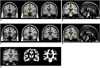
Despite the subcortical nuclei present in the pons, the pontine area was classified as a white matter image due to low intensity in the T1 MR images in the present study (Fig. 3E). The intensity of one voxel is composed of gray matter, white matter, and CSF intensities (the intensity of gray matter + white matter + CSF =1). If the neuronal density in the pons is low, the pontine area may appear to have higher WMC in T1 MR images. Thus, increased WMC in the pontine area may biologically reflect reduced neuronal density.
In conclusion, the present study demonstrates an abnormality of the hippocampal-thalamo-cortical network, GMV reduction in the anterior frontal lobe, a structural abnormality in the pontine area, and correlations between heterotopic neuron counts and the GMC and WMC in the white matter of the left anterior temporal lobe. However, future investigations should explore in more detail the neuronal correlation with the GMCs in the anterior temporal white matter.
 XML Download