

 PDF
PDF ePub
ePub Citation
 Print
Print


Bilateral pulmonary agenesis is a rare and lethal condition in which all of those affected die in utero or within the first hour after birth (1-4). Dozens of cases associated with complete agenesis of both lungs had been previously reported (1, 2). Although Vettraino et al. (2) recently authored a report emphasizing the prenatal sonographic appearance characteristics of bilateral pulmonary agenesis (BPA), most of the previous case reports have focused on the macro- and microscopic pathologic conditions (1). Herein, we report a case of BPA diagnosed prenatally with the aid of high-resolution ultrasonography and a fetal MRI.
CASE REPORT
A 28-year-old primigravida was referred to our hospital at 31 + 2 weeks of gestation for further evaluation as a result of abnormal findings on a fetal echocardiography. At 29 weeks of gestation, blindly ended vessels originating from the heart or transposition of the great arteries was first noticed. However, no comments were made regarding any abnormal findings of the fetal lung on a fetal ultrasonography. In addition, the medical and obstetric histories were unremarkable.
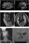
An ultrasound examination performed on the day of the first visit at our institute demonstrated an elevated diaphragm, raising the suspicion of associated lung abnormalities (Fig. 1A). The thoracic cavities, especially on the right, were reduced in size. The left thoracic cavity was largely occupied by the heart with a severely left-deviated axis, resulting from the remarkable displacement of the diaphragm cephalad. However, it was not evident whether any lung tissue was present, or whether there were any lung abnormalities or pulmonary veins connecting to the left atrium as well as the right and left branches of the main pulmonary artery. As a result, no specific structural abnormalities of the heart were identified.
For further evaluation of the lung abnormalities, a fetal MRI study was performed. Similar to the results of the ultrasound examinations, a significantly decreased thoracic volume associated with a bilateral elevation of the diaphragm was identified, thus resulting in an upward migration of the heart and rotation of the cardiac axis. Moreover, no definite lung was identified and the trachea ended blindly near the level of the cricoid cartilage without bronchial branching (Fig. 1B). Together, these findings strongly supported the diagnosis of BPA.
A male fetus weighing 2,530 g was delivered vaginally at 39 + 2 weeks of gestation after the induction of labor and in the presence of neonatologists. The Apgar scores at 1, 5, and 10 minutes after birth were 2, 2, and 2, respectively. Attempts at resuscitation were unsuccessful and after 15 minutes, the cardiac asystole persisted despite all efforts. The neonate died at 44 minutes after delivery. A postmortem infantogram was taken and the results of the study were consistent with the prenatal diagnosis (Fig. 1C). The karyotype of the neonate was 46, XY.
The body size of the fetus was appropriate for its gestational age. There were no congenital abnormalities noted on external inspection; however, multiple abnormalities were noted on an internal examination. The intact diaphragm was elevated bilaterally. A complete agenesis of both lungs was identified along with the absence of lung tissue and bronchial rudiments (Fig. 1D). In addition, neither pulmonary arteries nor veins were identified. An atretic trachea ended blindly at the level of the cricoid cartilage and esophageal stenosis was noted. The cardiac axis was rotated due to a mediastinal shift to the left. Four chambers of the heart had developed, however the main trunk of the pulmonary artery had no branches and joined the aorta at the site of the persistent ductus arteriosus. Lastly, a large atrial septal defect was present.
DISCUSSION
Embryologically, the lung develops as a pouch-like laryngotracheal diverticulum from the primitive foregut in the fourth to fifth week and then forms the lung buds. Both the lung buds and the encircling splanchnic mesenchyme, originating from the foregut, are crucial for the formation of the airways, vasculature, and lymphatic vessels. BPA results from the failure of lung or bronchial buds to develop and is associated with the absence of pulmonary veins and bilateral pulmonary arteries (2, 5-7). The exact etiology is not well known, although viral infections, genetic factors, and folic acid or vitamin A deficiencies have been suggested as contributing factors (2, 5, 7).
Among the reported cases of BPA, including our report, the absence of pulmonary vessels is common on autopsy findings. Color Doppler scans are of great value in detecting the pulmonary vasculature prenatally; even in the first trimester (6). The prenatal differentiation between BPA and other thoracic abnormalities, including congenital diaphragmatic hernia (CDH), congenital cystic adenomatoid malformation (CCAM), and pulmonary sequestration, is clinically important because the management and postnatal outcome are different; however, it is very difficult to do so because of sonographically-similar features, especially when the diaphragm cannot be visualized (2). This is particularly true in CDH cases, where the development of pulmonary blood vessels, as well as the lung parenchyma and airways is normal (8). Therefore, a systematic and meticulous approach using a standardized protocol, including the evaluation of the pulmonary vasculature, is critical to achieve a prenatal diagnosis of BPA.
Cardiac abnormalities (coarctation of the aorta, truncus arteriosus, right aortic arch and atrial septal defect [ASD]), as well as pulmonary abnormalities, were also detected in some cases (2). In the case reported herein, a large ASD was identified. These observations were consistent with the hypothesis that a teratogenic or vascular injury early in the embryonic period had led to developmental failure of not only the respiratory system but also the cardiovascular system because of near-field effects; although abnormalities of other foregut-related gastrointestinal and urinary systems can be associated.
In a previous report (9), a fetal MRI study offered no advantage over ultrasound examinations in the case of unilateral lung agenesis recognized by non-visualization of the corresponding lung vessels on the color Doppler scan. In general, however, fetal MRI study is of great value in the differential diagnosis of fetal thoracic abnormalities, including CDH, CCAM, pulmonary sequestrations, and esophageal atresia (10, 11). Moreover, another prenatal condition, when considering a differential diagnosis, is isolated diaphragmatic eventration, in which there is remaining lung tissue (12, 13). Therefore, to evaluate abnormalities of the thoracic cavity in detail, we recommend a fetal MRI study following the ultrasound examinations. In the current case, a fetal MRI was sufficiently beneficial to support the findings of the ultrasound examinations and more importantly, to identify an atretic trachea.
In conclusion, an elevated diaphragm and decreased thoracic volume on prenatal ultrasound examinations provide initial clues for BPA. However, it is essential to demonstrate the absence of the right and left branches of the pulmonary artery, as well as the absence of the pulmonary veins to enter the left atrium by using color Doppler scans. A detailed fetal echocardiography is helpful in detecting major cardiac anomalies and to trace the related vessels. To further differentiate BPA from other congenital abnormalities in the thoracic cage, we recommend fetal MRI as an additional imaging modality for suspected BPA cases from an ultrasound examination.
 XML Download