

 PDF
PDF ePub
ePub Citation
 Print
Print


Adventitial cystic disease of the vein is a rare condition. The arterial variety of adventitial cystic disease has often been described in the popliteal artery and this is characterized by a mucinous cyst located in the adventitia of the artery, with the contents of the cyst resembling the contents of a ganglion. In this report, we discuss the case of a 69-year-old man who presented with a swollen leg secondary to obstruction of the common femoral vein. We performed CT venography and ultrasound and this led to excision of a cyst and vein repair via a vessel graft. As a result, the patient made a full recovery. We also discuss the pathology and the diagnostic methods for this condition.
CASE REPORT
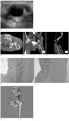
A 69-year-old man presented with a one month history of swelling of the left lower extremity. He had no specific past medical history and no history of trauma. No other abnormality was found on physical examination and we suspected that he suffered with deep vein thrombosis. Ultrasonography showed a cystic mass containing hypoechoic materials attached to the left common femoral vein (Fig. 1A). For further evaluation, we performed CT venography using 64 channel multi-detector computed tomography (Aquilion, Toshiba, Japan) with the patient in the supine position. A 22-gauge intravenous cannula was placed in the dorsal vein of the left foot, and a tourniquet was placed around the left knee. Forty milliliters of a contrast medium diluted with 60 milliliters of saline was automatically injected via a Y adapter into the left leg at a rate of 3 cc/sec. The optimal scan delay for the venous phase was determined by use of bolus tracking at the left popliteal vein. The axial and coronal reconstruction images showed a 1.7 cm sized cystic mass compressing the left common femoral vein along with dilated medial circumflex femoral and obturator veins that provided collateral circulation for the occluded left common femoral vein (Fig. 1B, C). A 3D volume rendering image showed an abrupt tapered disconnection of the left common femoral vein along with the formation of multiple collateral vessels (Fig. 1D). The patient underwent an operation because of his swollen leg. Surgical exploration showed an approximately 1.5 cm sized cystic mass over the anterior surface of the left common femoral vein. A longitudinal venostomy was performed at this location and a gelatinous material exuded from the mass and this gelatinous material could also be aspirated from the mass. A 10-cm long segment of the left common femoral vein/distal iliac vein was resected and a Gore-Tex graft was interposed. Microscopically, a cystic widened adventitial layer of the vein showed adventitial hemorrhage, fibrin deposition and focal aggregation of foamy histiocytes and focal myxoid degeneration (Fig. 1E, F). CT venography was performed again after the operation for a follow up evaluation. The cystic mass in the left inguinal areas was gone, as were the multiple collateral vessels (Fig. 1G). The final diagnosis was adventitial cystic disease.
DISCUSSION
Adventitial cystic disease is an unusual condition of an uncertain etiology in which a mucin-containing cyst forms in the wall of the artery or vein and this causes symptoms of intermittent claudication (1). Adventitial cystic disease was first reported in 1947 by Atkins and Key (2). They reported the case of a 40-year-old man with intermittent claudication associated with a palpable swelling above the inguinal ligament. At operation, a cyst was dissected from the external iliac artery. More than 200 case reports are currently available in the world literature with the majority of cases being associated with the popliteal artery (3, 4). Adventitial cystic disease has also been described in the external iliac artery, the femoral artery, the radial and ulnar arteries and in branches of the popliteal artery.
Fewer than 20 cases of adventitial cystic disease of the vein have been reported in the worldwide literature (5-7). Since the earliest reports, this disease has been considered equivalent with the adventitial cystic disease of the arteries (6). The cysts in both types of vessels show slow growth and a tendency to recur after treatment. However, adventitial cystic disease of the arteries is more frequent in men, it is predominantly located in the popliteal artery and it clinically presents with intermittent claudication. In contrast to arterial adventitial cystic disease, the venous variety rarely affects the popliteal segment. The venous variety occurs with an equal frequency in both sexes and it most often involves the common femoral vein and causes swelling of the affected limb.
The etiology of adventitial cystic disease remains unclear, but there are several theories (3, 5, 7-9). Repeated microtrauma, ectopic aganglionosis, degeneration of the adventitia due to connective tissue diseases and developmental theory have all been discussed in the medical literature. Histopathologically, the cyst may be uniloculated or multiloculated (2, 4). The disease process produces an expanding cyst that destroys the elastic tissue between the medium and the adventitia of the vessel wall, and the elastic tissue is replaced with fibrous connective tissue. There is usually no acute or chronic inflammation. The cyst is lined by fibrous connective tissue and the cyst contains an eosinophilic mucoid gel that consists of mucoproteins and mucopolysaccharides.
The diagnosis of adventitial cystic disease of the vein can be suspected on the basis of the patient history, the results of a physical examination and the image findings (6). Venography shows a smooth-walled stenosis that may be curvilinear with an hourglass or spiral configuration (1, 3, 4, 10). The important CT features in this case included eccentric compression of the lumen owing to a thin-walled cystic mass with an enhanced rim. Predictably, the mucinous cyst contents showed no enhancement and the contents had intermediate attenuation values between that of water and muscle. Ultrasonography may show a typical hypoechoic fluid filled cyst with a posterior acoustic window and this may allow ultrasound-guided treatment (5). Duplex US may show stenosis and blood flow changes in the prestenotic and poststenotic regions. Yet ultrasonography cannot display the distribution of the collateral vessels seen on CT and CT venography. Because of the collateral vessels, it is much more difficult to determine the exact anatomic relationships around the mass. Performing CT venography in patients with venous adventitial cystic disease can reveal the site and the extent of the obstruction, and it may show a classic scalloped appearance or hourglass narrowing caused by the extrinsic compression of the vessel lumen. CT venography is superior to traditional venography for making the diagnosis because the cystic mass can be directly observed regardless of the degree of obstruction. The involved vessel and also the structures such as collateral vessels can also be visualized during this procedure, and follow up evaluations are routine and not so difficult to perform.
In summary, adventitial cystic disease of the vein is a rare malady, but it should be suspected for patients with the symptoms of deep vein thrombosis, and especially when the diagnostic investigations indicate an extrinsic mass. CT venography is useful for demonstrating the lesion and increasing the specificity of the radiologic diagnosis, and this modality can be routinely used for the postoperative follow-up evaluation.
 XML Download