

 PDF
PDF ePub
ePub Citation
 Print
Print


Recent studies suggest that mesenchymal stem cells (MSCs) show significant potential as a novel cellular therapy for tissue regeneration (1). The development of new stem cell-based therapies requires a quantitative and qualitative assessment of stem cell distribution to the target organs, differentiation outcome, and engraftment (2, 3). In particular, the monitoring of cell trafficking in vivo and distinguishing whether cellular regeneration originated from an exogenous cell source is a key issue for developing successful stem cell therapies (4, 5). Although magnetic resonance imaging (MRI), radionuclide imaging, and optical imaging are all used to track the transplanted cells, MRI is progressing towards playing an important role in the development of stem cell therapy because its ability to demonstrate anatomic details with high contrast and exquisite spatial resolution, and can noninvasively follow stem cells for long periods of time (6, 7).
The detection of the transplanted cells by MRI requires cells to be labeled magnetically either by endocytic internalizing or cell surface attachment of magnetic nanoparticles (MNPs) (8-11). Recently, a variety of magnetic nanoparticles such as superparamagnetic iron oxide (SPIO) have been developed. Although native iron oxide particles are used as the currently preferred cell-labeling materials, they have low intracellular labeling efficiency and the exposed metal ion on the surface of nanoparticles can cause metal elemental toxicities in cells (12-14). The surface coating of magnetic nanoparticles with polystyrene, dextran, and dendrimers have been reported to improve the cellular-internalization of contrast agent, but the efficiency of internalization is still generally low and it requires long-term incubation time and high concentration of particles (13-15). It is therefore highly desirable to develop a nanoparticle that is nontoxic, biocompatible, efficient at intracellular labeling, and highly sensitive to detection. The current challenges also include the development of multimodality imaging for compensating the limited MRI sensitivity and extending its application by combining the strengths of different imaging modalities.
Bifunctional contrast agents with both optical and magnetic contrast were made using various methods and demonstrated to serve as good molecular imaging probes for multimodality imaging (16-18). By incorporating organic fluorescent dye molecules into the silica-coated magnetite core-shell, we synthesized uniform dual-functionalized nanoparticles which permit dual-modality detections (19, 20). Recently, several noninvasive, MR-based monitoring methods were used to tract MSCs transplant. However, the bifunctional magnetofluorescent nanoparticles as a single molecule have rarely been applied in MSCs tracking prior to this study. We investigated the feasibility of labeling human mesenchymal stem cells (hMSCs) with bifunctional nanoparticles and assessed their potential as imaging probes in the monitoring of hMSC transplantation.
MATERIALS AND METHODS
Preparation of Magnetic Nanoparticles
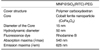
The bifunctional nanoparticles were synthesized using a previously reported method (19, 20). Cobalt ferrite magnetic nanoparticles (MNP, CoFe2O4) were coated with a silica shell (SiO2) of stable and biocompatible material to avoid the potential toxic effects on cells. To acquire additional fluorescent properties, an organic fluorescent dye (rhodamine B isothiocyanate, RITC) was incorporated into the silica shell. Thus, the MNP@SiO2(RITC)s have a bifunctional property which enables dual modality detection by MRI and optical imaging. The surface of the dual-functionalized silica-coated nanoparticles was modified with biocompatible polyethylene glycol (PEG) groups to render them biocompatible by preventing the nonspecific adsorption of proteins to the nanoparticles. Attachment of the PEG did not significantly change the size of the nanoparticles. The average diameter of MNP@SiO2(RITC)-PEG used in this study was about 50 nm. The characteristic properties of this compound are summarized in Table 1. The absorption (λmax) and emission maxima (λem) were observed at 540 and 625 nm, respectively.
Cell Culture
Human mesenchymal stem cells purchased from Cambrex (Cambrex, Walkersville, MD) were used for cell labeling. The cells were cultured in 75-cm2 plastic culture flasks (BD, USA) in 8 ml of medium at 37℃, in a humidified incubator at 5% CO2. The culture medium used was mesenchymal stem cell basal medium (MSCBM, Cambrex, Walkersville), which contains mesenchymal cell growth supplement (MCGS, Cambrex, Walkersville), 4 mM L-glutamine, and 100U Penicillin-Streptomycin. Next, the hMSCs were cultured under generally established cell conditions, transferred every three days, and split at a ratio of 1:5 upon confluence.
Determination of Cellular MNP@SiO2(RITC)-PEG Uptake by Confocal Laser Scanning Fluorescence Microscopy
For the confocal imaging experiments, hMSCs were seeded and cultured onto Lab-Tek Chambered Coverglass (Nalge Nunc, Naperville, IL) at a density of 3 × 10⁴ cells/cm2 for 24 hours prior to labeling and staining. Next, the cells were cultured at different concentrations of MNP@SiO2(RITC)-PEG for 2, 4, 6 hours in a 37℃ humidified incubator maintained at 5% CO2. After labeling, the cover slips were washed twice with cell culture medium and twice with phosphate buffer solution (PBS). The fluorescence images were obtained using a LSM 510 confocal microscope (Zeiss, Jena, Germany) equipped with Argon (458, 488, 514 nm) and HeNe (543, 633 nm) lasers for fluorescence. The exposure time for fluorescence was adjusted at each time point to localize the fluorochromes. Background fluorescence was determined by analyzing unlabeled cells. Image acquisition and analysis was performed using the LSM 5 PASCAL software.
Determination of Intracellular Distribution by Transmission Electron Microscopy (TEM)
MNP@SiO2(RITC)-PEG-labeled hMSCs were washed with PBS and fixed with 2.5% glutaraldehyde (in pH 7.4 and 0.1 M PBS) for two hours. Subsequently, the glutaraldehyde-fixed specimens were treated with 2% osmium tetroxide buffered in 0.1 mM cacodylate buffer for two hours. The specimens were dehydrated with graded ethanol (50% to 100% ethanol and propylene oxide) (EM Sciences). The samples were embedded in pure Epon resin at 60℃ for three days. Next, ultrathin sections were cut with glass knives and a Diatome diamond knife (Reichert-Jung, Vienna, Austria) on an ultramicrotome (RMC MTXL; Tucson, AZ), stained with lead citrate and uranyl acetate (both from EM Sciences), and observed by JEOL-JEM-100 CX Transmission Electron Microscopy (JEOL Ltd. Tokyo, Japan).
In Vitro Cytotoxicity and Cell Proliferation Assay
Cell viability was assessed by a hemocytometer-based trypan blue dye exclusion assay. To exclude the presence of free floating cells in the growth medium, the supernatant of the cells was controlled for floating cells prior to trypsination. The cells were cultured in 96 well plates. The 96 wells were incubated for two hours, four hours, and six hours at different MNP concentrations (0, 0.5, 1, 2, 4 mg/ml). After incubation, the cells were washed three times with PBS and dissociated using trypsin-EDTA. Next, the cells were resuspended with 10 ml of media. A total of 20 µl of the cell suspension was mixed with 30 µl of Trypan blue solution (GIBCO, Gland Island, NY). The fraction of dead cells was determined using a Neubauer's counting chamber.

Each value represents the mean ± standard error (SE) of three independent experiments performed in triplicate. The effects of the internalized particles on cell proliferation were studied by comparing the growth curves of MNP@SiO2(RITC)-PEG-labeled (4 hour incubation time with 4 mg of MNP@SiO2[RITC]-PEG/ml growth medium) and unlabeled hMSCs. Vision fields were counted under the microscope at 1, 2, 3, and 4 days after labeling. Each value represents the mean ± SE of three independent experiments performed in triplicate.
Magnetic Resonance Imaging
Measurement of T1 and T2 Relaxivity of MNP@SiO2(RITC)-PEG measured by a 1.5T and 3T MR scanner: MR imaging of the test tubes was performed using a 1.5T and 3T MR scanner (Signa, GE Medical Systems, Milwaukee, WI) and a standard knee coil (Clinical MR Solutions, Brookfield, WI). To avoid susceptibility artifacts from the surrounding air in the scans, all probes were placed in a water-containing plastic container at room temperature (20℃). The concentrations of MNP@SiO2(RITC)-PEG in each test tube were 0, 10, 30, 50, 100, 200, and 300 µg/ml. To measure the T1 relaxation times, axial spin echo sequences were obtained at multiple TR values of 200, 300, 500, 1,000, 3,000, 5,000, 8,000, 12,000 ms with a fixed TE of 12 ms at 1.5T and 3T. To measure the T2 relaxation times, axial T2-weighted SE images were obtained with a TR of 4,000 ms and increasing TEs of 12, 20, 50, 80, 150, 300, and 500 ms at 1.5T and 3T. All sequences were acquired at a field of view of 130×130 mm, a matrix of 256×256 pixels, a slice thickness of 5 mm, and one acquisition. The signal intensities of the test tubes, with contrast medium in solution at corresponding iron concentrations, were measured using custom written software for the region of interest analysis. The signal intensity for each pixel, as a function of time, was expressed as follows: SI = So(1-exp [-TR/T1]) and SI = So exp (-TE/T2). The T1 relaxivities (r1 =ΔR1/concentration; mM-1s-1) and T2 relaxivities (r2 =ΔR2/concentration; mM-1s-1) at 1.5 and 3T for free solution of MNP@SiO2(RITC)-PEG were deduced from the linear fit of the ΔR1 and ΔR2 values displayed as a function of the total iron concentration.
MR Phantom Studies and In Vivo MR imaging: For MR phantom imaging, hMSCs were cultured in MSCBM containing MNP of various concentrations (MNP concentrations of 0.5, 1, 2 and 4 mg/ml). After incubation, cells were washed three times with PBS, trypsinized, and collected in 50 ml tubes. Next, the samples were prepared by suspending 105 and 5×10⁵ cells in 350 µl 4% gelatin (Wako Pure Chemical Industries, Osaka, Japan). The cell suspensions were loaded onto 96 well plates and allowed to solidify at 4℃. The experimental MR imaging of a phantom of labeled hMSCs was performed with a 3T MR scanner (Signa, GE Healthcare, Milwaukee, WI) using the three-dimensional b-SSFP (balanced Steady-State Free Precession) pulse sequence (FIESTA; Fast Imaging Employing Steady State Acquisition, GE Healthcare, Milwaukee, WI) with a TR of 8.8 ms, TE of 4.1 ms, field of view of 100×100 mm, and a voxel size of 1×1×1 mm2. The region of interest for signal intensity (SI) measurement was 20 mm2. The percent change of SI was calculated using the following equation: ΔSI = (SIlabeled - SIunlabeled)/ SIunlabeled×100%, where SIlabeled and SIunlabeled were the SI of the labeled and unlabeled cells, respectively. For animal experiments, hMSCs were cultured in MSCBM containing 4 mg of MNP@SiO2(RITC)-PEG/ml media for four hours. After incubation and washing, the 0.5-1×10⁶ labeled cells/200 µl were injected into a kidney of SD (Sprague Dauley) rats and different numbers of MNP@SiO2(RITC)-PEG-labeled hMSCs (1×10⁴, 1×10⁵, 0.5×10⁶, 1×10⁶ mg/ml) were injected subcutaneously into nude mice. Six hours after injection of the labeled hMSCs, sequential MR imaging of SD rats and nude mice were obtained with a 3T MR scanner. Three pulse sequences were applied in the axial and coronal plane: 3D FIESTA (TR/TE/flip angle: 8.4/4.1/35), FSE (fast spin echo, TR/TE/flip angle: 3,500/89.8/90), and MGRE (multiple gradient-recalled echo, TR/TE/flip angle: 800/13.4/20).
In Vivo and Ex Vivo Fluorescence Imaging
For optical imaging, nude mice were injected subcutaneously with the different numbers of MNP@SiO2(RITC)-PEG-labeled hMSCs (1×10⁴, 1×10⁵, 0.5×10⁶, 1×10⁶ mg/ml) and 1×10⁵ unlabeled hMSCs for the controls. We performed in vivo fluorescence imaging using a Kodak Image Station 4000 mm digital imaging system. And also we excised the kidney with labeled hMSCs of SD rats after performing MRI and subsequently performed ex vivo fluorescence imaging to confirm the presence of the fluorescence signal coming from the transplanted labeled hMSCs.
RESULTS
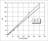
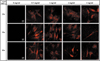
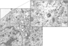
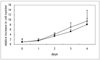
MNP@SiO2(RITC)-PEG showed both superparamagnetic and fluorescent properties. The r1 and r2 relaxivity values of the MNP@SiO2(RITC)-PEG were 0.33 and 398 mM-1 s-1 at 1.5T, respectively, and 0.29 and 453 mM-1 s-1 at 3T, respectively (Fig. 1). The effective internalization of MNP@SiO2(RITC)-PEG into the hMSCs was observed by confocal laser scanning fluorescence microscopy as shown in Figure 2. In these fluorescence images, the hMSCs cultured with MNP@SiO2(RITC)-PEG took up a substantial amount of MNP@SiO2(RITC)-PEG as clearly identified by intracellular fluorescence signals, whereas those cultured without MNP@SiO2(RITC)-PEG were not detected by confocal LSM. There are no differences in the rate of uptake with increasing incubation concentrations of MNP@SiO2(RITC)-PEG. Confocal LSM showed that MNP@SiO2(RITC)-PEG particles resided in the cytoplasm and outside of the nucleus of a cell. These results confirmed that the MNP@SiO2(RITC)-PEG particles exhibited a strong role in targeting hMSCs and that the cellular uptake of MNP@SiO2(RITC)-PEG particles can be visualized by fluorescence imaging at the cellular level. We examined the localization and morphology of MNP@SiO2(RITC)-PEG particles by electron microscopy. The characteristic granular morphology of these MNP@SiO2(RITC)-PEG particles was identified only in MNP@SiO2(RITC)-PEG -labeled hMSCs and never in unlabeled controls. The TEM images showed that MNP@SiO2(RITC)-PEG were indeed internalized into the cells and mainly resided in the cytoplasm (Fig. 3).
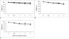
A trypan blue exclusion assay showed that there was no increase in the number of floating cells in the supernatant as determined by visual control under the microscope. In addition, the number of adherent cell was not significantly different between the controls and the labeled cells (Fig. 4). After incubating for two hours with 0.5 mg MNP@SiO2(RITC)-PEG/ml growth medium, we observed no significant difference in the viability between controls (unlabeled cells) and MNP@SiO2(RITC)-PEG-labeled cells. After incubating for two hours at different concentrations of MNP@SiO2(RITC)-PEG, the percentage of viable cells was greater than 96%. After incubating for four hours at different concentrations of MNP@SiO2(RITC)-PEG, cell viability decreased by 3.5% (0.5 mg/ml), 4.9% (1 mg/ml), 7.2% (2 mg/ml), and 9.8% (4 mg/ml). After incubating for six hours at different concentrations of MNP@SiO2(RITC)-PEG, cell viability decreased by 5.4% (0.5 mg/ml), 6.9% (1 mg/ml), 10.9% (2 mg/ml), and 14.4% (4 mg/ml). The proliferation of MNP@SiO2(RITC)-PEG-labeled cells did not significantly differ from the controls (Fig. 5). Moreover, the mean doubling time for the labeled and unlabeled cells was 26.8 hours.
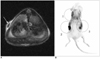
The MR images using the FIESTA sequence from the in vitro gelatin phantom of 5×10⁵ labeled and unlabeled MSCs of 2, 4, 6-hour culture, 1×10⁵ labeled and unlabeled MSCs of 2, 4, 6-hour culture were shown in Figure 6. The SI changes of the labeled cells from the 4-hour culture were -20%, -36%, -67%, and -80% at each incubation concentration of MNP@SiO2(RITC)-PEG (0.5, 1.0, 2.0, and 4.0 mg/ml, respectively). The SI decrease for the 5×10⁵ labeled cells at the 4 mg/ml incubation concentration was 71%, 80%, and 80% at 2, 4, and 6-hours of incubation, respectively. The SI decrease of 1×10⁵ labeled cells at the 4 mg/ml incubation concentration was 39%, 77%, and 60% at 2, 4, and 6-hours of incubation, respectively. The overall SI change for the in vitro gelatin plate MR phantom of the labeled hMSCs was maximal at the four hour incubation time and 4 mg/ml incubation concentration of MNP@SiO2(RITC)-PEG. The labeled hMSCs containing tubes revealed a color fluorescent signal in the tubes. In order to examine the ability of MRI to detect transplanted stem cells, we injected labeled hMSCs into the kidneys of SD rats. The animals were imaged in vivo at six hours after implantation of the labeled cells with several pulse sequences. The imaging revealed a significant drop in signal intensity at the area of cell implantation (Fig. 7). However, the fluorescence signal cannot be detected in the rats in vivo. The fluorescent signal coming from the transplanted labeled hMSCs was detected at the excised kidney. The fluorescence signal of the MNP@SiO2(RITC)-PEG-labeled hMSCs could be imaged in vivo with a subcutaneous injection of a variable number of the labeled hMSCs into nude mice (Fig. 8). The subcutaneously inoculated labeled hMSCs showed a fluorescent signal in nude mice. However, no fluorescent signal was observed when the labeled cells were deeply seated in the body of the mice. The depth of the subcutaneous injection and imaged sites or positions significantly influenced the detectability of the fluorescent signal coming from the injected labeled MSCs.
DISCUSSION
Superparamagnetic iron oxide nanoparticles and their composites are the most popular contrast agents for tracking and studying stem cells by MRI. Unfortunately, the internalizing efficiency of the iron oxide nanoparticles is generally low. The labeling of the cultured stem cells with superparamagnetic iron oxide nanoparticles requires long-term incubation; from approximately 10 hours to a few days in previous studies, despite the various coating material substitutes (14, 15, 21). Popular MRI contrast agents, such as Feridex (AMAG Pharmaceuticals Inc., Cambridge, MA), require the addition of a transfecting agent (internalization agent) for efficient cellular uptake (22). In this study, we demonstrated that the synthesized MNP@SiO2(RITC)-PEG could be efficiently internalized into hMSCs without the assistance of the transfecting agent. In addition, the incubation time used to label the hMSCs was short and the nanoparticles were substantially internalized during the first two hours of incubation. The maximal signal drop in MRI was seen at the four hour incubation time amongst the comparison of the 2, 4 and 6 hours incubation times. The improved labeling efficiency with the short incubation time and the independent transfecting agent might be contributed by the surface modification with PEG groups. PEG coatings of silica shells provides biocompatibility by preventing nanoparticle agglomeration and nonspecific adsorption of proteins to the nanoparticles, thus increasing the particle blood circulation time and the efficiency of their internalization by targeted cells when introduced in vivo (23). A recent study revealed that amorphous silica may present toxicity concerns at high doses (24). The cytotoxic effects of silica nanoparticles can be significantly reduced by surface modification of silica shells with chitosan or PEG (20, 24).
The surface properties of nanoparticles influence its uptake mechanisms. Further, there are different mechanisms for the cellular internalization of the magnetic nanoparticles. However, the best known is the clathrin-dependent endocytosis (25, 26). Kim et al. (26) showed that the uptake of magnetic nanoparticles into the cells is an energy-dependent process and it is mediated through endosomes. After entering the cytoplasm, the ingested nanoparticles are delivered to early endosomes and then transported to the lysosome. The ingested nanoparticles can be stored in different compartments (such as mitochondria) in the cytoplasm (25). The TEM images in this study showed the intracytoplasmic localization of the internalized nanoparticles with a granular appearance. The effective energy-dependent endocytic uptake of the nanoparticles may allow the simultaneous intracellular uptake of the drugs- or genes-of-interest by the formation of complex molecules when the surface of the nanoparticles is optimally modified (26).
We demonstrated that MNP@SiO2(RITC)-PEG was less toxic without affecting the cell viability and cell proliferation of hMSCs after MNP@SiO2(RITC)-PEG-treated cells were incubated with growth medium for various lengths of time. These facts indicated that MNP@SiO2(RITC)-PEGs are quite biocompatible. The noncovalent surface modification of the nanoparticles has a serious limitation for biological applications, because the exposed metal ion on the surface of nanoparticles can cause metal elemental toxicities in cells (in vivo model) (27). In our research, silica (SiO2) was selected for the surface modification of MNPs because it is a good biocompatible material and resistant to decomposition in vivo (20). Consequently, silica-coated core-shell nanoparticles have been extensively studied over the past decade (25), and were recently synthesized with a functionalized surface for bioconjugation through various simple methods that are applicable in biological systems. To improve the versatility of silica-coated core-shell nanomaterials, an organic fluorescent dye was incorporated into the silica shell. Thus, the magnetic and fluorescence properties enable the dual detection of the silica-coated core-shell magnetic nanoparticles (MNP@SiO2). The additional advantage provided by the incorporation of a fluorescent dye into the silica shell is the significant increase in photochemical stability, resulting in minimal photobleaching even after multiple exposures. This aforementioned topic is of great importance among researchers using confocal laser scanning microscopy (20).
Several imaging modalities are currently in use for the molecular and cellular imaging techniques. Unlike the previous approaches in stem cell research that required the processing of fixed tissue samples, these imaging modalities enable the imaging of living, intact transplanted cells in vivo. Since each imaging modality has its own merits and disadvantages, multi-modality imaging has emerged as a strategy that combines the strengths of different imaging methods. Multi-modality imaging can be achieved by the development of true hybrid systems that collect images under the same condition and by the development of sophisticated technologies for image registration and fusion. The development of multifunctional contrast agents can be another way to develop multimodal imaging. Recently, the synthesis of similar nanoparticles for dual modal imaging was reported by various authors (18, 23, 28, 29). They reported that the bifunctional nanoparticles can be used for magnetic resonance as well as the optical imaging of human stem cells, brain tumor, and sentinel lymph node. Despite the rapid advances in optical imaging, there are several challenges in the development of highly sensitive and photostable nanoparticles with a substantial penetration depth. We used organic dyes (RITC) to provide the magnetic nanoparticles with their fluorescent property. The use of organic dyes for stem cell imaging might be limited, since these dyes are often cleared rapidly and thus require a large quantity of dye for optical imaging. For this reason, the light in the near-infrared region of 650 nm-900 nm is most commonly used in practical applications (30). It is still necessary to develop suitable optical probes for deep tissue optical imaging.
The quality of a contrast agent for MRI is dependent on its relaxivity. In this study, the synthesized nanoparticles in an aqueous solution showed significantly higher r2 relaxivity (398 mM-1 s-1 at 1.5T and 453 mM-1 s-1 at 3T) and a higher r2/r1 relaxivity ratio compared to commercially available superparamagnetic iron oxide nanoparticles (ferumoxide and ferucarbotran). Thus, the T2 effect dominates and the MNP@SiO2(RITC)-PEG can be a good T2/T2* agent. The contrast agents exhibit magnetic and optical properties. This bifunctional property enables the simultaneous detection by MRI and optical imaging. We found that MRI and fluorescence imaging can facilitate the identification of labeled stem cells, both in vitro and in vivo and thus, provide an accurate estimate of cell delivery. However, fluorescence imaging showed limitations in penetration depth and consequently visualized the subcutaneously injected labeled hMSCs. However, the deep seated stem cells in small animal cannot be visualized. Although the biomedical application of optical imaging is still limited, the combination of MRI and optical imaging can extend their utilities for molecular and cellular imaging.
In the past decade, the therapeutic use of mesenchymal stem cells in tissue regeneration has been under intense investigation. To date, the true functional role of MSCs in vivo is not clear (31). MSCs may act by paracrine or even endocrine mechanisms related to the production of a broad array of mediators and growth factors with immunosuppressive, anti-inflammatory, antiapoptotic and proliferative effects (32). There is a general consensus that the field of MSC biology is significantly less developed than other stem cell fields. Therefore tracking the distribution and migration of the transplanted MSCs in the patients are important for the development of their therapeutic use. One innovative development for stem cell tracking is the multimodal imaging which might be a powerful method for in vivo visualization of transplanted stem cells by using the combined strengths of each modality. In this study, we demonstrated a bifunctional (magneto/optical) contrast agent using a synergistic integration of magnetic nanoparticles with fluorescent molecules to serve as a good imaging probe for stem cell research. Owing to its bifunctional property, dual modality detection was simultaneously achieved using a single material. The dual modal imaging using magneto/optical nanoparticles is expected to give good anatomical information via MRI, along with more detailed subcellular information via fluorescence imaging taken at the same point.
In this study, we have demonstrated that MNP@SiO2(RITC)-PEG could be efficiently internalized into hMSCs and was less toxic (no significant effect on cell viability and proliferation). As well, the hMSCs labeled with MNP@SiO2(RITC)-PEG were detected in vitro and in vivo, and noninvasively traced by both optical imaging and MRI. In conclusion, although further investigations are needed for stem cell tracking, this study suggests MNP@SiO2(RITC)-PEG is a useful contrast agent for stem cell imaging, which is suitable for a bimodal detection by MRI and optical imaging.


 XML Download