

 PDF
PDF ePub
ePub Citation
 Print
Print


Following the introduction of low-dose digital radiography at the Budker Institute of Nuclear Physics in the beginning of the 1980s, the demand for and the use of digital image acquisition, display, and archiving systems has greatly increased in the field of radiology. Recently, the slot-scan digital radiography (SSDR) method has been introduced for chest and breast imaging as a new scanning technology of digital radiography. The system uses slot scanning geometry to acquire the radiography without the use of an anti-scatter grid. The narrow fan-like beam is synchronized with the detector when scanning a patient. As a result, the image data are obtained from the detector as the patient is scanned via the time consuming method (1).
Some important advantages of the SSDR method include the limited number of detector elements, high spatial resolution, and the lack of a scatter grid (2). In comparison with the conventional computed radiography (CR), the SSDR can offer an improved dynamic range, low-contrast resolution, more rapid access to images, and the opportunity for dedicated image processing (2). Because the intrinsic features of the SSDR detector provide direct conversion, no dead zone, and no dead pixels, the SSDR method can prevent scattered X-rays as well as produce an arbitrary image size with low distortion (3, 4). Although the SSDR method has been primarily used to for chest imaging, it has also recently been used for the skeletal system of both humans and animals. There are several studies citing the use of the SSDR method for musculoskeletal imaging (5, 6). Some of the studies evaluated the long bone in animal models (3), whereas other studies evaluated the spine, hands and feet in human models (6-8). To the best of our knowledge, no studies have reported on the use of the SSDR method for the lower extremity region in human subjects. The purpose of this study was to evaluate the feasibility using the SSDR method for the lower extremity region by comparing the CR method with respect to the image quality and radiation exposure.
MATERIALS AND METHODS
Patients
We enrolled 54 patients (25 men and 29 women; mean age: 29.3 years; age range: 13-80 years) who underwent a CR, followed by a SSDR of the lower extremities at our hospital between January 2003 and March 2007. The CRs were performed between January 2003 and June 2006, whereas the SSDRs were performed between February 2007 and March 2007 (We started the SSDR method at our hospital in July 2006). The mean time interval between performing the CR and SSDR for each patient was 20 months (range: 2-75 months). The underlying diseases of the patients included previous trauma such as traffic accidents and fall injuries (n = 15), osteoarthritis of the knee joints (n = 11), previous infectious arthritis (n = 5), osteochondromatosis (n = 4), congenital vascular malformation (n = 3), Legg-Calvé-Perthes disease (n = 2), a tumor (n = 2), fragile X syndrome (n = 1), and unknown causes (n = 11).
Radiographic Techniques
The CRs were performed using the DHF-158H2 (Hitachi, Tokyo, Japan) and CXDI-40G (Canon Digital Radiography Systems, Tokyo, Japan) units (Table 1). The CR method yielded two image sets (standing anteroposterior radiography of the lower extremities [SAPL] and scanogram of the lower extremities [SL]) of the lower extremities.
The SAPL was performed to evaluate the alignment of the knee joints and to visualize the detailed anatomy of the bone and soft tissue. The SAPL was performed with the patients facing the radiographic tube and both patellae pointing forward. The minimum distance between the patient and the tube was 200 cm, and an image was obtained using 80 kVp, 200 mAs and 0.25 sec. The images were recorded on a CXDI-40G, which had a amorphous silicone flat sensor, using a vertical cassette holder with three individual 36 × 43 cm standard cassettes. The three images were then stitched together to form a composite image that was distributed to a picture achieving and communication systems (PACS) workstation (9).
The SL was performed in the supine position and its purpose was to measure the length of both legs and to compare the differences in the length. The lower extremities were positioned with both patellae pointing toward the ceiling and a radiopaque ruler taped on the table between both legs. The distance between the patient and the tube was 110 cm. Three separate anteroposterior images were obtained with the center over the hip, knee, and ankle joints using 3 separate standard-sized (36 × 43 cm) cassettes at 80 kVp and 50 mAs for the hips, 66 kVp and 16 mAs for the knees, as well as 60 kVp and 10 mAs for the ankles. The images were then distributed to the PACS workstation (9). The SL could provide the exact lengths of the lower extremities. The images of lower extremities performed on SAPL of CR are more magnified than a radiopaque ruler, since the distance between the ruler and cassette is shorter than the distance between the patient and the cassette (10).
The SSDR method was performed to evaluate the alignment of the knee joints as well as measure the lengths of lower extremities. To obtain an SSDR image, we used a Scan-DX (Advanced Digital Technology, Seoul, Korea) (Table 1). The position of the patient for SSDR was the same as for the SAPL of the CR method. The SSDR was obtained with a slot-scan in such a way that the X-rays were collimated into a narrow, horizontally oriented, and fan-shaped beam that closely matched the rectangular detector (2). The distance between the patient and the tube was 130 cm. The SSDR was performed using 110 kVp and 200 mAs at all levels, scanning from the anterior superior iliac spine to the foot, to cover the entirety of the lower extremity. Exposure time depended on the total leg length of each patient. Usually, 8 to 12 seconds were required to scan the lower extremities, including the pelvis (average scanning time per line of scan: 0.005 sec). We did not obtain a separate scanogram using the SSDR method because we could analyze the deformities of the lower extremities as well as simultaneously measure the exact lengths of lower extremities using the SSDR method exclusively without the average magnification of images (11). In this study, the digital radiography system was applied with a multi-channel ionization chamber (MIC) detector, which provided high quality imagery and very short image processing time. This was attributed to direct conversion, low radiation scatter, and high fill factor.
Image Evaluation
Two musculoskeletal radiologists evaluated all of the lower extremities images by consensus, using a PACS (Centricity 2.0, GE Healthcare, Milwaukee, WI), which displayed all the image data on two monitors (two monitors, 1,536 × 2,048 image matrices, 8-bit viewable gray scale, 60-foot-lambert luminescence). To avoid any recall bias, the SSDR and SAPL were evaluated in a different order and at two-week intervals. Before we evaluated each image in detail, we optimized the window width and level of each image on the PACS monitor to allow for optimal image evaluation.
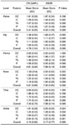
We evaluated the visibility of four features, including the outer margin of the cortex of the bone, the inner margin of the cortex, the trabeculae of the medullary bone (visibility of an individual trabeculae), and the intermuscular fat plane. In turn, this evaluation was performed at six different levels of the lower extremity region, including the pelvis, hip, femur, knee, tibia and ankle (4). A visibility grading scale was used for assessing the image quality of each feature (0: inadequately visualized, 1: sufficiently visualized, 2: well visualized). The visibility score was calculated as the sum of the grades for each of the four features. The maximum and minimum scores were eight and zero, respectively. We compared the mean grading scores for the SSDR and CR methods at each level and for each feature. Finally, we compared the mean of the overall scores for each method to determine whether one of the methods was a significantly superior diagnostic tool for evaluation of the lower extremities.
Radiation Exposure
We measured the entrance skin dose using an X-ray anatomical phantom (Victoreen, Moedling, Austria). The phantom was composed of three sections, including a hip (model 76-642), knee (model 76-675), and ankle (model 76-659). The unit consisted of radiopaque isocyanate rubber that had approximately the same absorption and secondary radiation-emitting characteristics as living tissue. Similarly, the unit was homogeneous and cast shadows similar to the shadows cast by tissue. No spongy portions were present in the phantom and bone marrow was simulated with tissue-equivalent material, thereby allowing for a critical detailed study of bone structure and sharpness comparison using X-rays.
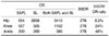
We evaluated the radiation exposure of the CR and SSDR methods at the hip, knee, and ankle on two occasions. In addition, we measured each entrance skin dose for SAPL and SL using the CR method, and then summated the doses of both SAPL and SL at each site. Last, we measured the entrance skin dose for the SSDR method at the same sites. The equipment used for dose measurement was a model Solidose 400 dosimeter (RTI Electronics, Mölndal, Sweden). We placed the dosimeter on a cassette for the CR method or a detector for the SSDR method, respectively. Before obtaining the images, the dosimeter was fixed to maintain the distance from the cassette or detector. Radiation dose was measured in µGy.
Statistical Analysis
The image quality score was analyzed using the paired t-test (SPSS, version 14; SPSS, Chicago, IL). We compared the mean scores of the features and overall scores for the six levels and for the two methods. A p-value of less than 0.05 indicated a statistically significant difference. The measured entrance skin dose between for each method was not statistically analyzed.
RESULTS
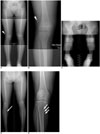
The SSDR method demonstrated a better image resolution and contrast when compared against the SAPL of the CR method (Fig. 1A-E). A comparison of the image quality using the two methods was shown in Table 2. The overall scores for the SSDR method, for all levels, were significantly higher than those obtained by the CR method (p < 0.05). However, the mean scores of outer margin of the cortex at the levels of the pelvis, femur, knee and tibia, as well as those of inner margin of cortex at hip, tibia and ankle, were not significantly different between the two methods.
The entrance skin dose of the SSDR method was less than half of the entrance dose of the SAPL for the CR method at the level of the hip and knee, but was similar at the level of the ankle (Table 3). Similarly, it is also lower (much lower in some cases) for the comparison of the entrance skin dose for SSDR method compared to the SL and the SAPL and SL combined of the CR method (Table 3).
DISCUSSION
Several studies pertaining to the advantages of the SSDR method with respect to other digital techniques have already been reported for chest imaging and mammography exist in the literature (1, 2, 12, 13). The SSDR method was determined to be advantageous over the full-field digital radiography based on significant scatter reduction, high detective quantum efficiency, and improved image quality (1). Further, the SSDR method has allowed for dose-efficient scatter rejection and the ability to use small detectors to produce a large-area image during a mammography (14). As a result, some studies have suggested that the SSDR method could be an alternative to other digital techniques (2). Some studies have also cited the advantages of using a MIC detector (15, 16). Since the MIC detector is a direct conversion type, it enables a high counting rate, high spatial resolution, low sensitivity to gas impurity, and low cost (16). In our study, we applied both a slot-scan type and MIC detector to obtain a digital radiography of better image quality with lower radiation dose of the lower extremities, compared to the CR method.
In terms of evaluating each level, most levels showed differences in image quality when comparing the CR and SSDR methods; however the images at the level of the tibia showed less of a difference in comparison to the other levels (Table 2). Reasons for this include a relatively shallower thickness of the lower extremities below the knees, which resulted in a higher spatial resolution below the knees, in comparison to the pelvis and thigh for the CR method (1). The image quality at the level of the pelvis and hip were relatively worse than at the other levels due to the presence of thick soft tissue and multiple pelvic organs (17).
The mean scores of the outer margin of the cortex at the levels of the pelvis, femur, knee, and tibia were not significantly different between the SAPL of the CR method and the SSDR method. The cortex is very radio-opaque on a plain radiography. The outer margin of the cortex has a higher contrast due to the surrounding soft tissue, which is relatively radiolucent as compared with the medullary bone. Therefore, despite its lower contrast resolution, the CR method imagery for the outer margin of the cortex is as good as the SSDR method.
The scanning time required to obtain the SSDR of the lower extremities was about 10 seconds. As a result, the SSDR method had a high probability of producing a motion artifact. However, a previous study demonstrated that motion in a brief period degraded only a small segment of slot scanner image, while it affects the entire portion of CR image (18). Continuous motion throughout the image acquisition period caused an artifact that was less apparent with the SSDR method than with the CR method. Actually, in our cases, none of the SSDR images had any significant motion artifacts leading to a difficulty in reaching the correct diagnosis. The lower extremities are not as affected by spontaneous body movement, which usually occur as a result of respiration, heartbeat, and bowel movement.
In this study, we measured the entrance skin dose instead of the effective radiation dose to evaluate radiation exposure. Generally, the effective radiation dose is used because it reflects the absorption of radiation (6). To obtain the effective radiation dose, a thermo-luminescent dosimeter-loaded phantom and commercially available software for calculation are required. However, we did not have access to such equipment; especially for the lower extremities. Therefore, we had no choice but to measure the entrance skin dose as an alternative method for measuring radiation exposure.
The entrance skin dose for the SSDR method was much lower than that of the CR method (Table 3). The largest difference in entrance skin dose was recorded at the level of the pelvis, which contained multiple pelvic organs (urinary bladder, bowel, and large arteries as well as the uterus in females), large bones (pelvic bones, sacrum and femoral heads) and a fair amount of soft tissue (subcutaneous and intrapelvic fat, muscles). As the body mass of a patient increases, the scatter fraction also increases (1). Therefore the entrance skin dose for both SAPL and the SL of the CR method was much larger than the entrance skin dose of the SSDR method. When the SL was performed, the patient was exposed to X-rays on three occasions. One of the three X-ray exposures was intended to obtain an image at the targeted level, whereas the other two X-ray exposures were caused unnecessarily by a scatter fraction to the other two levels.
Despite the conclusive results, the study had some inherent limitations; first, the images obtained by the CR and SSDR methods could not be obtained at same time. The mean time interval between first performing the CR, followed by the SSDR was 20 months with a maximum interval time of 75 months. Second, we measured the entrance skin dose to evaluate radiation exposure instead of the effective radiation dose, because we did not have access to a commercially available phantom for the lower extremity region.
In conclusion, the data suggest that the SSDR method can provide a better image quality than the CR method in the lower extremity region, as evidenced by a more detailed anatomy in the medullary bone and soft tissue of the lower extremity region. In addition, the SSDR method, for the most part, exposes the patient to a lower entrance skin dose, especially for imaging at the level of the hip and knee.

 XML Download