

 PDF
PDF ePub
ePub Citation
 Print
Print


The notochord is an embryonic tissue that induces the development of the vertebral column in fetal life, and completely disappears to form the nucleus pulposus of the intervertebral disc (1, 2). Residual notochordal tissue does not normally remain in the vertebral body and is only rarely evident in the intervertebral disc beyond an age of 10 years (3). Extraosseous macroscopic notochordal rests, termed ecchordosis physaliphora and found in 0.4 to 2% of autopsies, are well established entities and are only rarely symptomatic (4). However, vertebral notochordal rests are rarely found in adult autopsies and are then only in microscopic findings. A giant vertebral notochordal rest (GVNR) is a new and evolving term used to describe benign notochordal tissue that may produce a macroscopic vertebral lesion, with only seven previous cases reported in the literature (5-7). We here report magnetic resonance imaging (MRI) findings, including diffusion weighted imaging (DWI), of a patient with a GVNR, with an emphasis on its distinction from a chordoma.
CASE REPORT
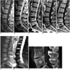
A 48 year-old woman sought medical attention for persistent lower back pain. Neurologic examinations were normal, as was a roentgenographic examination of the spine. An MRI of the lumbar spine was then performed, which clearly demonstrated a lesion of the fifth lumbar vertebra, within the confines of the vertebral cortex, having low T1- and high T2-weighted signal intensities, and showing no enhancement (Fig. 1A, B). There was no evidence of soft tissue extension. In the presence of only mild trabecular sclerosis and no bone destruction as seen on computed tomography (CT), the provisional diagnosis of a GVNR was deemed most likely and the patient was subject to close follow-up with bimonthly intervals. Repeat MRI, DWI and CT, performed one year after the patient first presented with symptoms failed to demonstrate any lesion growth (Fig. 1C-H). Based on the nonaggressive radiological pattern and the lack of morphological features associated with a chordoma, the patient was considered as having a GVNR and has been recommended to continue follow-up. Fourteen months after her first presentation, the patient is alive and well, and her back pain has benefited greatly from medical and physical therapy.
DISCUSSION
A chordoma is a rare tumor (0.2 cases per 100,000) of the axial skeleton found most commonly at the sacrococcygeal and spheno-occipital regions (6). The typical presentation is in the form of a large destructive bone lesion. The presence of soft tissue invasion and its location makes complete surgical excision difficult to perform and can lead to a poor prognosis.
The origin of chordomas from notochordal rests is widely accepted. Notochordal remnants are generally extraosseous in location. They have a predilection for the spheno-occipital and sacral regions, where branching of the notochord occurs and chordomas are also usually found (5). Intravertebral notochordal rests are rare autopsy findings (about 1% of autopsy series) and when found are microscopic in size (6, 7). The actual rate of transformation for these intravertebral vestiges into a chordoma is not known. However, when their incidence are compared the transformation seems to be a rare event (7).
The diagnosis of giant vertebral notochordal rest in the above presented case is certainly debatable. Until recently, a similar patient would have invariably undergone a vertebrectomy with a leading diagnosis of a chordoma. Currently, the concept that a benign notochordal rest, termed a GVNR could present similarly in a patient has been accepted following strong debate, with a total of seven cases published in the literature (5-8).
The distinction between these two entities relies mostly on imaging findings and is of obvious clinical importance since the management and prognosis of a GVNR and a chordoma is completely different. The lack of progression, bone destruction or soft tissue extension are important imaging criteria agreed by most investigators that suggest a radiological diagnosis of a GVNR (7). Although a chordoma typically grows slowly, it is always progressive in nature. It is also well known that at the time of diagnosis, a chordoma invariably shows bone destruction and, in almost all cases, has obvious soft tissue extension (6). In the above presented case, the lesion met all of the defined radiological criteria and showed no progression between follow-up, establishing the diagnosis of a GVNR.
Other possible benign vertebral lesions that can be considered in the differential diagnosis include an atypical hemangioma, a simple bone cyst, an osteoid osteoma and an osteoblastoma. Although both an atypical hemangioma and bone cyst might show similar MRI findings, they can readily be distinguished by their CT appearances. Apart from clinical presentation, both an osteoid osteoma and osteoblastoma can easily be differentiated by their intense contrast enhancement.
An important reproach can be made for not performing a percutaneous needle biopsy to confirm the diagnosis in the above presented case. However, due to the histological heterogeneity of a chordoma, its distinction from a notochordal rest may be impossible to determine when presented with material from a needle biopsy tissue alone, which shows only physaliphorous and univacuolated cells (6). In these circumstances an excisional biopsy in the form of a vertebrectomy is needed for final diagnosis. Considering the benign nature of a GVNR, this procedure is not always preferred, making its diagnosis very much dependent upon the radiological findings.
Considering its ability to detect altered water-proton mobility, there have been recent efforts to implement DWI in the evaluation of spinal disorders. DWI has been particularly useful for distinguishing acute benign osteoporotic from malignant vertebral compression fractures (9, 10). Increased diffusion of interstitial water is a common phenomenon and may be observed in acute posttraumatic edema of the bone resulting in low signal intensity on DWI (10). In contrast, densely packed tumor cells restrict the diffusion, resulting in a lower phase shift with high signal intensity on DWI with low signal intensity on apparent diffusion coefficient (ADC) maps (9). To the best of our knowledge there are no previous reports concerning DWI findings of a GVNR. In the above presented case, DWI performed with a b value of 600 sec/mm2 showed high signal intensity on both trace images and the corresponding ADC maps. A quantitative evaluation of ADC values failed to demonstrate any diffusion restriction. Based on these findings, we hypothesized that increased signal intensities on DWI and corresponding ADC maps are not related to diffusion alteration, but are greatly influenced by the T2 shine-through effect and reflect the non-infiltrative character of the lesion.
In conclusion, a GVNR is a new, benign entity, which can easily be confused with a vertebral chordoma. Differentiation between these two entities can readily be made radiologically, with DWI being a useful new supplementary imaging tool. Management of a GVNR found incidentally during radiological imaging should be conservative, with periodic follow-up imaging in order to detect possible transformation of the lesion into a chordoma.
 XML Download