

 PDF
PDF ePub
ePub Citation
 Print
Print


Malignant mixed tumors of the salivary glands are classified into three distinct histologic types: 1) carcinoma ex pleomorphic adenoma, 2) benign metastasizing pleomorphic adenoma, or 3) true malignant mixed tumor (carcinosarcoma) (1). Of these tumors, carcinoma ex pleomorphic adenoma represents approximately 99% of cases (1). A true malignant mixed tumor is a very rare tumor composed of both malignant epithelial and malignant mesenchymal elements. The most common malignant epithelial component is squamous cell carcinoma or adenocarcinoma, whereas the most common malignant mesenchymal component is chondrosarcoma, followed by fibrosarcoma, leiomyosarcoma, osteosarcoma, and liposarcoma (2). A true malignant mixed tumor represents only 0.04% to 0.16% of salivary gland tumors and 0.4% of malignant salivary gland neoplasms (2). About 65% of cases occur in the parotid gland but it has also been described in the submandibular and minor glands of the palate (3). Although they do develop de-novo, an association with pleomorphic adenoma has been reported in approximately one third of patients (4).
A true malignant mixed tumor is an aggressive high-grade malignancy and has a high propensity for both local and regional recurrence and metastasis (5). However, the incidence and imaging appearance of pulmonary metastasis from a true malignant mixed tumor of salivary glands have not previously been described in English literature sources. Herein we report a case of a true malignant mixed tumor of the parotid gland that developed pulmonary metastases with atypical radiologic appearance. The imaging appearance and histopathologic features of the atypical pulmonary metastases are discussed.
CASE REPORT
A 58-year-old male patient presented with a one-week history of bloody otorrhea from the right side. In the past 34 years, the patient had undergone four surgeries to remove the masses from his right parotid gland. Histopathologic examinations performed after the first three surgeries indicated the presence of benign pleomorphic adenomas. The latest surgery was performed nine months ago to remove a 3.3 cm mass from the parotid gland. Following this surgery however, a histopathologic examination revealed a true malignant mixed tumor (carcinosarcoma) composed of poorly differentiated carcinoma and osteosarcoma. Immunohistochemical studies were positive for P53, Ki67, and vimentin, weakly positive for cytokeratin, and negative for Actin and the S100 protein. The patient was given adjuvant radiotherapy after the latest surgery and was doing well except for symptoms associated with irradiation.
The patient underwent a follow-up high-resolution CT scan of the temporal bone for the presented symptoms and showed extensive opacification of the right mastoid air cells, posterior petrous bone, middle ear, and the external auditory canal. The imaging appearance raised the suspicion of a recurrent tumor. Excisional biopsy of the external auditory canal mass was performed and confirmed the diagnosis of a recurrent true malignant mixed tumor.
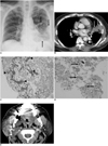
During this hospitalization, a chest radiograph indicated the presence of patchy opacities in his left lower lung field (Fig. 1A) not seen in previous chest radiographs; this warranted a CT scan for further characterization. A CT scan of the chest showed parenchymal consolidation with some amorphous calcifications (Fig. 1B); hence, the possibilities of fungal infection or granulomatous disease were considered initially. This patient had no fever and did not report associated symptoms of infectious or inflammatory respiratory tract diseases; therefore, a CT-guided biopsy of the pulmonary lesion was performed because of the concern of atypical pulmonary metastases. The biopsy generated two pieces of specimen that were used for histopathologic examination.
Microscopically, the specimen showed nearly total necrosis and the presence of atypical cells; within the mesenchymal element, some osteoid materials and foci of osteonecrosis were noted (Fig. 1C). Malignant osteoblastic cells surrounding osseous materials were also noted (Fig. 1D). Immunohistochemical studies were positive for vimentin, negative for TTF-1 (thyroid transcription factor-1) and cytokeratin. A review of the surgical specimen from the patient's right parotid gland nine months ago revealed similar findings. The specimen obtained during the lung biopsy showed no carcinomatous component, and did not demonstrate tumor emboli in pulmonary vessels. A radionuclide bone scan did not reveal increased activity at locations other than right neck and lung. The histopathologic analysis of the lung biopsy suggested a metastatic true malignant mixed tumor. Retrospective reading of his neck CT nine months ago clearly depicted calcification within the right parotid gland tumor (Fig. 1E).
DISCUSSION
The lung is a common site for metastases. Large autopsy series of patients with extrathoracic malignancies reveal pulmonary metastases in 20-54% of cases (6, 7). Typical radiologic findings of pulmonary metastases may include multiple round variable-sized nodules and a diffuse thickening of interstitium, representing hematogenous metastases and lymphangitic carcinomatosis, respectively (7, 8). In daily practice, however, atypical radiologic features of metastases are often encountered that make distinguishing between metastases and other nonmalignant pulmonary diseases difficult. Herein we describe a case of atypical pulmonary metastases from a true malignant mixed tumor of the parotid gland demonstrating calcification and an air-space pattern of the pulmonary metastases.
Calcification of a pulmonary nodule is usually suggestive of its benign nature, most commonly a granuloma and less commonly a hamartoma. However, calcification or ossification can also occur in metastases from a variety of tumors including osteosarcoma, chondrosarcoma, synovial sarcoma, giant cell tumor of the bone, carcinomas of the colon, ovary, breast, and thyroid (7). In our case, the mesenchymal component of the true malignant mixed tumor of the parotid gland was osteosarcoma. As a result, the lung metastases show calcification or ossification; this observation was confirmed with a CT-guided biopsy. The calcification is often depicted only with CT, as in our case.
Metastases from an extrapulmonary adenocarcinoma may spread into the lung along the intact alveolar walls (lepidic growth) in a fashion similar to a bronchioloalveolar carcinoma and exudative pneumonia (7, 8). This type of metastases may demonstrate an air-space pattern in radiologic studies. In one series, six of 65 patients with pulmonary metastases from an adenocarcinoma of the gastrointestinal tract had this pattern of metastasis (8). In addition, adenocarcinomas from the breast and ovary can also show this pattern of metastasis (9). The malignant epithelial component in our case was poorly differentiated carcinoma, and unusual scenario for the development pulmonary metastasis of air-space pattern. Rastogi et al. (10) described a variety of atypical locations and presentations of thoracic metastases from osteosarcoma in 16 patients; pulmonary metastasis of air-space pattern did not occur in their series. In our case, the extensive necrosis revealed by the lung biopsy may be one of speculative mechanisms of air-space pattern. Pulmonary infarction due to the presence of a tumor embolism may also demonstrate an air-space pattern (7). However, in our case, the limited pieces of specimen obtained at lung biopsy did not demonstrate foci of carcinomatous components and were negative for the presence of a tumor embolism. Although we could not explain the definitive mechanism of air-space pattern on the basis of limited histopathologic analysis, the current case demonstrated an extremely rare example of atypical pulmonary metastases from a true malignant mixed tumor of the parotid gland.
Although most cases of pulmonary metastases can be diagnosed radiologically on the basis of typical findings, atypical radiologic manifestations of pulmonary metastases make confident diagnosis difficult, as in our case. An awareness of the possibility of atypical pulmonary metastases in patients with known extrapulmonary malignancy was crucial to allow an accurate diagnosis. In cases displaying atypical radiologic features of pulmonary metastases, tissue diagnosis at bronchoscopy or percutaneous transthoracic needle aspiration biopsy is recommended.
 XML Download