Abstract
Acute obstructive cholangitis due to the migration of necrotized tumor fragment is a rare complication occurring after a transarterial chemoembolization. The percutaneous tumor removal procedure following percutaneous transhepatic biliary drainage is an appropriate treatment over endoscopic removal for the relief of acute cholangitis in this case. Following this serial management, no invasive hepatocellular carcinoma of the bile duct recurred after two years of follow-up.
Atranscatheter arterial chemoembolization (TACE) is the ubiquitous modality of choice for the treatment of hepatocellular carcinoma (HCC). Following a TACE, some infrequent side effects and complications have occurred and have been reported. The most common side effects are transient fever and pain at one or two weeks after the TACE, the so-called post-embolization syndrome. Other more infrequent complications, such as hepatic infarction and liver abscess or biloma have also been reported. However, acute obstructive cholangitis, due to the migration of completely necrotized tumor fragment after a TACE with obstruction of the distal common bile duct (CBD), is rare. Following these serial management, no recurred invasive HCC of the bile duct was observed over two years of follow-up. We report a case of a successfully treated acute obstructive cholangitis after TACE, due to the pathologically diagnosed, a total a total necrotic migrating tumor fragment on a CBD, after an interventional percutaneous removal procedure.
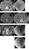
A 62-year-old male visited the Department of Endocrinology to control his hyperglycemia. The patient complained of intermittent fatigue, general aches, and chills over the previous two months. Further, the patient had a history of HBV carrier status, diabetes mellitus, and alcoholism over the last 20 years. The relevant laboratory results at the time of admission were as follows: glucose, 313 mg/dl; aspartate aminotransferase, 96 IU/K; alanine transferase, 164 mg/dl; serum albumin, normal range; alkaline phosphatase, 843 IU/dl (normal range, 103-335 IU/L); total serum bilirubin, 2.3 mg/dl (normal range 0.2-1.2 mg/dl); HBsAg, positive; HAV Ab-IgG, positive; and HBeAg, positive. The serum level of the alpha-fetoprotein was 182.9 ng/ml (normal range, 0.0-8.1 ng/ml). An initial abdominal computed tomography (CT) showed round, highly enhanced nodules within a dilated proximal common hepatic duct (CHD) and left hepatic duct (LHD) at segments 3 and 4 without ductal dilatation of the right lobe (Fig. 1A). These laboratory and imaging findings were consistent with intraductal invasive HCC. On the patient's 14th day of hospitalization, the first TACE was performed. During the TACE, an ill-defined curvilinear tumor staining was shown at segments 3 and 4, which extended along the left main portal vein on a selective left hepatic angiography (Fig. 1B). After the superselection of a main feeding artery from the left hepatic artery with a 3 Fr microcatheter (Terumo Co., Tokyo, Japan), a continuous infusion with 50 mg of cisplastin (Il-dong, Seoul, Korea) was performed for 15 minutes. Next, 10 ml of a mixture comprising 7 cc of iodized oil (Lipiodol; Laboratoire Andre Guerbet, Aulnaysous Bois, France) and 40 mg of doxorubicin hydrochloride (Adriamycin; Il-dong) were infused until the compact lipiodol uptake of the tumor was complete. In addition, the left hepatic artery, which was the main feeder, was occluded by 500 EA of gelfoam sponge particle (Gelfoam; Johnson & Johnson, Skipton, England), which was soaked in a mixture of doxorubicin hydrochloride (10 mg) and mitomicin-C (2 mg) (Mitomicin; Il-dong, Seoul, Korea). Finally, the scout image after the TACE showed compact curvilinear lipiodol uptake along left portal vein area.
Two days after the TACE, the patient complained of a fever, cough, and abdominal pain. The laboratory findings demonstrated an acute marked elevated alkaline phosphatase (1,015 IU/L), erythrocyte sedimentation rate (103 mm/hr), and serum total bilirubin (3.3 mg/dl). An abdominal CT performed one week after the first TACE showed multiple compact lipiodol deposits within proximal CHD and LHD (Fig. 1C). In spite of the supportive management for fever and abdominal pain over several days, the patient suffered from persistent abdominal pain and jaundice. A follow-up abdominal CT two weeks after the TACE showed an acutely dilated CHD and proximal CBD which had acutely increased since the previous CT examination. Moreover, the lipiodol deposits were not visualized at the same level (Fig. 1D). In addition, an obstructive dense lipiodolized tumor fragment (1 cm) was observed in the distal CBD (Fig. 1E). The percutaneous transbiliary drainage (PTBD) was performed for relief of obstructive jaundice two days after the CT. As a result, the cholangiography of the PTBD showed an ovoid large filling defect in the distal CBD with complete obstruction of the distal passage of contrast material (Fig. 1F). We planned the percutaneous removal of the migrated tumor fragment via the PTBD tract rather than endoscopic removal due to the patient's dyspnea. Three days after the PTBD, we performed the percutaneous removal of the tumor fragment in the distal CBD using a 1 cm biliary stone basket (Cook, Bloomington, IN) via an 8 Fr biliary sheath. During the procedure, the small part of the tumor fragment was removed percutaneously since most parts of the tumor fragment were not captured and removed by the stone basket due to the toothpaste-like character of the tumor fragment. An 8F aspiration catheter was then used for aspiration of the tumor fragment from the distal CBD. Subsequent to these procedures, a final cholangiography revealed complete disappearance of the tumor fragment in the distal CBD, followed by the restoration of normal contrast passage via the ampulla of Vater (Fig. 1G). We suspected that the tumor fragment was crushed after capture in the stone basket, which facilitated aspiration by the aspiration catheter. Following the tumor aspiration, the patient's symptoms including the jaundice, fever, cough, and abdominal pain subsided immediately and the laboratory values indicative of cholangitis gradually improved. The PTBD tube was removed seven days after the procedure and the patient was subsequently discharged. At the time of discharge, the laboratory values were as follows: alkaline phosphatase, 605 IU/L; and total serum bilirubin, 1.2 mg/dL. The pathologic diagnosis of the removed tumor fragment showed a total necrotic HCC.
The presence of jaundice in a patient with intraductal invasive HCC is an uncommon manifestation at the time of diagnosis which usually occurs in later stages (1-3). HCC may involve the biliary tract in several different ways: tumor thrombi, hemobilia, tumor compression, or diffuse tumor infiltration. Infrequently, jaundice may also result from the external compression of the major bile ducts by direct tumor encasement or by the metastatic lymphadenopathy at the porta hepatis. Histopathologically, intraductal tumor invasion shows features resembling the viable HCC in the liver parenchyma (4-6).
A few cases have reported the incidence of a necrotic tumor detaching from the bile duct wall, which migrated into the distal CBD and caused obstructive jaundice (2, 7). Further, the successful management of this with the interventional procedure was rare.
In our case, a subsequent abdominal CT scan and cholangiography of the PTBD revealed lipiodolized migrating necrotic tumor fragments in the distal CBD due to an apparent ischemic effect of lipiodol and gelfoam particles after a TACE for bile duct invasive HCC.
A TACE induces marked ischemic necrosis in HCC; however, its effect is apparently not limited to the tumoral vascular bed, as shown by the importance of bile duct injuries observed in an autopsy study of cirrhotic subjects treated by TACE (8). Recently, several reports have demonstrated the clinical efficacy and significant prolonged survival rate of patients after hepatic arterial chemotherapy, combined with 5-FU, adriamycin, and cisplastin in inoperable HCC patients (9, 10)
Patients with primary liver cancer and jaundice due to tumor invasion in the bile duct may benefit from a surgical resection. The goals of the surgical intervention include biliary decompression with removal of tumor debris or tumor-containing blood clots, and if possible, curative resection of the hepatic tumor. The commonly used operative methods are a lobectomy, hepatectomy plus a thrombectomy, choledochotomy with Y-tube drainage alone, or biliary diversion (11, 12).
Several non-surgical methods exist for treatment of migrated tumor fragments in the bile duct, caused obstructive jaundice in inoperable patients. Between the PTBD and endoscopic biliary drainage methods, the latter is usually performed as an initial treatment. However, the difference in survival between the two methods in the treatment of obstructive jaundice in HCC patient, is still a matter of debate (13, 14). The necessity of removing a migrating tumor fragment without symptoms is debatable because there is no existing comprehensive report of gross and pathologic features for a migrating tumor fragment in the CBD.
In our case, we performed a right PTBD and subsequent percutaneous removal using a biliary stone basket and aspiration catheter. During the removal procedure, the effect of the stone basket on the tumor fragment was highly effective in terms of maceration and fragmentation, which was helpful for the aspiration of tumor fragments and spontaneous expulsion into the duodenum. As a result, biliary obstruction was relieved and the intraductal lesion disappeared. During two years of follow-up, the patient showed no evidence of tumor recurrence or recurrent bile duct dilatation on abdominal CT.
As a general guideline, the results of this study bring to light the possible complications of treating an intraductal invasive HCC by TACE which should be made aware to the interventional radiologist for consideration. Nevertheless, subsequent interventional treatment, including PTBD with percutaneous removal, may be helpful in the management of obstructive cholangitis after a TACE. Most patients will have satisfactory palliation as well as the occasionally cure and long-term survival if the appropriate procedures are selected and executed safely.
Figures and Tables
 | Fig. 1Acute obstructive cholangitis after transarterial chemoembolization in 62-year-old male.
A. Initial abdominal CT scan shows ovoid-shaped, highly enhancing invasive intraductal hepatocellular carcinoma in proximal common hepatic duct and left hepatic duct with ductal dilation (black arrow).
B. Selective left hepatic angiography performed during initial transcatheter arterial chemoembolization showed ill-defined, tumor staining along proximal common hepatic duct and left hepatic duct (black arrows). Tumor staining occurred in identical location of bile duct invasion on CT image.
C. Follow-up abdominal CT performed one week after initial transcatheter arterial chemoembolization showed multifocal, dense lipiodol deposits within proximal common hepatic duct and left hepatic duct after initial transcatheter arterial chemoembolization (black arrow). We also found small, faint, parenchymal lipiodol deposits in peripheral portion of left lobe.
D. Follow-up abdominal CT performed two weeks after initial transcatheter arterial chemoembolization showed acutely dilated proximal common bile duct and left hepatic duct without visualization of previous lipiodol deposits at same level (black arrow).
E. Same abdominal CT showed distally migrated, dense lipiodolized tumor fragments in distal common bile duct (black arrow), which was completely obstructed in distal common bile duct and resulted in dilatation of proximal common bile duct, left hepatic duct, and gallbladder.
F. Percutaneous transhepatic biliary drainage was performed for treatment of obstructive jaundice. Cholangiography of percutaneous transbiliary drainage showed ovoid, large filling defect in distal common bile duct with complete obstruction of distal passage of contrast (black arrow).
G. Final cholangiography performed after these procedures showed complete disappearance of tumor fragment in distal common bile duct, restoration of contrast passage into duodenum, and decompressed biliary tree (black arrow).
|
References
1. Qin LX, Tang ZY. Hepatocellular carcinoma with obstructive jaundice: diagnosis, treatment and prognosis. World J Gastroenterol. 2003. 9:385–391.
2. Hiraki T, Sakurai J, Gobara H, Kawamoto H, Mukai T, Hase S, et al. Sloughing of intraductal tumor thrombus of hepatocellular carcinoma after transcatheter chemoembolization causing obstructive jaundice and acute pancreatitis. J Vasc Interv Radiol. 2006. 17:583–585.
3. Wang HJ, Kim JH, Kim JH, Kim WH, Kim MW. Hepatocellular carcinoma with tumor thrombi in the bile duct. Hepatogastroenterology. 1999. 46:2495–2499.
4. Murata K, Shiraki K, Kawakita T, Yamamoto N, Okano H, Sakai T, et al. Hepatocellular carcinoma presenting with obstructive jaundice: a clinicopathological study of eight cases. Hepatogastroenterology. 2003. 50:2057–2060.
5. Tantawi B, Cherqui D, Tran van Nhieu J, Kracht M, Fagniez PL. Surgery for biliary obstruction by tumour thrombus in primary liver cancer. Br J Surg. 1996. 83:1522–1525.
6. Satoh S, Ikai I, Honda G, Okabe H, Takeyama O, Yamamoto Y, et al. Clinicopathologic evaluation of hepatocellular carcinoma with bile duct thrombi. Surgery. 2000. 128:779–783.
7. Spahr L, Frossard JL, Felley C, Brundler MA, Majno PE, Hadengue A. Biliary migration of hepatocellular carcinoma fragment after transcatheter arterial chemoembolization therapy. Eur J Gastroenterol Hepatol. 2000. 12:243–244.
8. Kobayashi S, Nakanuma Y, Terada T, Matsui O. Postmortem survey of bile duct necrosis and biloma in hepatocellular carcinoma after transcatheter arterial chemoembolization therapy: relevance to microvascular damages of peribiliary capillary plexus. Am J Gastroenterol. 1993. 88:1410–1415.
9. Ando E, Tanaka M, Yamashita F, Kuromatsu R, Yutani S, Fukumori K, et al. Hepatic arterial infusion chemotherapy for advanced hepatocellular carcinoma with portal vein tumor thrombosis: analysis of 48 cases. Cancer. 2002. 95:588–595.
10. Hwang JY, Jang BK, Kwon KM, Chung WJ, Park KS, Cho KB, et al. Efficacy of hepatic arterial infusion therapy for advanced hepatocellular carcinoma using 5-fluorouracil, epirubicin and mitomycin-C. Korean J Gastroenterol. 2005. 45:118–124.
11. Peng SY, Wang JW, Liu YB, Cai XJ, Deng GL, Xu B, et al. Surgical intervention for obstructive jaundice due to biliary tumor thrombus in hepatocellular carcinoma. World J Surg. 2004. 28:43–46.
12. vanSonnenberg E, Ferrucci JT Jr. Bile duct obstruction in hepatocellular carcinoma (hepatoma)--clinical and cholangiographic characteristics. Report of 6 cases and review of the literature. Radiology. 1979. 130:7–13.
13. Matsueda K, Yamamoto H, Umeoka F, Ueki T, Matsumura T, Tezen T, et al. Effectiveness of endoscopic biliary drainage for unresectable hepatocellular carcinoma associated with obstructive jaundice. J Gastroenterol. 2001. 36:173–180.
14. Lee JW, Han JK, Kim TK, Choi BI, Park SH, Ko YH, et al. Obstructive jaundice in hepatocellular carcinoma: response after percutaneous transhepatic biliary drainage and prognostic factors. Cardiovasc Intervent Radiol. 2002. 25:176–179.


 PDF
PDF ePub
ePub Citation
 Print
Print


 XML Download