

 PDF
PDF ePub
ePub Citation
 Print
Print


Percutaneous vertebroplasty (PVP) has been widely used for the management of osteoporotic vertebral fractures. However, the percutaneous approach to the upper or middle thoracic vertebrae is difficult due to the small size of the pedicle and vertebral anatomy in the thoracic vertebrae (1-3). In a lateral view under fluoroscopy, it is very difficult to confirm the precise location of the pedicle of the upper thoracic spine due to interference from the shoulders. The use of a computed tomography (CT) scan during vertebroplasty provides excellent imaging and the possibility of exact positioning of the vertebroplasty needle. Potential polymethylmethacrylate (PMMA) leakage can be detected early in the procedure with the use of an axial CT scan. Therefore, the amount of leakage can be reduced and optimal filling of the vertebral body can be achieved (2).
CASE REPORT
Clinical and Radiological Findings
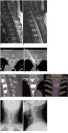
A 51-year-old woman was hospitalized due to posterior neck pain and left arm weakness after a traffic accident while riding a bicycle. On admission, there was a four-fifths decrease in motor function of the left arm. The patient had a paresthesia on the left C6-7 dermatome. Plain radiographs of the cervicothoracic spine revealed compression fractures of C7-T3 with anterior subluxation of facets at C6-7. CT and magnetic resonance imaging (MRI) showed anterior wedging of the C7 vertebral body, ligamentous disruption at the C6-7 level and acute compression fractures at T1-3 (Fig. 1A, B). In general, vertebral compression fractures have been treated mainly with bed rest, the use of analgesics, braces and various physical therapies. However, unlike lumbar compression fracture patients, the patient was uncomfortable in the supine position and even lying down in bed. The patient had to keep her posture in the sitting or a semi-Fowler position all day, and the patient had slight difficulty in deep breathing. The cervicothoracic junction was unstable due to injury of the posterior ligamentous complex. The patient underwent CT-guided vertebroplasty at T1-3 and an anterior discectomy and instrumented interbody fusion at C6-7.
Procedural Technique
The procedure was performed in strictly sterile manner in the CT (Somatom Sensation Open scanner, Siemens Medical Solutions, Erlangen, Germany) room. After the patient was placed on the CT table, blood pressure and pulse oximetry were monitored continuously. The area to be treated was prepared in a strictly sterile manner and then CT topography for the upper thoracic spine, including the symptomatic lesion, was checked. On this topographic image, the T3 vertebral body was identified and the course of needle insertion was determined. Axial CT scanning was performed for the T3 vertebral body and a sectional image for needle insertion was selected. The image mode was converted to CT fluoroscopy, and the image size was magnified for precise monitoring. With the administration of local anesthesia with 1% lidocaine, a 10-gauge vertebroplasty cannula was inserted to the affected vertebral body via the unilateral transpedicular route. The tip of the cannula was placed in the anterior half of the vertebral body (Fig. 1C, D). A Total of 3 cc PMMA was applied to the T3 vertebral body. During the procedure, needle advancement, venous leakage, the injection process of PMMA and extravasation out of the vertebral body were carefully monitored by real-time CT fluoroscopy. Compression fractures of the T1 and T2 vertebral bodies were managed under the same conditions. Six days after the procedure, the patient underwent an anterior discectomy and instrumented interbody fusion and interspinous wiring with a Songer cable at C6-7.
Postoperative Course
Postoperative radiographs and CT images demonstrated complete reduction of the C7 vertebral body and a good position of instruments (Fig. 1E). Posterior neck and shoulder pain were significantly decreased following PVP. A Philadelphia collar was used for postoperative neck immobilization. The patient was discharged six days after surgery with significant improvement in the left arm weakness and neck pain. However, paresthesia on the left arm still remained. At a 5-month follow-up examination, the patient reported no neck pain or limb weakness. A radiological study of the cervical spine performed five months after surgery revealed evidence of solid bone fusion and no cervical instability (Fig. 1F).
DISCUSSION
Percutaneous vertebroplasty was developed by Deramond and colleagues (4) in 1987 and has been widely used in the therapy of vertebral compression fractures. The procedure provides benefits in terms of pain relief, increased activity and decreased analgesic drug consumption in patients with an osteoporotic compression fracture. The use of PMMA leads to stabilization and immobilization of compressed vertebral bodies and the heat effect of cement on nerve endings plays a role in pain reduction (4, 5).
The average transverse diameter of the pedicle at T1 vertebral body is 6.4 mm in women and 7.3 mm in men. At T3, however, the mean diameter is 3.4 mm in women and 3.9 mm in men (3). The thoracic vertebral arch encloses a small, round spinal canal that will not admit the tip of an index finger. For this reason, the percutaneous transpedicular approach to the upper thoracic vertebrae is difficult and even a small amount of cement leakage can cause serious neurological complications.
Cho et al. (1) have used the posterolateral percutaneous vertebral body access technique (PVBA) to avoid the pedicle and provide direct access to the vertebral body in a middle thoracic compression fracture. This technique at the thoracic level is very safe in that direct injury to the nervous system is prevented as the needle passes through the costotransverse facet, rib head and lateral margin of the pedicle. This is further from the spinal canal than is the case with the transpedicular approach. However, Cho and colleagues were concerned about the potential risk of pneumothorax due to the posterolateral trajectory of the use of the PVBA technique. It is also very difficult to visualize the upper thoracic spine in a lateral view under fluoroscopy because of shoulder overlap. With the use of our CT guided technique, however, we could obtain good access to the high thoracic vertebra with monitoring of the full passage of needle advance from the skin to the vertebral body.
Inadvertent leakage of PMMA into spinal canal is the most serious complication, and can cause a major postprocedural neurological deficit (4-8). Ryu et al. (9) have advocated dose-dependent epidural leakage of PMMA after PVP in patients with osteoporotic vertebral compression fractures. With the help of the use of CT fluoroscopy, we could monitor the volume of PMMA injected until the optimal filling of the vertebral body, thus avoiding the risk of epidural leakage.
The use of CT fluoroscopy can help detect unintended PMMA leakage at an early stage of the procedure (2, 6, 8, 10). This is another advantage of the use of the CT guidance procedure as compared with the use of a conventional fluoroscopic guide. Early detection of PMMA leakage can help prevent a serious neurological complication and a pulmonary embolism. The PMMA leakage rate is variable as reported in the literature (range, 8-87.5%) (2, 4-10). However, the majority of the extravasations were asymptomatic. In our case, no PMMA leaking to the epidural space or paravertebral area occurred as seen on CT monitoring.
In spite of the feasibility and benefits, the use of CT fluoroscopic guidance has several limitations. One of the limitations is that the radiation dose associated with CT fluoroscopy is much higher than that of C-arm fluoroscopy (2). Therefore, the operator should avoid radiation exposure during the procedure by use of lead gloves or other protective devices. Another limitation is the inability to monitor intraoperative venography as the image of CT fluoroscopy is confined to an axial two-dimensional view. However, the necessity of venography during PVP in osteoporotic compression fractures has recently been questioned. In addition, the risk of contamination is relatively high due to the small working space, so operators should pay close attention to the possibility of postoperative infection.
In conclusion, we have presented a case of upper thoracic compression fracture treated with CT-guided PVP. This procedure might be a good treatment option for compression fractures, especially at the upper thoracic level. The procedure can overcome the problem of shoulder interference in the lateral X-ray image by providing a clear and precise trajectory toward the target point of the upper thoracic spine and can help to prevent a serious neurological complication by early detection of unintended PMMA leakage.
 XML Download