

 PDF
PDF ePub
ePub Citation
 Print
Print


Arc-welders pneumoconiosis is caused by chronic inhalation of inorganic dust fumes during the welding procedure. The principal component of the inhaled dust is iron-oxide; thus, the condition is also known as welders' siderosis (1-4). After the first description in 1936 by Doig and McLaughlin (1), a number of studies of the radiographic findings of the disease follewed (2, 3). The thin-section CT findings of 21 cases of arc-welders' pneumoconiosis have been described by Akira et al. (4) in a collation of uncommon pneumoconiosis.
The purpose of this study was to describe the thin-section CT findings of arcwelders' lung.
SUBJECTS AND METHODS
Subjects
Between April 1994 and January 1995, 85 arc-welders working in shipbuilding yards or assembly plants were referred to our hospital for thin-section CT scanning aimed at evaluating alleged lung abnormalities. Among them, 74 underwent CT because of respiratory symptoms without overt abnormal chest radiographic findings, and 11 because chest radiographs performed at other institutions showed positive findings of pneumoconiosis. Eighty-two patients were men, and three were women, and their age range was 25-57 (mean, 44) years. A history of exposure to dust fumes was available for 53 individuals, and the duration of this ranged from three to 30 (mean 15) years.
In the study group of arc-welders, we had no pathologic proof of the existence of pneumoconiosis, and because of this, and because smoking may effect their CT findings, we included in this study, for comparative purposes, a group of smokers with a smoking history of 10 pack-years or more. It consisted of 43 men, 29-71 (mean, 55) years old, who between February and August 1995 underwent thin-section CT for the following reasons: evaluation of lung mass or nodule (n = 16), suspicion of active tuberculosis (n = 10), chronic obstructive lung disease (n = 10), pneumothorax (n = 3), hemoptysis (n = 2), suspicion of pulmonary metastasis (n = 1), and Loeffler's syndrome (n = 1). Patients whose CT scans revealed diffuse parenchymal abnormalities were not included in this group. The range of their smoking history was 10-50 (mean, 25) pack-years.
Clinical findings and pulmonary function tests
Among welders, documents regarding clinical findings were available for 53 individuals who answered a questionnaire designed to elicit clinical information including the presence of cough, sputum production, dyspnea (classified as one of four grades, according to the New York Heart Association), longevity of occupational exposure history, and smoking history (pack-year). All 53 underwent pulmonary function tests which included measurement of forced vital capacity (FVC), and forced expiratory volume at 1 second (FEV1). FEV1/FVC was expressed as a percentage.
Imaging studies
All subjects in both the welders' (n = 85) and the smokers' (n = 53) group underwent thin-section CT, performed using either a HiSpeed Advantage scanner (GE Medical Systems, Milwaukee, WI) (n = 98) or a Somatom Plus-S scanner (Siemens Medical Systems, Erlangen, Germany) (n = 40). Scans were obtained with 1-mm collimation at full inspiration and at five designated levels: 4-cm above the carina, at the carina, 4 cm and 8 cm below the carina, and at the diaphragmatic dome. In no case was intravenous contrast medium administered. A high-spatial-resolution algorithm (bone algorithm) was used for image reconstruction, and images were photographed with window widths and levels appropriate for visualization of the pulmonary parenchyma (window width, 1500 H; window level, -700H).
Imaging analysis
Images of thin-section CT scans were interpreted retrospectively by two radiologists experienced in thoracic imaging and decisions were reached by consensus. Findings were classified as either poorly-defined centrilobular micronodules, branching linear structures, or ground-glass attenuation. Poorly-defined centrilobular micronodules were defined as those showing fuzzy centrilobular round opacity no greater than 7 mm in diameter, and 2-3 mm apart from the interlobular septae or pleural surface (5); branching linear structures as poorly-defined X-or Y-shaped centrilobular densities which did not taper peripherally as normal bronchovascular structures; ground-glass attenuation as poorly-defined opacity greater than 7mm in diameter which did not obliterate the parenchymal architecture. In each patient, the predominant pattern as well as its vertical and horizontal distribution was analyzed. Vertical distribution referred to whether the findings were observed predominantly in the upper lung zone (above the carina) or the lower (below the carina). Horizontal distribution referred to whether they were noted predominantly in the peripheral lung zone (less than 3 cm from the parietal pleura) or the central (more than 3 cm from the parietal pleura).
Using a scale graduated in multiples of ten percent, the extent of abnormalities was visually assessed; 20-30%, for example, indicated that micronodules or ground-glass attenuation occupied 20-30% of the area of lung parenchyma seen in the images. Using Spearman's correlation, the extent of abnormalities was statistically correlated with the results of pulmonary function tests (FEV1/FVC% and FVC) and the degree of dyspnea (NYHA class 0 to 4). If the findings of thin-section CT were positive, these were correlated with smoking history (Spearman's correlation).
RESULTS
Among 53 welders who answered to the questionnaire, 42 (79%) were smokers with a 1 to 30 (mean, 13) pack-year history, and 46 had symptoms such as dyspnea (n = 40), sputum (n = 33), and cough (n = 25).
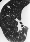
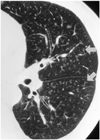
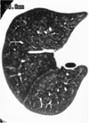
In 54 of 85 welders (64%), the findings of thin-section CT scanning were positive. The most common finding was poorly-defined centrilobular micronodules (n = 30) (Fig. 1), followed by branching linear structures (n = 18) (Fig. 2), and ground-glass attenuation (n = 6) (Fig. 3). While many cases of poorly-defined micronodules (17/30) and branching linear structures (10/18) showed slight upper zonal predominance, and were less commonly randomly distributed (7/30 for poorly-defined micronodules, 6/18 for branching linear structures), ground-glass attenuation was (in 5/6 cases) randomly distributed between upper and lower lung zones, As far as central or peripheral distribution was concerned, centrilobular micronodules and branching linear structures showed neither central nor peripheral predominance. Ground-glass attenuation was distributed more centrally (4/6). The extent of abnormalities was 0-10% in 29 cases, 10-20% in 11, 20-30% in seven, 30-40% in four, and more than 40% in three.
Other thin-section CT findings in welders included localized stable tuberculous lesions (n = 10), bronchial dilatation or wall thickening (n = 8), localized emphysema (n = 5), noncalcified mediastinal lymphadenopathy (n = 2), subsegmental atelectasis (n = 2), or tracheal dilatation (n = 1). In particular, in no patient was the appearance of emphysema compatible with so-called focal dust emphysema.
In the smokers' group, thin-section CT findings in six patients (14%) were positive. The predominant finding was poorly-defined micronodules in four, branching linear structures in one, and ground-glass attenuation in one. In all subjects, the extent of abnormalities was 0-10%. Other findings included localized bronchiectasis (n = 12), stable tuberculosis (n = 11), nodules or mass (n = 11), localized emphysema (n = 10), pleural thickening (n = 7), bulla (n = 4), consolidation (n = 3), fungus ball (n = 2), or lobar collapse (n = 1).
Among 53 welders who answered the questionnaire, the rate of positive thin-section CT findings was 57.1% for smokers (24/42) and 54.5% for non-smokers (6/11). The extent of abnormalities seen on thin-section CT showed no significant correlation with the results of pulmonary function tests (for FEV1/FVC, r = -0.045, p = .747; for FVC, r = 0.082, p = .55, respectively), with the degree of dyspnea (r = 0.023, p = 1), or with longevity of smoking (r = .0123, p = .949).
DISCUSSION
Arc-welders' pneumoconiosis is caused by the deposition of iron oxide, Fe2O3. Since the original description of the condition in 1936, it has been generally assumed that the disease is a benign one, not associated with respiratory symptoms or fibrosis. Some authors, however, reported respiratory difficulty associated with arc-welding (6), or histologic findings of emphysema or pulmonary parenchymal fibrosis in arc welders (7, 8). Here, the rare occurrence of parenchymal fibrosis has been attributed to ingredients other than iron (mostly silica) in welding fumes (9), based on cases where heavy deposition of iron in lung parenchyma was not associated with pulmonary fibrosis in either a human (10) or in experimental animal model (11). Indeed, in none of our cases with extensive ground-glass attenuation, which possibly represented the accumulation of large amount of iron oxide, was emphysema observed on thin-section CT scans. Likewise, we strongly doubt that in a study by Akira et al. (4), the emphysema revealed by thin-section CT scanning of seven welders with a smoking history was work related. Other thin-section CT findings in their study coincided with those in ours.
In our study, the predominant finding was centrilobular microndules, and this can be explained by reference to previous studies in which histological examinations were performed (13-14). They described the presence of pigmented macrophages in air spaces and interstitium close to the center of the acinus, with occasional fibrosis around pigmented deposits in these areas.
Our findings appeared similar to those of smokers' lung (14), and this similarity may be due to similarities in the pathologic findings of both entities. Small-airway lesions in patients with histories of exposure to nonasbestos dusts consist of deposits of fibrous tissue, often accompanied by pigment, in the walls of membranous and respiratory bronchioles and alveolar ducts (15, 16). In addition, the fact that 79% of the welders were smokers might call it into question whether the findings in our study truly represented exposure to welding and were not the results of smoking. On the basis of the following facts, however, we suggest that the findings in our subjects are mostly due to exposure to arc-welding. First, while there was a significant difference in the positive rate of CT findings between the welders (64%) and the smokers (14%), no such difference was seen in the positive rates for welders who smoked (57.1%) and those who did not (54.5%); second, among welders, there was no statistically significant dose-response relationship between smoking history and the extent of abnormalities.
Our study suffers from certain limitations. For example, those who interpreted the CT findings were not blinded as to whether a particular scan belonged to the welders' or the smokers' group. Thus, we admit that image interpretation for the latter could have been subject to prejudice, though not to the extent of significantly affecting the results of our study. Another limitation is that because most of the patients in our study sought compensation, they may have exaggerated the extent of their clinal symptons, and this may have diminished the statistical accuracy of our findings.
In conclusion, thin-section CT findings of arc-welder's lung include poorly-defined centrilobular micronodules, branching linear structures, and ground-glass attenuation. Some of our cases in which extensive abnormalities were not associated with the development of emphysema seem to indicate that this condition of emphysema is not caused by inhalation of arc-welding fumes. Since the CT findings in welder's lung can sometimes be very extensive, we believe that in order not to mistakedly perceive evidence of more serious conditions other than actually existing arc-welders' lung, cautious interpretation is required.
 XML Download