

 PDF
PDF ePub
ePub Citation
 Print
Print


Angiolipoma is a rare benign tumor composed of mature adipose tissue and blood vessels, and located mainly in the subcutaneous tissues of the trunk and extremities (1). Uncommon locations cited in the literature, however, have included the orbit, breast, parotid gland, palate, rib, mandible, mediastinum, intramedullary and epidural spine, and brain (2). Few cases involving the posterior mediastinum, with extension into the spinal canal, have been reported, and we report a case of posterior mediastinal angiolipoma with intraspinal extension.
CASE REPORT
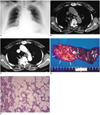
A 67-year-old woman presented with abnormality incidentally detected by chest radiography. She had no neurological symptoms or signs but radiographs demonstrated a round mass with a well-defined margin in the right upper paraspinal area (Fig. 1A). CT scanning of the chest revealed a 2 × 2.5-cm-sized lobulated mass in the right paravertebral area (Figs. 1B and 1C). The CT attenuation number of its most hypodense portion, as depicted on a unenhanced scan, was -31.0 H. Further scanning, performed after the injection of intravenous contrast media, demonstrated a highly enhanced area of tubular appearance (105-160 H) extending to the spinal canal (Fig. 1C), and also an area of moderate enhancement (46.2 H). Surgery revealed the presence of a 1.2 × 2.5 cm-sized hypervascular mass with a multifocal yellowish portions in the right 4th and 5th intercostal spaces. The mass extended through the T4-T5 intervertebral foramen to the right anterolateral epidural space.
Pathologic examination demonstrated mature, highly vascular adipose tissue, consistent with angiolipoma (Figs. 1D and 1E).
DISCUSSION
In 1960, Howard and Helwig (3) established angiolipomas as an anatopathological entity containing vascular and mature adipose elements, and Lin et al.(1) later defined the characteristics of the tumor as follows: (1) gross evidence of tumor formation with or without a capsule; (2) microscopic evidence of a tumor population of at least 50% mature lipocytes; (3) microscopic evidence of angiomatous proliferation inside the tumor. Microscopically, the essential diagnostic feature is the presence of a complex and tortuous capillary formation together with numerous fibrin thrombi.
Angiolipomas account for 5-17% of all lipomas (1, 3) and their common clinical denominator is pain and tenderness. They are located in the subcutis, most commonly that of the trunk and extremities. They usually occur in adolescents or young adults, though their occurrence in patients up to 84 years of age has been reported (1).
Angiolipomas arising from the posterior mediastinum are extremely rare, and to our knowledge, only one such case, confined to the paraesophageal area, has been reported (2). About 60 cases of spinal angiolipoma, mostly located in the dorsal epidural space of the thoracic spine and usually occurring in middle-aged patients (with a female predominance), have been described in the literature (4).
In our case, the mass exhibited focal fat attenuation on unenhanced CT scans and inhomogeneous areas of extremely high enhancement on ennanced scans. The ratio of adipose to vascular tissue found in angiolipomas varies, ranging from a neoplasm that is predominantly lipomatous with a small angiomatous focus, to one in which dense vascular and stromal elements have replaced most of the fat (5). This explains why the CT-demonstrated hypodensity of an angiolipoma, and the amount of contrast medium it absorbs, both vary.
The differential diagnosis in this case included a neurogenic tumor. The CT attenuation values of such tumors demonstrated after the administration of contrast medium vary, but except for paragangliomas, in which enhancement is exuberant, the enhancement observed is rarely as strong as that of the dense vascular tissue comprising an angiolipoma (6).
In summary, we report a case of posterior mediastinal angiolipoma extending to the spinal canal in which both fat and angiomatous features were revealed by CT.
 XML Download