

 PDF
PDF ePub
ePub Citation
 Print
Print


Primary malignant lymphoma of the lung may develop from bronchus-associated lymphoid tissue (BALT) (1, 2). Previously reported computed tomographic (CT) features of BALT lymphoma are the presence of consolidation or nodules in the lungs (1-5). To our knowledge, however, there has been no report of BALT lymphoma which manifested on CT as a mosaic pattern of inhomogeneous attenuation. The purpose of this report is to present a case of BALT lymphoma which on thin-section CT scan manifested as a mosaic pattern of inhomogeneous attenuation, and to correlate these findings with the histological findings.
CASE REPORT
A 33-year-old housewife with primary Sjögren's syndrome was admitted for the evaluation of dyspnea and general weakness which had persisted for two years. The patient claimed to have no history of smoking, allergies or occupational exposure to noxious agents. Physical examination revealed coarse breathing sounds with inspiratory crackle extending over the whole of the both lung fields. However, the results of blood analysis were normal.
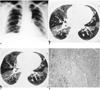
Chest radiographs demonstrated the presence of bilateral, diffuse, reticulonodular densities in both lungs (Fig. 1A). Thin-section CT scan showed diffusely distributed mosaic pattern of inhomogeneous attenuation extending over the entirety of lung zones, without zonal predominance. There was no difference in the extent of mosaic attenuation between inspiratory (Fig. 1B) and expiratory thin-section CT scans (Fig. 1C). As compared with more highly attenuated areas of the lung, however, the caliber and number of pulmonary vessels in areas of the lower attenuation were reduced. There were several foci of bronchial wall thickening, and these were more frequently found in areas of lower attenuation. In the right lower paratracheal area and subcarinal area, the presence of several small lymph nodes less than 1 cm in short axis diameter was noted.
Flexible fiberoptic bronchoscopy revealed no remarkable finding, though transbronchial multiple biopsies obtained from the right anterior segmental bronchus showed some lymphocytic infiltration. Cytologic analysis showed that the composition of bronchoalveolar lavage fluid was 45% lymphocytes, 1% eosinophils, and 54% macrophages. Open lung biopsy was performed in areas of both higher and lower attenuation. Histologic examination disclosed focal collections of lymphoid cells around bronchioles and extension of lymphoma cells from bronchiolar epithelium toward alveolar space. Immunocytochemically, the neoplastic cells reacted positively for CD 20 antigen and were focally positive for UCHL 1 antigen. Monoclonal antibody against cytokeratin indicated multiple lymphoepithelial lesions. The histologic diagnosis was consistent with low grade marginal zone B-cell lymphoma originating in BALT.
DISCUSSION
Primary malignant non-Hodgkin's lymphomas arising in mucosa-associated lymphoid tissue (MALT) develop most frequently in the stomach, but also in the bowel, salivary glands, larynx, thyroid gland, and lung (1, 2). Accordingly, when located in the lung, this lymphoma appears to arise from bronchus-associated lymphoid tissue (BALT) (1, 2). BALT lymphoma represents a low-grade B-lymphocyte lymphoma composed of monotonous small lymphocytes with focal plasmacytoid features (6). Since lymphomas at this site tend to be localized until late in the course of the disease, prognosis is considerably better than for nodal tumors of similar histologic grade (6).
Meanwhile, lymphoid infiltration of extranodal sites is a prominent feature of Sjögren's syndrome, and lymphoid proliferation in the respiratory system may take various forms: lymphocytic bronchitis and bronchiolitis, lymphocytic interstitial pneumonitis, pseudolymphoma, or malignant lymphoma (7, 8). Moreover, the risk of occurrence of lymphoma is much greater in such patients than in the general population (7). MALT lymphomas are also known to be associated with Sjögren's syndrome, AIDS, dysgammaglobulinemia, and collagen vascular diseases (1).
Previously reported CT features of BALT lymphoma are the presence of consolidation with poorly defined margins and air bronchograms (1, 4, 5). Other findings include nodules, diffuse bilateral air space consolidation, and segmental or lobar atelectasis (1-5). Pulmonary lymphoma associated with Sjögren's syndrome may appear as a diffuse interstitial process or as multiple nodular infiltrates (8). To our knowledge, however, BALT lymphoma manifesting on thin-section CT scan as a mosaic pattern of inhomogeneous attenuation has not been reported.
A mosaic pattern of inhomogeneous attenuation is non-specific and may be seen on thin-section CT scans of the lungs when various airway, vascular, or infiltratire lung diseases are present (9, 10). In cases of infiltrative lung disease, the mosaic pattern of inhomogeneous attenuation results from patchy areas of ground-glass attenuation and interposed normal lung parenchyma (9). Moreover, the size of vessels within the areas of ground-glass attenuation is usually similar to that seen in uninvolved lung, permitting distinction from airway or vascular diseases (9, 10). In cases involving such diseases, however, areas of increased attenuation have relatively large vessels, while areas of decreased attenuation have small vessels (9, 10). In cases of airway disease, the mosaic pattern of inhomogeneous attenuation results from reflex vasoconstriction secondary to hypoventilation of alveoli distal to airway obstruction and blood flow redistribution to adjacent normal areas of the lung (9). Moreover, bronchial dilatation and air trapping in abnormal lung may exist in cases of airway disease (9, 10). Though air trapping might not be present in cases of infiltrative or vascular lung disease (10).
In this case, the histologic diagnosis of the area of higher attenuation was low grade marginal zone B-cell lymphoma of BALT. Extension of lymphoma cells from bronchiolar epithelium toward alveolar space was noted, and bronchiolar lymphoepithelial lesion and infiltration of lymphoma cells within this space thus contributed to the increased attenuation of the lesion. There was, moreover, no difference in the extent of mosaic pattern of inhomogeneous attenuation between inspiratory and expiratory thin-section CT scan. Meanwhile, the histologic finding in the area of lower attenuation was focal collections of lymphoid cells around bronchioles. As compared with areas of higher attenuation, the caliber and number of pulmonary vessels in those of lower attenuation were reduced, being more pronounced on CT scan obtained at suspended full expiration. There were several foci of bronchial wall thickening, and these were more frequently found in areas of lower attenuation. The mechanism of airway disease causing mosaic pattern of inhomogeneous attenuation thus contributed to the decreased attenuation of the lesion. BALT lymphoma may, therefore, manifest on thin-section CT scan as a mosaic pattern of inhomogeneous attenuation. This is due to both small airway and infiltrative disease, as in cases of hypersensitivity pneumonitis.
 XML Download