Journal List > Korean J Physiol Pharmacol > v.22(2) > 1026237

Han, Kim, Kim, Choi, Park, Lee, Lee, Kim, Park, Kang, Oh, and Na: Oxytocin produces thermal analgesia via vasopressin-1a receptor by modulating TRPV1 and potassium conductance in the dorsal root ganglion neurons

Abstract

Recent studies have provided several lines of evidence that peripheral administration of oxytocin induces analgesia in human and rodents. However, the exact underlying mechanism of analgesia still remains elusive. In the present study, we aimed to identify which receptor could mediate the analgesic effect of intraperitoneal injection of oxytocin and its cellular mechanisms in thermal pain behavior. We found that oxytocin-induced analgesia could be reversed by d(CH2)5[Tyr(Me)2,Dab5] AVP, a vasopressin-1a (V1a) receptor antagonist, but not by desGly-NH2-d(CH2)5[DTyr2, Thr4]OVT, an oxytocin receptor antagonist. Single cell RT-PCR analysis revealed that V1a receptor, compared to oxytocin, vasopressin-1b and vasopressin-2 receptors, was more profoundly expressed in dorsal root ganglion (DRG) neurons and the expression of V1a receptor was predominant in transient receptor potential vanilloid 1 (TRPV1)-expressing DRG neurons. Fura-2 based calcium imaging experiments showed that capsaicin-induced calcium transient was significantly inhibited by oxytocin and that such inhibition was reversed by V1a receptor antagonist. Additionally, whole cell patch clamp recording demonstrated that oxytocin significantly increased potassium conductance via V1a receptor in DRG neurons. Taken together, our findings suggest that analgesic effects produced by peripheral administration of oxytocin were attributable to the activation of V1a receptor, resulting in reduction of TRPV1 activity and enhancement of potassium conductance in DRG neurons.

INTRODUCTION

Oxytocin is a neuropeptide produced in the paraventricular nucleus of the hypothalamus and primarily associated with parturition [1]. Recently, accumulating studies have illustrated the involvement of oxytocin in modulation of nociception [2]. The immerging roles of oxytocin as endogenous and exogenous analgesic has been reported in several clinical reports and basic researches using animal models [345]. A clinical study reported that patients suffering from back pain displayed reduced self-reported pain score after receiving oxytocin via intrathecal administration[3]. As oxytocin does not cross blood brain barrier [6], many animal studies aimed to elaborate the spinal mechanisms of oxytocin-mediated analgesia [789]. Previous research has demonstrated the distribution of oxytocin binding receptors in the dorsal horn of spinal cord [10]. Oxytocin has been demonstrated to inhibit transmission of nociceptive signals by enhanced inhibitory GABA signals from interneurons [8], and by modulating the potassium conductance in spinal dorsal horn neurons [9].
Another clinical study reported that patients with irritable bowel syndrome exhibited lowered threshold of visceral discomfort by intravenous administration of oxytocin [4]. Thereafter, recent studies have focused on elucidating cellular mechanisms of analgesia induced by peripheral administration of oxytocin. A study with knockout mice demonstrated that intraperitoneal (IP) injection of oxytocin induces analgesia via vasopressin-1a (V1a) receptor [11]. Oxytocin decreases depolarization-induced calcium transients in the dissociated dorsal root ganglion (DRG) neurons of rat model of neuropathic pain [12], suggesting the possible involvement of calcium channels in oxytocin-mediated peripheral analgesia. Oxytocin inhibits the activity of acid-sensing ion channels in the DRG neurons via V1a receptor [13].
Given that we still lack a detailed cellular mechanism of oxytocin-induced peripheral analgesia, we examined in the present study how the IP administration of oxytocin produces thermal analgesia. We found that V1a receptor in DRG neurons indeed mediates oxytocin-induced thermal analgesia, and its regulations of transient receptor potential vanilloid 1 (TRPV1) and potassium conductance in the DRG neurons via V1a receptor are likely to contribute to the thermal analgesia.

METHODS

Animals

Male Sprague-Dawley rats (Orient Bio, Korea), aging 6-8 weeks, were used in this study. All the experiments were approved by Institutional Animal Care and Use Committee at Seoul National University (SNU-160216-4) and Institutional Animal Care and Use Committee at Korea University College of Medicine (KOREA-2016-0022). Animals were housed under a 12-h light/dark cycle. Food and water were available ad libitum.

Drugs

Oxytocin, atosiban, and capsaicin were purchased from Sigma-Aldrich (USA). Selective oxytocin receptor antagonist (desGly-NH2-d(CH2)5[D-Tyr2,Thr4]OVT, OTRA) and V1a receptor antagonist (d(CH2)5[Tyr(Me)2,Dab5]AVP, V1aRA) were generously donated from Dr. M. Manning at the University of Toledo College of Medicine (Toledo, OH, USA). All drugs were diluted to the final concentrations from stock solutions on the day of the experiment. The solutions used in behavioral test were dissolved in normal saline. The solutions used in electrophysiological recordings and calcium imaging were dissolved in external solutions of each experiment except capsaicin which were dissolved in ethanol as a stock solution and further diluted in the extracellular solution when applied.

Thermal nociception assay

We adopted Hargreaves' test for thermal nociception assay [14]. As previously described [15], thermal pain threshold was measured using radiant heat test. Rats were placed on a glass surface at 23-25℃. A heat source was focused on the mid-third hindpaw of a rat standing on the glass plate with all four feet aground, and paw withdrawal latency was assessed using Series 8, Model 390 (IITC Life Science, US).

Preparation of DRG neurons

Modified from the previous study [16], rats were anesthetized with isoflurane and then decapitated. The DRGs were taken out from L1-L6 and placed immediately in 4℃ HEPES buffered HBSS at pH 7.4. After removing the surrounding connective tissues, the DRGs were minced with fine spring scissors under a microscope and the ganglions were transferred to a 15 ml tube containing 2 ml HEPES buffered HBSS in which 100 mg/ml collagenase and 2.4 U/ml dispase had been dissolved, and incubated at 37℃ in a water bath for 40 min. The cells were washed in DMEM containing 10% FBS and 0.5% Penicillin and Streptomycin and triturated with a flame-polished Pasteur pipette to separate cells and remove processes. The dissociated cells were placed on 1 mg/ml poly-Dlysine (Sigma-Aldrich, US)-coated glass cover slips (25 mm in diameter), then maintained at 37℃ in an incubator. The dissociated neurons were kept for at least another 4 h before electrophysiological recordings or calcium imaging, and used for the experiments within 36 h. The neurons selected for electrophysiological experiments and single cell RT-PCR were 15-35 µm in diameter. More than 3 rats were sacrificed for the collection of dissociated DRG neurons in each experiment.

Single cell reverse transcription polymerase chain reaction (scRT-PCR)

The cells were harvested into a micropipette with a tip diameter of about 20 µm, put into a tube containing reagents solution, and reverse-transcription was performed using EasyScript Two-Step RT-PCR Kit (ABM Inc, Canada) according to the manufacturer's instruction. Subsequent cDNA product was divided into aliquots of 2 µl and the each was used in separate reactions. All PCR amplifications were performed with nested primers (Table 1) using 2X PCR Taq Plus MasterMix (ABM Inc, Canada) according to manufacturer's protocol. Briefly, the first round of PCR was performed in 50 µl of reaction buffer containing 25 µl MasterMix, 2 µl cDNA, 2 µl “outer” primers. The second round reaction buffer (25 µl) contained 12.5 µl MasterMix, 2 µl product from the first round, and 1 µl “inner” primers. The protocol included a 5 min initial denaturation step at 94℃ followed by 40 cycles for the first round or 30 cycles for the second round of 30 s denaturation at 94℃, 30 s annealing at 55-60℃ depending on primers, 60 s elongation at 72℃. The reaction was completed with 5 min of final elongation. A negative control was acquired from the pipette that did not collect any cell contents, but were submerged in the extracellular solution. The PCR products were displayed on Loading-STAR (DYNEBIO, Korea) stained, 2% agarose gels. Primers were shown in Table 1.

Immunocytochemistry

Coverslips cultured 24 h with rat DRG neurons were fixed in 4% formaldehyde solution for 20 min. The DRG neurons were washed with 0.01 M phosphate-buffered saline (PBS) and immunostained with following antibodies. The primary antibodies were rabbit anti-V1a receptor polyclonal antibody (1:400; Alomone Labs, UK) and guinea pig anti-TRPV1 polyclonal antibody (1:500; Novus biologicals, USA), diluted in PBS containing 0.3% Triton X-100, 1% normal donkey serum (Jackson ImmunoResearch Co, USA), incubated overnight at 4℃ then incubated for 1 h with donkey anti-guinea pig IgG conjugated with cyanine dye 3 (Cy3, 1:200; Jackson ImmunoResearch Co, USA) and donkey anti-rabbit IgG conjugated with fluorescein-isothiocyanate (FITC, 1:200; Jackson ImmunoResearch Co, USA) diluted in the same buffer as the primary antibody. All rinses before and after the incubations were performed with PBS. Coverslips were photographed using confocal microscope (LSM-700; Zeiss, Germany).

Electrophysiological recordings

Modified from the previous study [17], whole-cell currentand voltage-clamp recordings were performed to measure action potentials and voltage-gated potassium current respectively. The signals from neurons amplified by Axoclamp-700A amplifier (bandwidth filter set at 10 kHz for current-clamp and 1 kHz for voltage-clamp recordings) were digitized and sampled at 50 µs intervals (Digidata 1440, pClamp 10.3; Molecular Devices). Patch pipettes were pulled from borosilicate capillaries (P-97; Sutter Instruments, USA). The resistance of the pipettes was 3-5 MΩ when filled with the intracellular solution composed of (mM): 140 K-gluconate, 10 HEPES, 2 MgCl2, 2 CaCl2, 11 EGTA, 4 K2-ATP and 0.3 Na2-GTP adjusted to pH 7.2-7.3 with KOH, osmolarity 280-285 mOsm. Recording chamber was continuously perfused (1-2 ml/min) with the warm (32-35℃) extracellular solution composed of (mM): 150 NaCl, 4 KCl, 2.5 CaCl2, 10 glucose, 2 MgCl2, 10 HEPES, and 0.5 tetrodotoxin (TTX, applied for voltage-clamp experiments), adjusted to pH 7.4 with NaOH, osmolarity 290-300 mOsm. Na+-free extracellular solution contained 150 mM Tris instead of Na+ and was adjusted to pH 7.4 with HCl [18]. Cadmium chloride (300 µM) was added to the Na+-free extracellular solution for blockade of calcium channels [19]. A small patch of cell membrane underneath the tip of the pipette was aspirated to form a gigaseal. After gigasealing, negative pressure was applied to rupture the membrane for a whole-cell configuration. In voltage-clamp experiments, the membrane voltage was maintained at –60 mV. At the holding potential, the membrane potential was stepped to –110 mV for 200 ms and then a depolarizing ramp pulse to 0 mV at a rate of 110 mV/s (step-and-ramp pulse) was applied. In current-clamp experiments, action potentials were generated by 1000 ms depolarizing current with 100-500 pA. Only cells with a stable resting potential (more negative than –40 mV) were used in this study.

Calcium imaging

As described in the previous study [16], DRG neurons were loaded with 3 µM of the fluorescent calcium indicator dye Fura-2-AM (dissolved in DMSO, F1201, Thermo Fisher Scientific) for 40 min in an incubator at 37℃ and then washed. After washing, the cells on a coverslip were placed on a chamber with a perfusion system (4 channel gravity ALA-VM4, Charles Austen Pump, Dynamax 30) in dark surroundings at room temperature. Regions of interest were defined on a computer connected to a CCD camera and the recording area of which were viewed through an inverted microscope. Fura-2 fluorescence of the cells was recorded at 510 nm during shifting excitation at 340 and 380 nm at 1Hz using Lamda DG4. The ratio of emission at 510 nm from excitation at 340 nm to at 380 nm was analyzed. Only cells with a 340/380 fluorescence ratio difference higher than 0.3 were analyzed. Since our experimental protocol of repeated treatment of capsaicin produced TPRV1 desensitization, Phorbol 12-myristate 13-acetate (PMA) was mixed to the 200 nM capsaicin to the final concentration of 10 µM before the experiment to reduce capsaicin-induced calcium-dependent desensitization of TPRV1 [20]. The cells whose 340/380 ratio increased more than 0.3 in response to capsaicin were considered as capsaicin responsive cells. The cells showing 20% decrease in the second capsaicin response in comparison to the first capsaicin response were considered as oxytocin responsive cells.

Statistical Analysis

Data were expressed as mean±SEM. Behavioral and electrophysiological data were analyzed using ANOVA followed by posthoc Bonferroni's test. Differences with a p<0.05 were considered significant.

RESULTS

Oxytocin-induced thermal analgesia was revered by V1a receptor, but not by oxytocin receptor

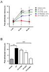
We aimed to determine the specific receptor that mediates the oxytocin-induced thermal analgesia in vivo by using Hargreaves' test. Thus, we examined whether the analgesic effect produced by IP injection of oxytocin (1 mg/kg), would be affected by coadministration with OTRA (1 mg/kg), atosiban (1 mg/kg) and V1aRA (1 mg/kg). Paw withdrawal latency after thermal stimuli was significantly increased at 30 and 60 min after the injection of oxytocin (Fig. 1A). This thermal analgesia induced by oxytocin was significantly reversed by co-injection of 1 mg/kg of V1aRA, vasopressin-1a receptor antagonist, but not with 1 mg/kg of OTRA, oxytocin receptor antagonist (Figs. 1A and B). Co-application of atosiban, oxytocin and vasopressin receptor antagonist, also showed trends for the reversing effect although not statistically significant (Fig. 1B; p>0.01, posthoc Bonferroni's test). These results suggest that oxytocin induces thermal analgesia via V1a receptor.

Predominant expression of V1a receptor in TRPV1-positive DRG neurons

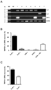
Given that TRPV1 is the primary transducer of thermal nociception in DRG neurons [21], we then examined mRNA expression pattern for oxytocin binding receptors in relation with TRPV1 by using scRT-PCR. We only collected small to medium-sized DRG neurons for scRT-PCR (Fig. 2A, Supplementary Fig. 1A). Amongst 54 GAPDH expressing neurons, mRNA transcripts of V1a receptor (n=35/54, 65%), vasopressin-1b receptor (V1b, n=3/54, 6%) and oxytocin receptor (n=1/54, 2%) were expressed in DRG neurons. Vasopressin-2 (V2) receptor mRNA was not detected (n=0/54, 0%) (Fig. 2B). To examine whether the samples were contaminated by glial cells, we investigated the expression of glial fibrillary acidic protein (GFAP) mRNA and GFAP mRNA was not detected in the V1a receptor expressing neurons (n=0/14, 0%) (Supplementary Fig. 1B). We also found that 67% (n=23/34) of TRPV1-expressing DRG neurons were also co-expressed with V1a receptor. (Fig. 2C). Our result also demonstrated that the co-expression of TRPV1 and V1a receptor proteins in the DRG neurons (Supplementary Fig. 2). These findings suggest that V1a receptor activated by oxytocin might transact with TRPV1 to induce thermal analgesia.

Oxytocin decreased capsaicin-induced calcium transient in the DRG neurons via V1a receptor

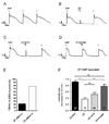
To verify the functional interaction between oxytocin and TRPV1, we examined effects of oxytocin on capsaicin-induced calcium transients in DRG neurons. A series of repetitive capsaicin application elicited reproducible calcium transients in DRG neurons with PMA although we observed small desensitization (Fig. 3A). Our results demonstrated that bath application of 5 µM oxytocin significantly decreased calcium transients induced by capsaicin (Figs. 3B and F). The co-application of 5 µM oxytocin with either 20 µM atosiban (Figs. 3C and F) or 5 µM V1aRA (Figs. 3D and F) reversed the reducing effect of oxytocin on capsaicin-induced calcium transients. We found that 78% (55/70) of capsaicin responsive DRG neurons also responded to oxytocin application (Fig. 3E). These results suggested that oxytocin-induced thermal analgesia might be attributed to the reduction of TRPV1 activity by oxytocin via V1a receptor.

Oxytocin decreased action potential firing frequency of DRG neurons through V1a receptor

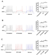
We also investigated effects of oxytocin on the membrane excitability in the small to medium-sized DRG neurons. While oxytocin (5 µM) for 60 s lowered the number of action potential in response to a 300 pA and 1000 ms current injection, the number of action potentials evoked by the current injection returned almost to the control value after 5 min-washout (Fig. 4A). It is noteworthy that action potentials were observed only at the onset and before the offset of the current pulse, suggesting activation of voltage-gated potassium channels rather than other voltage-gated ion channels. Consistent with our results above, inhibitory effects of oxytocin on the action potential firings were also reversed by atosiban (20 µM, Fig. 4B) and V1aRA (5 µM, Fig. 4C). These results again suggest that oxytocin reduce membrane excitability of DRG neurons via V1a receptor.

Oxytocin elicited outward potassium currents and small hyperpolarization-induced inward current in DRG neurons via V1a receptor

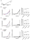
As reduction of the number of action potentials indicated regulation of voltage-gated potassium channels, we performed voltage clamp recordings with the small to medium-sized DRG neurons in the presence of 0.5 µM TTX. At a holding potential of –60 mV, the membrane potential was stepped to –110 mV for 200 ms and then a depolarizing ramp pulse to 0 mV at a rate of 110 mV/s (step-and-ramp pulse) was applied (Fig. 5A). Effects of oxytocin on the membrane current response to the ramp pulse were evaluated as a change in the I-V relationship that was obtained by subtraction of the control response from the response obtained after oxytocin application. Application of oxytocin (5 µM) for 60 s markedly enhanced the outward current activated at membrane potentials more positive than –40 mV (Fig. 5B, left and middle) while it caused a slight increase in hyperpolarization-activated inward current at –110 mV (Fig. 5B, left and middle). The outward current was recorded in Cd2+ containing Na+-free external solution (Supplementary Fig. 3B). The mean peak current density of the DRG neurons at 0 mV also significantly increased by oxytocin (Fig. 5B, right). In agreement with our previous results, not only atosiban (20 µM, Fig. 5C) but also V1aRA (5 µM, Fig. 5D) inhibited the currents elicited by oxytocin, which would indicate that oxytocin induce the prominent outward potassium current and modest hyperpolarization-induced inward current via V1a receptor.

DISCUSSION

In the present study, we demonstrated that oxytocin reduced TRPV1 activity and increased potassium conductance in small to medium-sized DRG neurons via V1a receptor. These might contribute to the thermal analgesic effect produced by peripheral administration of oxytocin. We also found co-expression of TRPV1 and V1a receptor mRNA, and predominant mRNA expression of V1a receptor over other oxytocin binding receptors such as oxytocin, V1b, and V2 receptors in DRG neurons.
Oxytocin binds to not only oxytocin receptor, but also various vasopressin receptors such as V1a, V1b and V2 receptors [122]. Oxytocin has been reported to exert its analgesic effects via oxytocin receptor in spinal modulation [723]. However, in Hargreaves' test, we found that oxytocin-induced thermal analgesia was only reversed by V1a receptor antagonist, but not by oxytocin antagonist and oxytocin/vasopressin receptor antagonist. Consistent with our results, previous studies also showed that oxytocin exerts its analgesic effect through V1a receptor rather than oxytocin receptor by using knock-out mice [1113]. Drug doses from our study were determined based on the previous study illustrating the dose dependent analgesic effects of intraperitoneal injection of oxytocin [5]. It was interesting to note that the effect of V1a receptor antagonist is much larger than that of oxytocin/vasopressin receptor antagonist (Fig. 1). This might be due to the difference in efficacy between drugs. As V1aRA was four times more efficient than atosiban as anti-vasopressor [24], V1aRA blocked the analgesic effect of oxytocin more efficiently than the same dose of atosiban.
In parallel with results in mouse DRG neurons [11], mRNA expression of V1a receptor was predominant over other oxytocin-binding receptors such as oxytocin receptor and the other vasopressin receptors in small to medium-sized rat DRG neurons. In addition, mRNA of V1a receptor was also found in two-third of the primary afferent neurons expressing TRPV1, which has been reported to convey the thermal nociception of Hargreaves' test and is expressed in more than fifty percent of DRG neurons and mostly nociceptive C-fibers [212526]. However, recently, oxytocin receptor has been reported to colocalize with calcitoningene related peptide or isolectin B4in the DRG neurons [27]. This discrepancy is attributed to two possible scenarios. First, although we pulled a considerable amount of DRG neurons (n=54), the number of collected DRG neurons still lacked for unbiased analysis of scRT-PCR. The other is that immunostaining plausibly revealed characteristics of a small population of DRG neurons. Taken together with the observation that oxytocin elicited analgesic effects on thermal pain behavior via V1a receptor, these results suggest that V1a receptor plays a role in thermal analgesia by oxytocin.
To determine whether oxytocin could modulate TRPV1 activity in vitro, we performed calcium imaging. Since our experimental protocol of repeated treatment of capsaicin produced TPRV1 desensitization, we applied PMA to reduce capsaicin-induced calcium-dependent desensitization of TPRV1 [20]. Similar to the results of mRNA transcript analysis by scRT-PCR, we found that three-fourth of the DRG neurons responding to capsaicin also functionally responded to oxytocin treatment in calcium imaging study. Capsaicin-induced depolarization activated various calcium channels other than TRPV1, which possibly contributed to long-lasting calcium responses in the DRG neurons. Reduction of the depolarization by oxytocin might affect the activation of the other calcium channels and subsequently reduce the long-lasting calcium responses. Concentration of oxytocin was determined based on a previous report which showed dose-dependent activity of oxytocin for inhibiting acid-sensing ion channels in vitro [13].
Many ligands of G-protein coupled receptor (GPCR) such as bradykinin, neurokinin and prostaglandins enhance TRPV1 activity in primary afferent neurons and increase pain sensitivity [282930]. However, from calcium imaging experiment, we found that oxytocin reduces TRPV1 activity via V1a receptor, a GPCR, in the DRG neurons. In parallel with our results, resolvin D1 attenuated calcium influx through TRPV1 by modulating GPCR [31]. Modulation of TRPV1 activity was reported to reduce peripheral nociceptor activity and pain [32]. These implicate that GPCR-mediated reduction of TRPV1 activity in the DRG neurons might elicit analgesic effects.
V1a receptor employs not only G-proteins including Gs and Gq but also β-arrestins as intracellular signaling pathways [3334]. Activation of V1a receptor induced calcium influx markedly in smooth muscle cells using IP3 intracellular signaling pathway. This would indicate Gq-mediated vasopressin-1 receptor signaling is associated with marked calcium influx. However, in the present study, oxytocin did not evoke any significant calcium transients in DRG neurons (Fig. 3). One possibility is that V1a receptor may adopt β-arrestin dependent receptor internalization for intracellular signaling. A previous research has revealed that β-arrestin-2desensitize TRPV1 in primary sensory neurons of rats using siRNA knockdown [35]. Furthermore, it has been reported that arrestin activation modifies conductance of potassium channels without efficiently activating G-proteins signaling [36]. Thus, further experiments are required to determine intracellular signaling pathways that mediate the analgesic effect of V1a receptor.
We also found that oxytocin modulates membrane excitability of small to medium-sized DRG neurons. In current-clamp recordings, oxytocin reduced the number of action potential firings evoked by current injection via V1a receptor. In details, oxytocin predominantly inhibited action potentials during the current pulse and firings except at its onset or before its offset. This indicated that the reduction in the number of action potentials was associated with inactivating potassium channels rather than with persistent potassium channels [3738]. Next, to confirm whether oxytocin indeed modulates activity of potassium channels, we performed voltage ramp test in the presence of TTX. Oxytocin markedly enhanced potassium outward current and slightly enhanced hyperpolarization-activated inward current in response to the ramp pulse through V1a receptor. Several lines of evidence have addressed the roles of inactivating potassium channels in modulation of nociception [394041]. Moreover, the previous study has also reported that potassium channel is involved in the antinociceptive effect of oxytocin in neurons of the spinal cord [9]. Taken together, our results show that oxytocin-induced enhancement of outward potassium currents via V1a receptor is associated with its anti-nociceptive action in DRG neurons.
In conclusion, oxytocin reduces TRPV1 activity and enhances potassium conductance via V1a receptor in DRG neurons, leading to analgesic effect produced by IP injection of oxytocin. These results suggest that peripheral administration of oxytocin might directly produce thermal analgesia, and V1a receptor might be a potential target for peripheral therapeutics to treat certain types of pathological pain conditions with thermal pain.

Figures and Tables

Fig. 1

Reversal of oxytocin-induced thermal analgesia via V1a receptor, not oxytocin receptor.

(A) Thermal paw-withdrawal latency was measured and compared before the injection with 30 min and 60 min after the IP injection of vehicle, OT (1 mg/kg) and OT with OTRA (A selective oxytocin receptor antagonist 1 mg/kg), atosiban (an oxytocin and vasopressin receptor antagonist, 1 mg/kg) and V1aRA (a selective vasopressin 1a receptor antagonist, 1 mg/kg) in Hargreaves' test. **p<0.001, ***p<0.0001, Two-way RM ANOVA followed by posthoc Bonferroni's test. Error bars represent SEM (n=6, each group). (B) Nocifensive responses at 60 min after IP co-injection of antagonists were compared to the OT injection. **p<0.001, ***p<0.0001, One-way ANOVA followed by posthoc Bonferroni's test. Error bars represent SEM. (n=6, each group). OT, oxytocin; V1aRA, vasopressin-1a receptor antagonist; OTRA, oxytocin receptor antagonist; ns, not significant.
kjpp-22-173-g001
Fig. 2

The expression patterns of oxytocin receptors and TRPV1 in DRG neurons revealed by single cell RT-PCR analysis.

(A) Representative gels show expression patterns of oxytocin binding receptors and TRPV1 from seven individual DRG neurons. Predicted size for selected markers are oxytocin receptor (158 bp), V1a receptor (151 bp), V1b receptor (194 bp), V2 receptor (V2, 153 bp), TRPV1 (330 bp) and GAPDH (268 bp). (B) The percentage of oxytocin binding receptors-positive cells in 54 GAPDH-positive DRG neurons. (C) The percentage of V1a receptor-positive cells within TRPV1 expressing DRG neurons. MM, molecular marker; NC, negative control.
kjpp-22-173-g002
Fig. 3

Oxytocin-induced decrease in capsaicin-induced calcium transients in the DRG neurons mediated via V1a receptor.

Representative traces of intracellular calcium responses of cultured sensory neurons to 3 repetitive application of (A) 200 nM CAP for 10 s, (B) repetitive CAP application with pre-application of 5 µM of oxytocin for 120 s, (C) CAP application with 5 µM of oxytocin mixed with 20 µM of atosiban, and (D) 5 µM of oxytocin mixed with 5 µM of V1aRA before the second capsaicin application. (E) The proportion of oxytocin responsive cells within the DRG neurons responding to capsaicin in calcium imaging. (F) Summary of normalized ratio of capsaicin responses by without OT (n=21), OT alone (n=70), and OT with its antagonists, ATO (n=66) and V1aRA (n=70), relative to peak amplitude of 1st CAP transient. Before CAP application, 10 µM PMA was mixed with CAP solution. *p<0.05, One-way ANOVA followed by posthoc Bonferroni's test. Error bars represent SEM. CAP, capsaicin; PMA, Phorbol 12-myristate 13-acetate; OT, oxytocin; ATO, atosiban; V1aRA, vasopressin-1a receptor antagonist; ns, not significant.
kjpp-22-173-g003
Fig. 4

Decrease of action potential firings by oxytocin in small to medium-sized DRG neurons mediated via V1a receptor.

(A, left panel) Representative recording illustrating effect of 5 µM oxytocin on action potential firing. The effect of oxytocin was reversed after 5-min washout. (A, right panel) Oxytocin decreased the mean number of action potential evoked by current injection (n=12). (B) 20 µM of Atosiban (n=9) and (C) 5 µM of V1aRA (n=9) reversed oxytocin-induced reduction of the number of action potentials in small to medium-sized DRG neurons. *p<0.01, ***p<0.0001, repeated measured-ANOVA followed by posthoc Bonferroni's test. Error bars represent SEM. OT, Oxytocin; V1aRA, vasopressin-1a receptor antagonist; ns, not significant.
kjpp-22-173-g004
Fig. 5

Enhancement of outward potassium currents by oxytocin in small to medium-sized DRG neurons mediated via V1a receptor.

(A) Depolarizing (from –110 mV to 0 mV) ramp protocol for voltage-clamp experiments (B, left panel) Representative trace showing effects of oxytocin (5 µM) on I-V relationship response to ramp depolarization. (B, middle panel) Representative trace illustrating markedly enhanced a voltage-activated outward current and slightly enhanced a hyperpolarization-activated inward current (B, right panel) Oxytocin increased the mean peak current density at 0 mV by inducing outward potassium current in small to medium-sized DRG neurons (n=20). (C) 20 µM of atosiban (n=24) and (D) 5 µM of V1aRA blocked oxytocin-induced the voltage-activated outward potassium current and the hyperpolarization-activated inward current. TTX (0.5 µM) were treated in solution of all experiments (n=13). *p<0.01, **p<0.001, repeated measured-ANOVA followed by posthoc Bonferroni's test. Error bars represent SEM. OT, Oxytocin; V1aRA, vasopressin-1a receptor antagonist; ns, not significant.
kjpp-22-173-g005
Table 1

scRT-PCR primers

kjpp-22-173-i001

ACKNOWLEDGEMENTS

This research was supported by the Public Welfare & Safety research program through the National Research Foundation of Korea (NRF) funded by the Ministry of Education, Science and Technology (NRF-2014M3C8A5030612), (NRF-2016M3A9B6021209) and supported by The Chung Yang, Cha Young Sun, M.D. and Jang Hi Joo Yeu Sa Memorial Fund. We would like to thank Dr. Morris Manning for generous donation of the neuropeptide receptor antagonists.

Notes

Author contributions H.S.N. and S.B.O. conceived the idea, obtained funding for the study and guided the project; H.T.H. performed hargreaves' test, single-cell RT-PCR and whole-cell patch clamp and H.B.K. performed calcium imaging, whole-cell path clamp, immunostaining; Y.B.K. performed and analyzed whole-cell patch clamp; K.M.C. performed single-cell RT-PCR; G.Y.P. and P.R.L. performed DRG preparation and calcium imaging, respectively; J.H.L. and H.Y.K. performed and analyzed single-cell RT-PCR; C.K.P. conceived the idea and analyzed the results. Y.N.K analyzed the data; H.T.H., H.B.K., Y.N.K., S.B.O. and H.S.N. wrote the manuscript. All authors gave final approval and agree to be accountable for all aspects of the work.

CONFLICTS OF INTEREST The authors declare no conflicts of interest.

References

1. Vrachnis N, Malamas FM, Sifakis S, Deligeoroglou E, Iliodromiti Z. The oxytocin-oxytocin receptor system and its antagonists as tocolytic agents. Int J Endocrinol. 2011; 2011:350546.
crossref
2. González-Hernández A, Rojas-Piloni G, Condés-Lara M. Oxytocin and analgesia: future trends. Trends Pharmacol Sci. 2014; 35:549–551.
crossref
3. Yang J. Intrathecal administration of oxytocin induces analgesia in low back pain involving the endogenous opiate peptide system. Spine (Phila Pa 1976). 1994; 19:867–871.
crossref
4. Louvel D, Delvaux M, Felez A, Fioramonti J, Bueno L, Lazorthes Y, Frexinos J. Oxytocin increases thresholds of colonic visceral perception in patients with irritable bowel syndrome. Gut. 1996; 39:741–747.
crossref
5. Lundeberg T, Uvnäs-Moberg K, Agren G, Bruzelius G. Anti-nociceptive effects of oxytocin in rats and mice. Neurosci Lett. 1994; 170:153–157.
crossref
6. Kang YS, Park JH. Brain uptake and the analgesic effect of oxytocin−its usefulness as an analgesic agent. Arch Pharm Res. 2000; 23:391–395.
7. Juif PE, Poisbeau P. Neurohormonal effects of oxytocin and vasopressin receptor agonists on spinal pain processing in male rats. Pain. 2013; 154:1449–1456.
crossref
8. Breton JD, Veinante P, Uhl-Bronner S, Vergnano AM, Freund-Mercier MJ, Schlichter R, Poisbeau P. Oxytocin-induced antinociception in the spinal cord is mediated by a subpopulation of glutamatergic neurons in lamina I-II which amplify GABAergic inhibition. Mol Pain. 2008; 4:19.
crossref
9. Breton JD, Poisbeau P, Darbon P. Antinociceptive action of oxytocin involves inhibition of potassium channel currents in lamina II neurons of the rat spinal cord. Mol Pain. 2009; 5:63.
crossref
10. Wrobel L, Schorscher-Petcu A, Dupré A, Yoshida M, Nishimori K, Tribollet E. Distribution and identity of neurons expressing the oxytocin receptor in the mouse spinal cord. Neurosci Lett. 2011; 495:49–54.
crossref
11. Schorscher-Petcu A, Sotocinal S, Ciura S, Dupré A, Ritchie J, Sorge RE, Crawley JN, Hu SB, Nishimori K, Young LJ, Tribollet E, Quirion R, Mogil JS. Oxytocin-induced analgesia and scratching are mediated by the vasopressin-1A receptor in the mouse. J Neurosci. 2010; 30:8274–8284.
crossref
12. Hobo S, Hayashida K, Eisenach JC. Oxytocin inhibits the membrane depolarization-induced increase in intracellular calcium in capsaicin sensitive sensory neurons: a peripheral mechanism of analgesic action. Anesth Analg. 2012; 114:442–449.
13. Qiu F, Qiu CY, Cai H, Liu TT, Qu ZW, Yang Z, Li JD, Zhou QY, Hu WP. Oxytocin inhibits the activity of acid-sensing ion channels through the vasopressin, V1A receptor in primary sensory neurons. Br J Pharmacol. 2014; 171:3065–3076.
14. Hargreaves K, Dubner R, Brown F, Flores C, Joris J. A new and sensitive method for measuring thermal nociception in cutaneous hyperalgesia. Pain. 1988; 32:77–88.
crossref
15. Han RT, Lee H, Lee J, Lee SB, Kim HJ, Back SK, Na HS. Brief isolation changes nociceptive behaviors and compromises drug tests in mice. Pain Pract. 2016; 16:749–757.
crossref
16. Kim YH, Park CK, Back SK, Lee CJ, Hwang SJ, Bae YC, Na HS, Kim JS, Jung SJ, Oh SB. Membrane-delimited coupling of TRPV1 and mGluR5 on presynaptic terminals of nociceptive neurons. J Neurosci. 2009; 29:10000–10009.
crossref
17. Kim YB, Kim YS, Kim WB, Shen FY, Lee SW, Chung HJ, Kim JS, Han HC, Colwell CS, Kim YI. GABAergic excitation of vasopressin neurons: possible mechanism underlying sodium-dependent hypertension. Circ Res. 2013; 113:1296–1307.
18. Achilles K, Okabe A, Ikeda M, Shimizu-Okabe C, Yamada J, Fukuda A, Luhmann HJ, Kilb W. Kinetic properties of Cl uptake mediated by Na+-dependent K+-2Cl cotransport in immature rat neocortical neurons. J Neurosci. 2007; 27:8616–8627.
19. Díaz D, Bartolo R, Delgadillo DM, Higueldo F, Gomora JC. Contrasting effects of Cd2+ and Co2+ on the blocking/unblocking of human Cav3 channels. J Membr Biol. 2005; 207:91–105.
20. Mandadi S, Numazaki M, Tominaga M, Bhat MB, Armati PJ, Roufogalis BD. Activation of protein kinase C reverses capsaicin-induced calcium-dependent desensitization of TRPV1 ion channels. Cell Calcium. 2004; 35:471–478.
crossref
21. Caterina MJ, Leffler A, Malmberg AB, Martin WJ, Trafton J, Petersen-Zeitz KR, Koltzenburg M, Basbaum AI, Julius D. Impaired nociception and pain sensation in mice lacking the capsaicin receptor. Science. 2000; 288:306–313.
crossref
22. Manning M, Stoev S, Cheng LL, Wo NC, Chan WY. Design of oxytocin antagonists, which are more selective than atosiban. J Pept Sci. 2001; 7:449–465.
crossref
23. Eliava M, Melchior M, Knobloch-Bollmann HS, Wahis J, da Silva Gouveia M, Tang Y, Ciobanu AC, Triana Del Rio R, Roth LC, Althammer F, Chavant V, Goumon Y, Gruber T, Petit-Demoulière N, Busnelli M, Chini B, Tan LL, Mitre M, Froemke RC, Chao MV, Giese G, Sprengel R, Kuner R, Poisbeau P, Seeburg PH, Stoop R, Charlet A, Grinevich V. A new population of parvocellular oxytocin neurons controlling magnocellular neuron activity and inflammatory pain processing. Neuron. 2016; 89:1291–1304.
crossref
24. Chini B, Manning M. Agonist selectivity in the oxytocin/vasopressin receptor family: new insights and challenges. Biochem Soc Trans. 2007; 35:737–741.
crossref
25. Dubin AE, Patapoutian A. Nociceptors: the sensors of the pain pathway. J Clin Invest. 2010; 120:3760–3772.
crossref
26. Tan CH, McNaughton PA. The TRPM2 ion channel is required for sensitivity to warmth. Nature. 2016; 536:460–463.
crossref
27. González-Hernández A, Manzano-García A, Martínez-Lorenzana G, Tello-García IA, Carranza M, Arámburo C, Condés-Lara M. Peripheral oxytocin receptors inhibit the nociceptive input signal to spinal dorsal horn wide-dynamic-range neurons. Pain. 2017; 158:2117–2128.
crossref
28. Zhang H, Cang CL, Kawasaki Y, Liang LL, Zhang YQ, Ji RR, Zhao ZQ. Neurokinin-1 receptor enhances TRPV1 activity in primary sensory neurons via PKCepsilon: a novel pathway for heat hyperalgesia. J Neurosci. 2007; 27:12067–12077.
29. Moriyama T, Higashi T, Togashi K, Iida T, Segi E, Sugimoto Y, Tominaga T, Narumiya S, Tominaga M. Sensitization of TRPV1 by EP1 and IP reveals peripheral nociceptive mechanism of prostaglandins. Mol Pain. 2005; 1:3.
30. Premkumar LS, Ahern GP. Induction of vanilloid receptor channel activity by protein kinase C. Nature. 2000; 408:985–990.
crossref
31. Bang S, Yoo S, Yang TJ, Cho H, Kim YG, Hwang SW. Resolvin D1 attenuates activation of sensory transient receptor potential channels leading to multiple anti-nociception. Br J Pharmacol. 2010; 161:707–720.
crossref
32. Caires R, Luis E, Taberner FJ, Fernandez-Ballester G, Ferrer-Montiel A, Balazs EA, Gomis A, Belmonte C, de la Peña E. Hyaluronan modulates TRPV1 channel opening, reducing peripheral nociceptor activity and pain. Nat Commun. 2015; 6:8095.
crossref
33. Neves SR, Ram PT, Iyengar R. G protein pathways. Science. 2002; 296:1636–1639.
crossref
34. Terrillon S, Barberis C, Bouvier M. Heterodimerization of V1a and V2 vasopressin receptors determines the interaction with beta-arrestin and their trafficking patterns. Proc Natl Acad Sci U S A. 2004; 101:1548–1553.
35. Por ED, Bierbower SM, Berg KA, Gomez R, Akopian AN, Wetsel WC, Jeske NA. β-Arrestin-2 desensitizes the transient receptor potential vanilloid 1 (TRPV1) channel. J Biol Chem. 2012; 287:37552–37563.
crossref
36. Gurevich VV. Arrestins-pharmacology and therapeutic potential. Heidelberg: Springer;2014.
37. Everill B, Rizzo MA, Kocsis JD. Morphologically identified cutaneous afferent DRG neurons express three different potassium currents in varying proportions. J Neurophysiol. 1998; 79:1814–1824.
crossref
38. Gold MS, Shuster MJ, Levine JD. Characterization of six voltage-gated K+ currents in adult rat sensory neurons. J Neurophysiol. 1996; 75:2629–2646.
39. Du X, Gamper N. Potassium channels in peripheral pain pathways: expression, function and therapeutic potential. Curr Neuropharmacol. 2013; 11:621–640.
crossref
40. Tsantoulas C, McMahon SB. Opening paths to novel analgesics: the role of potassium channels in chronic pain. Trends Neurosci. 2014; 37:146–158.
crossref
41. Duan KZ, Xu Q, Zhang XM, Zhao ZQ, Mei YA, Zhang YQ. Targeting A-type K+ channels in primary sensory neurons for bone cancer pain in a rat model. Pain. 2012; 153:562–574.
SUPPLEMENTARY MATERIALS

Supplementary data including three figures can be found with this article online at http://pdf.medrang.co.kr/paper/pdf/Kjpp/Kjpp022-02-07-s001.pdf.

Supplementary Fig. 1

Collection of isolated DRG neurons and GFAP mRNA expression in the collected samples.

Supplementary Fig. 2

Coexpression of TRPV1 and V1a receptor proteins in DRG neurons.

Supplementary Fig. 3

Enhancement of outward potassium currents by oxytocin in Cd2+ containing Na+-free external solution.
TOOLS
Similar articles