

 PDF
PDF ePub
ePub Citation
 Print
Print


INTRODUCTION
Ischemia is defined as the reduction or complete interruption of blood flow to tissues because of various reasons, thus resulting in the tissues being deprived of oxygen. In tissue ischemia, a series of chemical events occurs progressively until cellular dysfunction and cell necrosis. Therefore, the first intervention in the case of ischemic tissue is to restore blood supply (reperfusion) [1]. However, abundant oxygen supply to ischemic tissue in reperfusion causes the overproduction of free oxygen radicals [2]. Known as reperfusion mediators, these free oxygen radicals oxidise cellular membrane lipids and forms toxic products from lipids, such as aldehyde and malondialdehyde (MDA) [3]. Thus, reperfusion creates more severe damage than that caused by ischemia alone [1]. Testicular ischemia reperfusion (I/R) damage, resulting from detorsion of the torsioned testis through surgical intervention, is a pathological event [4]. The correction of a torsioned testicle with detorsion, which supplies a generous amount of oxygenated blood to the ischemic testicular tissue, leads to severe oxidative stress in the testes [5]. Shih et al. [6] demonstrated the role of increased levels of oxidants, interleukin one beta (IL-1β) and infiltration of activated polymorph nuclear leukocytes (PNL) in torsion-detorsion damage in rats. Proinflammatory cytokines, such as tumour necrosis factor alpha (TNF-α) and IL-1β, have been reported to be released from activated PNLs [78]. This information from the literature indicates that antioxidant and anti-inflammatory treatment is inevitable before and after surgical intervention in testicular torsion. Delay in diagnosis and treatment may result in loss of the testes and infertility. Nevertheless, the testicles can be spared by 100% in detorsion cases within the first 6 h, by 20% after 12 h and by 0% after 24 h [9]. Etoricoxib (5-chloro-2-[6-methyl pyridin-3-yl]-3-[4-methylsulfonylphenyl] pyridine), which is used to test the testicular I/R damage model in this study, is a second-generation selective cyclooxygenase-2 inhibitor non-steroidal anti-inflammatory agent [10]. Etoricoxib has antioxidant, anti-inflammatory, analgesic and antipyretic activities [1112]. Yapca et al. [13] reported that etoricoxib significantly prevented oxidative ovarian damage induced by I/R. This finding indicates that etoricoxib may be effective in testicular torsion and in the damage that may occur following the torsion. No information has been found in the literature about the protective effect of etoricoxib on testicular damage in rats induced with I/R. Thus, this study aimed to investigate whether or not etoricoxib has a preventive effect against oxidative damage induced by torsion-detorsion in rat testes. I/R injury was created with torsion-detorsion in rat testes. The effect of etoricoxib was evaluated by histopathological examination and oxidant/antioxidant status, and the proinflammatory cytokines were measured.
Go to : 
METHODS
Animals
In this study, 40 Wistar albino male rats weighing 215~225 g and supplied by Ataturk University Medical Experimental Application and Research Centre were used. The animals were housed and fed in groups at normal room temperature (22℃) before the experiment. Animal experiments were performed in accordance with the National Guidelines for the Use and Care of Laboratory Animals and approved by the local animal ethics committee of Ataturk University, Erzurum, Turkey (Date: 27. 11. 2015, meeting no: 9, decision number: 182).
Chemical agents
Ketamine and etoricoxib used in the experiment were supplied by Pfizer (Turkey) and Merck Sharp & Dohme (England), respectively.
Animal groups
The experimental animals were divided into groups administered with torsion-detorsion in the testicles (TTD), 50 mg/kg etoricoxib + testicular torsion-detorsion (ETD-50), 100 etoricoxib mg/kg + testicular torsion-detorsion (ETD-100) and sham operation (SOG).
Experimental procedure
The ETD-50 group (n=10) was given 50 mg/kg etoricoxib, and the ETD-100 group (n=10) was given 100 mg/kg etoricoxib as a single oral dose with gavage to the stomach. The TTD and SOG rat groups were administered with distilled water in the same way. After 1 h of drug administration, the animals were anaesthetised and surgical procedures were initiated.
The surgical interventions were performed in a proper laboratory setting under sterile conditions with 50 mg/kg intraperitoneally ketamine anaesthesia. The animals were given a whiff of xylazine with adequate intervals. Scrotum regions of the rats in the TTD, ETD-50, ETD-100 and SOG groups were cleaned with 10% povidone iodine solution. Vertical skin and subcutaneous incision at 2 cm was applied on the midline of the scrotum in all subjects. In the scrotal cavity, the right testicle was dissected together with the tunica vaginalis and spermatic cord from the gubernaculum with a blunt dissection and taken out. Conversely, the testes of the SOG group were inserted into the scrotum again without subjecting them to any procedure. The testes of animals in the TTD, ETD-50 and ETD-100 groups were torsioned at 720° for 4 h. At the end of this period, the testes were detorsioned to provide reperfusion for 4 h. The incision site was closed with sterile sponge soaked in a physiological solution following each process. All the rat groups were then killed with high-dose anaesthesia, and their testes were removed. Biochemical, gene expression and histopathological examinations were performed on the removed testicular tissues. The results obtained from the ETD-50, ETD-100 and TTD groups were evaluated in comparison with those of the SOG group.
Biochemical procedures
Preparation of the samples
Minced testes tissues were added to 2 ml in a 1.15% potassium chloride solution for determination of MDA; a pH=6 potassium phosphate buffer containing 0.5% HDTMAB (0.5% hexa-decyl tri-methyl ammonium bromide) for determination of MPO; and pH=7.5 potassium phosphate for other measurements, and homogenized on ice. Then the mixtures were centrifuged at 10,000 rpm for 15 min at +4℃. The supernatant portion was used as the analysis sample.
Malondialdehyde analysis
The malondialdehyde measurement was based on the method described by Ohkawa et al. [14]. This method is based on the spectrophotometric measurement of the absorbance of the pink colour complex formed by thiobarbituric acid (TBA) and MDA at a high temperature (95℃), at 532 nm wavelength. The homogenates were centrifuged at 5,000 g for 20 min, and these supernatants were used to determine the amount of MDA. Two hundred and fifty µl of homogenate, 100 µl of 8% sodium dodecyl sulfate (SDS), 750 µl of 20% acetic acid, 750 µl of 0.08% TBA and 150 µl of distilled water were pipetted into covered test tubes and vortexed. The mixture was incubated at 100℃ for 60 min, 2.5 ml of n-butanol was added to it, and spectrophotometrical measurements were carried out. The amount of red colour, that was formed, was read at 532 nm using 3 ml cuvette and the MDA amounts in the samples were determined utilizing the standard chart, which was created using a prepared MDA stock solution, taking into account dilution coefficients.
Determination of myeloperoxidase activity
A pH=6 potassium phosphate buffer containing 0.5% hexa-decyl trimethyl ammonium bromide (HDTMAB) was prepared to determine MPO levels in the testicular tissue homogenates. The mixture was then centrifuged at 10,000 rpm for 15 min at +4℃ and the supernatant portion was used as the analysis sample. An oxidation reaction with MPO mediated hydrogen peroxide involving 4-amino antipyrine/phenol as a substrate was used in the determination of MPO activity [15].
Total glutathione analysis
The amount of GSH in the total homogenate was measured according to the method of Sedlak and Lindsay, with some modifications [16]. The sample was weighed and homogenized in 2 ml of 50 mM Tris-HCl buffer containing 20 nM ethylenediaminetetraacetic acid and 0.2 mM sucrose at a pH of 7.5. The homogenate was immediately precipitated with 0.1 ml of 25% trichloroacetic acid, and the precipitate was removed after centrifugation at 4,200 rpm for 40 min at 4℃. The supernatant was used to determine the GSH level. A total of 1,500 µl of measurement buffer (200 mM Tris-HCl buffer containing 0.2 mM EDTA at a pH of 7.5), 500 µl of supernatant, 100 µl 5,5-Dithiobis 2-nitrobenzoic acid (DTNB) (10 mM) and 7,900 µl methanol were added to a tube and vortexed and incubated for 30 min in 37℃. Next the DTNB was used as a chromogen, and it formed a yellow-coloured complex with sulfhydryl groups. The absorbance was measured at 412 nm using a spectrophotometer (Beckman DU 500, U.S.A.). The standard curve was obtained by using reduced glutathione.
Glutathione reductase analysis
Glutathione reductase activity was determined spectrophotometrically by measuring the rate of nicotinamide adenine dinucleotide phosphate (NADPH) oxidation at 340 nm, according to the Carlberg and Mannervik method [17]. After tissue homogenization, the supernatant was used for the GSHRd measurement. After the NADPH and Glutathione disulfide addition, the chronometer was turned on, and absorbance was measured for 5 min by 30-min intervals at 340 nm using spectrophotometrical methods.
IL-1β and TNF-α quantitative analysis
Tissue homogenate IL-1β and TNF-α concentrations were measured using the rat-specific sandwich enzyme-linked immunosorbent assay Rat Interleukin 1β ELISA Kit (Cat no: YHB0616Ra, Shanghai LZ) and Rat Tumour Necrosis Factor α ELISA Kit (Cat no: YHB1098Ra, Shanghai LZ).
Determination of IL-1β and TNF-α gene expression
A total of 200.0 µl of the extract obtained from the fragmented tissue was placed in a MagNA Pure Compact automatic RNA isolation device (Roche). Then, a 50.0 µl of the RNA sample was obtained through RNA isolation using the MagNA Pure Compact RNA isolation kit (Roche).
Stage of cDNA synthesis
The concentration of the RNA obtained was measured. Based on the measured DNA concentration, the RNA was either diluted or undiluted to yield 15~20 ng of cDNA. Then, 10.0 µl of each calibrated sample, 2.0 µl of random primer and 1.0 µl of distilled water from the Transcriptor First Strand cDNA Synthesis Kit (tube No. 6) were transferred into the 0.2 PCR tube. Denaturation was then conducted in the reverse-transcription PCR instrument at 65℃ for 10 min. The mixture was added to the denatured RNA to form cDNA. The quantities of the substances included in the mixture used for each sample were as follows (from the Transcriptor First Strand cDNA synthesis kit): 4.0 µl of the reaction buffer (No. 2), 5.0 µl of RNAase (No. 3), 2.0 µl of the deoxynucleotide mix (No. 4) and 0.5 µl of the reverse transcriptase (No. 1). After the addition of 7.0 µl of the mixture to the denatured RNA, the tube was placed in the reverse-transcription PCR instrument and subjected to an appropriate PCR program.
Gene expression analysis
Gene expression analysis was performed with real-time PCR reactions at a final volume of 5.0 µl cDNA, 8.0 µl distilled water, 10.0 µl Light Cycler 480 Probes Master and 2.0 µl primer-probe set. The following thermal cycling conditions were applied using the Light Cycler 480 II instrument: enzyme activation and denaturation at 95℃ for 10 min, 45 cycles of amplification (95℃ for 10 s and 60℃ for 30 s) and signal detection at 72℃ for 1 s with detection and cooling at 40℃ for 30 s.
Histopathological analysis
Testicular tissues obtained from the rats were fixed in 10% formalin solution for 24 h. After routine tissue processing, 4 µm-thick sections were cut from the paraffin blocks and stained with hematoxylin & eosin. All sections were examined under an optic microscope (Olympus BX 52, Tokyo, Japan) by a pathologist who was not aware of the treatment protocols under an optic microscope (Olympus BX 52, Tokyo, Japan).
Statistical analysis
The results obtained from the experiments were expressed as mean±standard error of mean (Mean±SEM). Significance of difference among the groups was determined using one-way ANOVA followed by Fisher's post-hoc least significant difference test. All statistical analyses were performed with SPSS Statistics version 18, and p values <0.05 were considered significant.
Go to :
RESULTS
Biochemical results
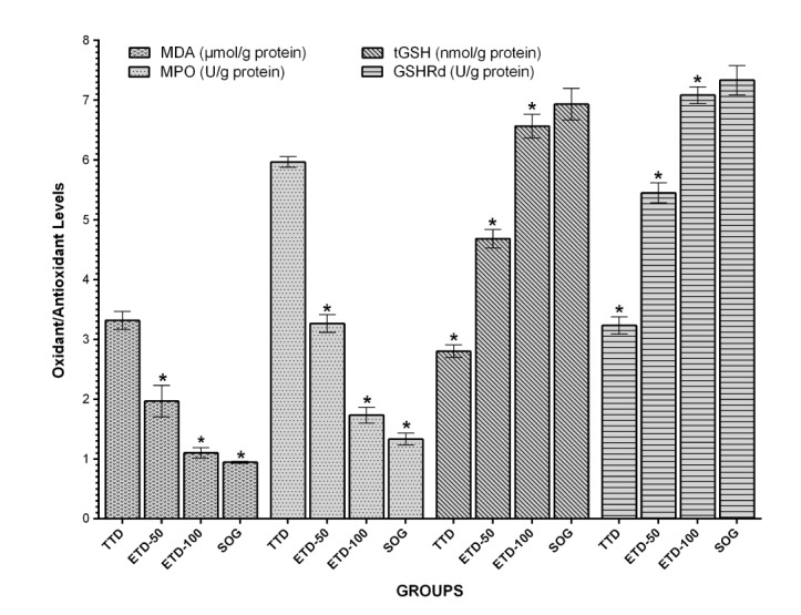
As shown in Fig. 1, the amount of MDA was found to be 0.94±0.04 µmol/g protein in the testicular tissue of the SOG group. The amount of MDA increased to 3.31±0.15 µmol/g protein in the TTD group subjected to torsion-detorsion, and that in the rat groups given 50 and 100 mg/kg of etoricoxib decreased to 1.8±0.1 and 1.0±0.06 µmol/g protein, respectively. The torsion-detorsion process also caused an increase in MPO activity. MPO activity, which increased up to 6.0±0.08 U/g protein after the torsion-detorsion process, decreased to 3.3±0.14 in the group given 50 mg/kg of etoricoxib and to 1.7±0.13 in the group given 100 mg/kg of etoricoxib. MPO activity was found to be 1.3±0.09 U/g protein in the SOG group (Fig. 1). The amount of tGSH was found to be lower (2.8±0.10 nmol/g protein) in the testicular tissue administered with torsion-detorsion than in the SOG group (6.9±0.26 nmol/g protein). The amount of tGSH was elevated to 4.7±0.15 and 6.6±0.19 in the groups administered with 50 and 100 mg/kg of etoricoxib, respectively (Fig. 1). However, the torsion-detorsion process led to a decrease in GSHRd activity in the testicular tissue aside from tGSH. The GSHRd activity was found to be 3.2±0.14 U/g protein in the group administered with torsion-detorsion, and this value was determined as 5.4±0.16 and 7.1±0.14 U/g protein in the groups administered with 50 and 100 mg/kg if etoricoxib, respectively (Fig. 1).
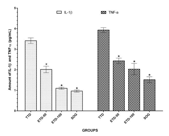
The amount of IL-1β was measured as 0.97±0.06 pg/ml in the SOG group that was not applied with torsion-detorsion. The amount of IL-1β increased to 3.4±0.14 in the group applied with torsion-detorsion. IL-1β levels decreased to 2.0±0.16 pg/ml and 1.1±0.05 pg/ml in the groups administered with 50 and 100 mg/kg of etoricoxib, respectively (Fig. 2). The torsion-detorsion process led to a significant increase in the amount of TNF-α in testis tissue. As a result of torsion-detorsion, the amount of TNF-α increased to 3.9±0.12 pg/ml. It decreased to 2.4±0.13 and 2.0±0.27 pg/ml by administering etoricoxib at 50 and 100 mg/kg doses, respectively. In the SOG group, the amount of TNF-α was 1.5±0.14 pg/ml (Fig. 2).
IL-β and TNF-α gene expression results
The IL-1β gene expression level was elevated to 4.7±0.13 pg/ml in the testicular tissue administered with torsion-detorsion, whereas this value decreased to 2.3±0.11 and 1.3±0.10 pg/ml in the groups administered with 50 and 100 mg/kg of etoricoxib, respectively. The IL-1β gene expression level was 1.1±0.06 pg/ml in the SOG group (Fig. 3). Additionally, TNF-α gene expression level was measured in the testicular tissue of all groups. TNF-α gene expression level was 1.8±0.10 pg/ml in the SOG group. After the torsion-detorsion process, the TNF-α gene expression level increased to 5.5±0.15 pg/ml in the TTD group. The TNF-α gene expression level was significantly low in the ETD-50 and ETD-100 groups at 3.7±0.14 and 2.2±0.22 pg/ml, respectively (Fig. 3).

Histopathological findings
Fig. 4A shows the tunica albuginea, Leydig cells, interstitium lumen, Sertoli cells, spermatogonium, primary spermatocyte and spermatid in the testicular tissue of the SOG group. The seminiferous tubule destruction, interstitial tissue damage and diffuse edema were monitored in the TTD control group (Fig. 4B). Coagulative necrosis was determined in the seminiferous tubule of the TTD group (Fig. 4C). The destructed seminiferous tubule containing germ cell damage and the diffused Leydig cell damage in the interstitium were remarkable (Fig. 4D). In comparing the ETD-50 group administered with 50 mg/kg of etoricoxib with the TTD group, dilated congested blood vessels in the interstitium (Fig. 5A), mild disorganisation in the seminiferous tubules and sporadically continuing edema (Fig. 5B), and focally decreased spermatid were found in the seminiferous tubules despite the significant improvement in the ETD-50 group (Fig. 5C). The histopathological findings in the ETD-100 group were similar to those of normal testicular tissue although dilated congested vessels still appeared (Fig. 5D).
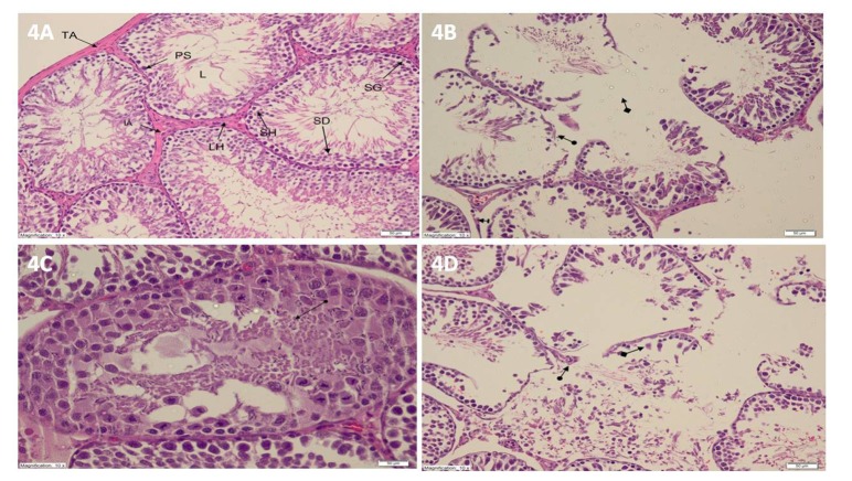 | Fig. 4Histological appearance of the testicular tissue in SOG and TTD groups.(A) Normal histological appearance of the testicular tissue in SOG group. The section shows TA-Tunica albuginea, IA-Interstitium, LH-Leydig cell, L-Lumen and stages of germ cells (SG-Spermatogonium, SH-Sertoli cell, PS-Primary spermatid, SD-Spermatid) in the seminiferous tubules (Magnification, ×10; HE&200). (B) Histopathological appearance of TTD control group: the section shows seminiferous tubule destruction (circle-arrow), interstitial tissue damage (line-arrow) and diffuse edema (square-arrow) (Magnification, ×10; HE&200). (C) Histopathological appearance of TTD control group: The section shows seminiferous tubule structure containing coagulative necrosis (arrow) (Magnification, ×10; HE&400). (D) Histopathological appearance of TTD control group: The section shows destructed seminiferous tubule involving diffuse germ cell damage (square-arrow) and diffuse leydig cell damage in the interstitium (circle-arrow) (Magnification, ×10; HE&200). Scale bars, 50 µm.
|
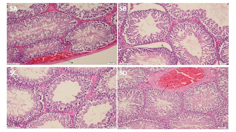 | Fig. 5Histopathological appearance of ETD-50 and ETD-100 groups.(A) Histopathological appearance of ETD-50 group. Dilated congested blood vessel (arrow) in the interstitium (Magnification, ×20; HE&200; Scale bar, 20 µm). (B) Histopathological appearance of ETD-50 group: The section shows mild disorganization and sporadic edema in the seminiferous tubule (arrow) (Magnification, ×20; HE&200; Scale bar, 20 µm). (C) Histopathological appearance of ETD-50 group: focal decreased spermatid in some seminiferous tubules (arrow) (Magnification, ×20; HE&200; Scale bar, 20 µm). (D) Histopathological appearance of ETD-100 group: The section shows dilated congested vessel structure (arrow) (Magnification, ×10; HE&200; Scale bar, 50 µm).
|
Go to :
DISCUSSION
In this study, we investigated the effect of etoricoxib on oxidative I/R damage that was developed in rat testicles, which were subjected to 720° torsion for 4 h followed by reperfusion for another 4 h. Generally, testicular torsion is among the most important conditions requiring emergency intervention because of its implications [18]. If not treated, it leads to irreversible testicular necrosis [19]. Heindel et al. found that 360° torsion did not lead to any change but 720° or greater torsion caused decreased fertility [20]. In addition, the severity of damage due to torsion was reported to be associated with duration [21]. In their experimental study, Turner et al. reported that 360° and 1 h torsion caused only acute vascular changes, whereas 720° and 4 h torsion resulted in complete ischemia [22]. In the present study, the levels of MDA and MPO showed a significant increase in the testicular tissue of the TTD group subjected to 720° and 4 h torsion and reperfused with 4 h detorsion compared with those in the healthy controls.
Additionally, the levels of tGSH and GSHRd were found to be low in the TTD group, which had high levels of MDA and MPO. In the literature, ischemia has been reported to cause oxidative stress [12]. Mertoglu et al. reported that I/R significantly increased the amount of MDA in rat testicle [23]. Another study demonstrated elevated MDA in testicular tissue damaged by torsion-detorsion [24]. This finding indicates that our experimental results are consistent with those in the literature. Aside from MDA, torsion-detorsion was reported to cause a significant increase in MPO activity in tissue [25]. I/R is a pathologic process that expands with inflammation [12]. Increased MPO activity in the TTD group suggests that the inflammatory reaction was developed in the testicular tissue of this group. A PNL-specific enzyme, MPO, is considered a marker of reperfusion damage [26].
Moreover, the decreased tGSH and GSHRd in the TTD group indicates that the oxidant-antioxidant balance changed in favour of the oxidants. Impairment of the oxidant-antioxidant balance in favour of the oxidants indicates oxidative stress [27]. In a study by Ozturk et al., tGSH was reported to be low in testicular tissue subjected to I/R [28]. The amount of tGSH in tissues is kept constant by GSHRd [29]. GSHRd was found to play a crucial role as an antioxidant in the protection of cellular integrity against oxidative stress [30].
In the present study, the gene expression level and the amount of proinflammatory cytokines, such as IL-1β and TNF-α, in the testicular tissue increased in the TTD group. The functional roles of IL-1β and TNF-α in the testicles have been documented in the literature [31]. Studies have suggested that IL-1β and TNF-α, which are known proinflammatory cytokines, show an increase in testicular I/R damage [32]. Recent studies have reported that the gene expression level of IL-1β and TNF-α increased in the testicular tissue subjected to detorsion following 720° torsion [33].
Our experimental results demonstrated that etoricoxib prevented the increase in the levels of MDA, MPO, IL-1β and TNF-α in testicular tissue administered with torsion-detorsion in a dose-dependent manner. No studies were found in the literature examining the effect of etoricoxib on oxidants, antioxidants and cytokines in testicular tissue administered with the I/R process. However, recent studies have reported that etoricoxib protects the liver tissue against I/R damage by preventing the increase in the levels of MDA and MPO, and the decrease in the amounts of tGSH and GSHRd [34]. Etoricoxib was found to decrease MDA and MPO and to prevent the decrease in the level of tGSH in ovarian I/R damage [13]. Furthermore, etoricoxib was revealed to inhibit IL-1β and TNF-α gene expressions in the brain [35]. Aside from their cytotoxic effect, IL-1β and TNF-α are known to have an important role in inflammatory reaction and regulation [3637]. TNF-α and IL-1β form in the early period of inflammation and have many functions, such as the oxidative burst of neutrophils through similar common signal molecules and the release of free radicals [38].
The biochemical and gene expression results obtained from all the groups included in this study are consistent with the histopathological findings. Such pathological findings as destructed seminiferous tubules, coagulative necrosis, germ cell damage, severe damage and edema in the interstitium and Leydig cell that were observed in the TTD group, which had the highest amount of oxidant and cytokines, were not found in the ETD-100 group. However, dilated congested blood vessels in the interstitium, mild disorganisation in the seminiferous tubules, decreased spermatid and edema were found in the ETD-50 group, which suppressed oxidants and cytokines milder than those in the ETD-100 group. Torsion-detorsion has been experimentally demonstrated to lead to pathological changes, including disorganisation in the seminiferous tubules, necrosis of the germinal cells, decreased spermatozoa [39] and interstitial edema [4]. In their study, Minutoli et al. histopathologically showed severe damage in the testes caused by I/R [40]. Oxidants and cytokines were found to play a crucial role in the pathogenesis of histological disruptions in testicular tissue caused by torsion-detorsion [2241].
In conclusion, the biochemical, gene expression and histopathological findings showed that the torsion-detorsion process created oxidative stress in testicular tissue. The torsion-detorsion process in the testis tissue led to an increase in oxidants and proinflammatory cytokines, and a decrease in antioxidant parameters. Moreover, the torsion-detorsion process caused severe interstitial tissue damage, seminiferous tubule destruction, diffuse edema, germ cell damage and diffused Leydig cell damage. The dose of 100 mg/kg etoricoxib prevented the histopathological damage due to torsion-detorsion better than the dose of 50 mg/kg. Etoricoxib may prevent testicular oxidative damage induced by torsion-detorsion.
Therefore, etoricoxib may be useful in the reduction of damage that may occur during the repurfusion of torsioned testicles with detorsion.
Go to :
 XML Download