Journal List > Korean J Physiol Pharmacol > v.21(3) > 1026177

Nam, Kim, Kim, Chin, Kim, Bae, Nam, and Kim: Activation of transient receptor potential vanilloid 3 by the methanolic extract of Schisandra chinensis fruit and its chemical constituent γ-schisandrin

Abstract

Transient receptor potential vanilloid 3 (TRPV3) is a non-selective cation channel with modest permeability to calcium ions. It is involved in intracellular calcium signaling and is therefore important in processes such as thermal sensation, skin barrier formation, and wound healing. TRPV3 was initially proposed as a warm temperature sensor. It is activated by synthetic small-molecule chemicals and plant-derived natural compounds such as camphor and eugenol. Schisandra chinensis (Turcz.) Baill (SC) has diverse pharmacological properties including antiallergic, anti-inflammatory, and wound healing activities. It is extensively used as an oriental herbal medicine for the treatment of various diseases. In this study, we investigated whether SC fruit extracts and seed oil, as well as four compounds isolated from the fruit can activate the TRPV3 channel. By performing whole-cell patch clamp recording in HEK293T cells overexpressing TRPV3, we found that the methanolic extract of SC fruit has an agonistic effect on the TRPV3 channel. Furthermore, electrophysiological analysis revealed that γ-schisandrin, one of the isolated compounds, activated TRPV3 at a concentration of 30 µM. In addition, γ-schisandrin (~100 µM) increased cytoplasmic Ca2+ concentrations by approximately 20% in response to TRPV3 activation. This is the first report to indicate that SC extract and γ-schisandrin can modulate the TRPV3 channel. This report also suggests a mechanism by which γ-schisandrin acts as a therapeutic agent against TRPV3-related diseases.

INTRODUCTION

Transient receptor potential (TRP) cation channel, subfamily vanilloid (V), member 3 (TRPV3), which belongs to the TRP family of ion channels, shares 43% sequence homology with TRPV1 [1]. TRPV3 is a thermosensitive channel that is activated at temperatures above 30~33℃ and is modestly permeable to calcium ions (PCa/PNa permeability ratio=~10) [1]. Although TRPV3 was initially proposed as a thermosensor in the human body, it is also activated by synthetic small molecules such as 2-aminoethyl diphenyl borate (2-APB), analogs of 2-APB such as diphenylboronic anhydride, and drofenine [12]. Moreover, like other thermosensitive TRP channels, TRPV3 is also activated by natural plant-derived compounds such as camphor, carvacrol, and eugenol thymol [34]. Camphor is a terpenoid with a strong aroma produced by the wood of the camphor laurel (Cinnamomum camphora) and was the first natural compound found to activate TRPV3 [4]. Carvacrol, eugenol, and thymol are monoterpenoid compounds. They are major components of plants such as oregano, savory, clove, and thyme, and can activate TRPV3. In addition, TRPV3 is more sensitive to them than it is to camphor [5]. In mammals, TRPV3 is expressed in keratinocytes, adipocytes, neurons, lymphocytes, and epithelial cells in parts of the alimental canal such as the oral cavity, tongue, larynx, and distal colon [1678]. TRPV3 expression is highest in the skin. Thus, extensive researches are being performed to understand skin physiology, such as skin barrier formation or hair growth, and skin pathophysiology, such as skin inflammation, wound healing, and itching sensation [91011]. In keratinocytes, TRPV3 activation is required for transglutaminase activity, which is essential for the formation of the cornified envelope in the epidermis [12]. In addition, TRPV3 activation has been shown to result in an increase in the proliferation of oral epithelial cells; therefore, TRPV3 agonists can be used to promote epithelial wound healing [13]. It has also been shown that TRPV3 channels in adipocytes can regulate adipogenesis since chronic treatment with a TRPV3 agonist prevented adipogenesis and weight gain in mice fed high-fat diets [6]. Therefore, there is a need to identify TRPV3 agonists that can be used in studies to understand the role of TRPV3 in mammalian physiology. As well, TRPV3 agonists can be used to treat TRPV3-related human disorders and diseases. The dried fruit of Schisandra chinensis (Turcz.) Baill (SC) is an important component of herbal medicines and is used a food additive in China, Japan, Korea, and Russia [1415]. The fruit of SC is extensively used in the treatment of various diseases such as diarrhea, cough, asthma, phlegm, jaundice, insomnia, palpitation, and diabetes [15]. Several studies have demonstrated the diverse pharmacological activities of SC, which include antiseptic [16], antiallergic [17], anti-inflammatory [18], wound healing [15], and antiobesity [19] effects.
As part of our ongoing research on using compounds from natural sources as modulation agents of ion channels [202122], we investigated the effects of SC fruit extract on TRPV3. We also investigated the effects of chemical compounds isolated from the SC fruit by bioassay-guided fractionation on TRPV3. To the best of our knowledge, this is the first report on the modulatory effects of SC fruit extract and chemical compounds isolated from the SC fruit on TRPV3 ion channels that are related to skin barrier formation and inflammation.

METHODS

Chemicals

All the reagents used in the study, except 2-APB, were purchased from Sigma-Aldrich (St. Louis, MO, USA). 2-APB was purchased from Tocris (Bristol, UK).

Preparation of SC seed oil and methanolic and ethanolic extracts of SC fruits

The SC seed oil used in the present study was provided by a local company (Hyojongwon, Goyang, Gyeonggi-do, Korea). SC fruits (100 g) were refluxed with MeOH or EtOH for 2 h and evaporated in vacuo. The crude methanolic or ethanolic extract was then lyophilized.

Chemical isolation

Dried SC fruits (5.8 kg) were macerated in MeOH, after which the liquid portion of the mixture was separated. The procedure was repeated three times and the pooled extract obtained was evaporated in vacuo. Afterward, the extract (1.53 kg) was suspended in H2O and partitioned with CHCl3, EtOAc, and n-butanol successively to obtain a CHCl3-soluble extract (180 g), an EtOAc-soluble extract (132 g), a butanol-soluble extract (434 g), and a water-soluble extract (759 g). Next, 35 g of the CHCl3-soluble extract (SCC) was separated by silica column chromatography (5×90 cm, 600 g) using the gradient method. Hexane-EtOAc (30:1~2:1) and CHCl3-MeOH (20:1~1:1) were used as the mobile phase in increasing polarity. The resultant eluent was fractionated into 17 sub-fractions (SCC-1–SCC-17). The SCC-3 fraction (42.4 mg) was separated on a Sephadex LH-20 column (2×90 cm; GE Healthcare, PA, USA), using CHCl3–MeOH (1:1) as the mobile phase in gradient elution mode, and fractionated into three sub-fractions (SCC-3A–SCC-3C). The SCC-5 fraction (162.3 mg) was subjected to high-performance liquid chromatography (HPLC) using a Phenomenex Luna C18 column (250×10 mm, 5 µm) for chromatographic separation, and MeCN and H2O as the mobile phase. The flow rate of the mobile phase was set at 3 mL/min. The mobile phase was run as follows: MeCN and H2O (70:30) for 34 min, followed by 100% MeCN for 6 min. The retention times (tRs) of γ-schisandrin (6.5 mg) and gomisin N (21.2 mg), which were obtained from the HPLC, were 30.25 and 33.10 min, respectively. The SCC-12 fraction (2062.5 mg) was purified by medium-pressure liquid chromatography (MPLC) using a reverse-phase (RP) column and a MeOH-H2O (5:90~90:10) mixture to give 14 sub-fractions (SCC-12A–SCC-12N). The SCC-12K fraction (56.5 mg) was purified by MPLC using an RP column and a MeOH-H2O (40:60~80:20) mixture to give five sub-fractions (SCC-12K1–SCC-12K5). The SCC-12K4 fraction (36.3 mg) was subjected to HPLC using the Phenomenex Luna column for chromatographic separation and MeCN and H2O as the mobile phase (flow rate, 3 mL/min). The mobile phase was run as follows: MeCN-H2O (60:40) for 20 min, followed by 100% MeCN for 6 min. This yielded schisandrin (10.5 mg) and angeloyl gomisin H (12.5 mg) with tRs of 10.55 and 16.45 min, respectively. The structures of the isolated compounds were confirmed by comparing their nuclear magnetic resonance and mass spectrometry data with those reported in literature (schisandrin [23], γ-schisandrin [24], gomisin N [25], and angeloyl gomisin H [26]).

Cell culture

HEK293T cells (catalog no. CRL-3216; American Type Culture Collection, Manassas, VA, USA) were subcultured onto a 25-cm2 culture flask (Thermo Fisher, Waltham, MA, USA) with Dulbecco's Modified Eagle's Medium containing 10% heat-inactivated fetal calf serum, 100 units/mL penicillin, and 100 µg/mL streptomycin (Thermo Fisher) at 37℃ in a 10% CO2 humidified incubator.

DNA constructs and transient transfection of hTRPV3

Human TRPV3 (pReceiver-M02) was purchased from Gene-Copoeia (Rockville, MD, USA) and subcloned into pcDNA 3.1 (Thermo Fisher). One day before transfection, HEK293T cells are subseeded onto a 35-mm diameter cell culture dish at 60% confluence. The following day, the cells were transfected with Lipofectamine Plus reagent (Thermo Fisher) according to the manufacturer's instructions. In all the transfection studies, we cotransfected pEGFP-N1 plasmid, which can express a green fluorescence protein, with hTRPV3 plasmid to allow for selecting transfected cells based on the green fluorescence signal. pEGFP-N1 was cotransfected into HEK293T cells at a ratio of 9 (hTRPV3):1 (EGFP).

Electrophysiology

Whole-cell current recordings were performed using an Axopatch 200B amplifier (Molecular Devices, Sunnyvale, CA, USA) with a microglass electrode (patch pipette) which had a resistance of 3~5 MΩ at room temperature (~25℃). The acquired data were digitalized (Digidata 1440A, Molecular Devices) at a sampling rate at 10 kHz and then digitally filtered through a low-pass filter at 1 kHz using pCLAMP 10.4 software (Molecular Devices). All voltage and current trace data were analyzed using Clampfit software 10.4 and stored on a desktop computer. For the measurement of hTRPV3 current (ITRPV3), a mixture of 140 mM CsCl, 10 mM 4-2-hydroxyethyl)-1-piperazineethanesulfonic acid (HEPES), 10 mM ethylene glycol-bis (β-aminoethyl ether)-N,N,N′,N′-tetraacetic acid (EGTA), 4.85 mM CaCl2, 3 mM MgATP, and 10 mM HEPES adjusted to pH 7.2 with CsOH was used as the internal solution. The bath solution for ITRPV3 contained 139 mM NaCl, 5 mM KCl, 10 mM HEPES, 3 mM BaCl2, 2 mM MgCl2, 1 mM EGTA, and 10 mM glucose adjusted to pH 7.4 with NaOH.

Intracellular free calcium concentration ([Ca2+]i) measurements

Transient transfected TRPV3-HEK293T cells were harvested and centrifuged twice at 1000 rpm for 1 min with HEPES-buffered ringer solution (at 4℃; 145 mM NaCl, 3.6 mM KCl, 1.3 mM CaCl2, 1 mM MgCl2, 5 mM D-glucose, and 10 mM HEPES adjusted to pH 7.4 with NaOH). Next, the cell suspensions were loaded with 5 µM fura-2-acetoxymethyl ester (Thermo Fisher) at room temperature (~25℃). After 30 min, the cells were washed with HEPES-buffered ringer solution at 4℃. The cells were maintained at 4℃ during the experiments. Fluorescence was measured in water-jacketed quartz microcuvettes (37℃) with continuous stirring. The microcuvettes contained 1 mL of cells (~1 million cells) in HEPES-buffered ringer solution. Fluorescence was monitored with a fluorescence spectrophotometer (Photon Technology Instruments, Birmingham, NJ, USA) at excitation and emission wavelengths of 340 and 380 nm and 510 nm, respectively. Fluorescence intensities were recorded every 0.2 sec.

Statistical analysis

Data were analyzed by one-way analysis of variance and post-hoc tests using the Bonferroni procedure. All data are expressed as mean±standard error of the mean. N refers to the number of experiments conducted at each experimental condition. Probability values less than 0.05 were considered statistically significant. We used Prism 6.0 (GraphPad, La Jolla, CA, USA) and Origin 8.0 (Microcal, Northampton, MA, USA) for the statistical analyses.

RESULTS

Methanolic extract of SC can activate ITRPV3
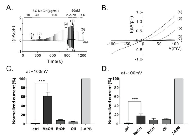
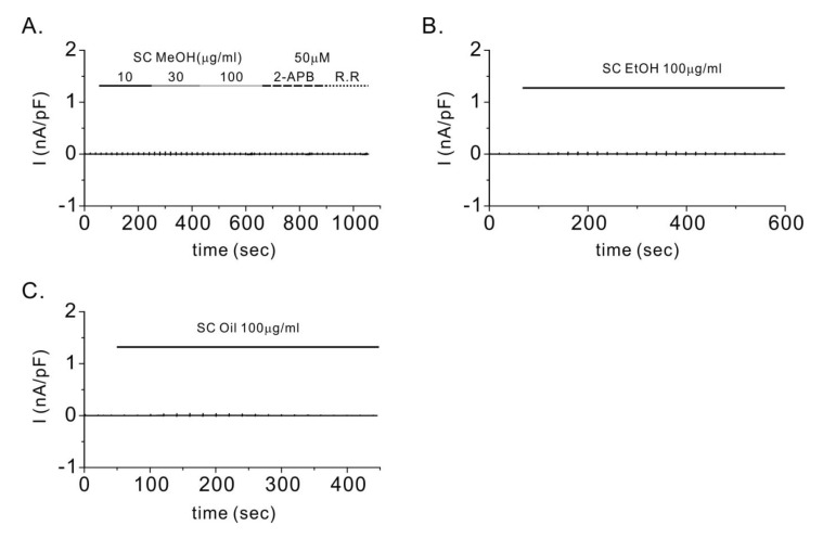
To elucidate whether SC fruit extracts or SC seed oil could activate TRPV3 channel activity, we prepared SC seed oil and two SC fruit extracts. To compare the agonistic effects of the SC extracts on TRPV3, we performed whole-cell patch clamp recording using HEK293T cells that were transiently transfected with hTRPV3 (hTRPV3-HEK293T). After confirming there was no baseline current, we assessed which SC extract showed agonistic effects on TRPV3 channel. As shown in Fig. 1A, we applied 10, 30, or 100 µg/mL of the methanolic extract of dried SC fruits (SCMeOH) to the bath solution. After SCMeOH treatment, we serially applied 50 µM 2-APB (a potent agonist of TRPV3) and ruthenium red (R.R.) (an inhibitor of TRPV3) to determine the ITRPV3 maximum current of each cell and the ITRPV3 inhibition. Current-voltage relationship curves (I~V curves) for ITRPV3 were obtained in the presence of the following: 10 (1), 30 (2), or 100 (3) µg/mL of the SC fruit extracts; 50 µM 2-APB (4); and 10 µM R.R (5) (Fig. 1B). From the results, SCMeOH at concentrations of 10, 30, and 100 µg/mL dose-dependently activated ITRPV3 by 1.67±0.41, 2.55±0.43, and 17.84±4.65%, respectively, at −120 mV. However, the ethanolic extract of SC (SCEtOH) and the SC seed oil (SCseed oil) did not show any effects on the hTRPV3-HEK293T cells. In Fig. 1C and D, we compare the normalized activation rates of ITRPV3 when 100 µg/mL of each extract was applied at −100 and 100 mV clamp voltages. SCMeOH only showed statistically significant effects on ITRPV3 (p<0.01, n=5). The summarized data for each concentration of the SC fruit extract (10, 30, and 100 µg/mL) are presented in Table 1. At the same conditions, non-selective cationic current by TRPV3 activation was not observed by the SC extracts in mock-transfected HEK293T cells (Fig. 2).

Agonistic effect of γ-schisandrin on ITRPV3

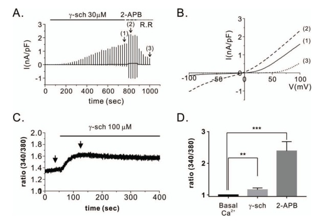
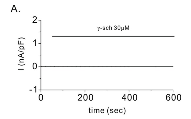
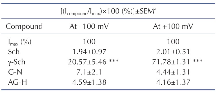
To identify the compounds in the SC fruit extract that were responsible for the observed agonistic effects of the extract on ITRPV3, we isolated and examined the following four compounds from the dried fruits: schisandrin, γ-schisandrin, gomisin N, and angeloyl gomisin H (Fig. 3). We then performed whole-cell patch clamp experiments on the isolated compounds using the same protocol stated in section 3.1 to examine whether the compounds activate ITRPV3. Because of the limited amounts of the isolated compounds, we used each chemical at 30 µM in the bath solution and assessed its effect. As shown in Table 2, one potent TRPV3 activator, γ-schisandrin (γ-Sch), was identified among the four isolated compounds. γ-Sch at a concentration of 30 µM showed statistically significant agonistic effects on TRPV3 at both positive and negative voltage ranges. Fig. 4A and B show representative traces and their related I-V curves for γ-Sch. Treatment of the cells with 30 µM γ-Sch activated ITRPV3 at a positive voltage about three times more than it did at a negative voltage. This phenomenon was similar to that observed for the SCMeOH treatment, which also activated ITRPV3 at a positive membrane voltage more than it did at a negative membrane voltage. To confirm whether ITRPV3 activation was due to non-specific cationic current activation, we treated mock-transfected HEK293T cells with 30 µM γ-Sch; however, the γ-Sch treatment did not result in current activation (Fig. 5). ITRPV3 activation at a negative voltage is much more important than that at a positive voltage. This is because the activation can result in Ca2+ influx through TRPV3 to generate Ca2+ signaling. Therefore, we measured intracellular Ca2+ concentrations [Ca2+]i in fluorimetry experiments by using a Fura-2 calcium sensitive fluorescence dye. As shown in Fig. 4C, treatment with 100 µM γ-Sch resulted in increased intracellular Ca2+ concentrations in the TRPV3-HEK293T cells by about 13±2.6% more than the basal Ca2+ level. We also compared the agonistic effects of γ-Sch and 2-APB, each at 100 µM (Fig. 4D). Data obtained from the patch clamp experiments showed that 2-APB generated a stronger inward current than γ-Sch did (Fig. 4B). In addition, 2-APB efficiently increased [Ca2+]i in the TRPV3-HEK293T cells more than γ-Sch did. Taken together, the results support that γ-Sch can activate ITPRV3 and generate intracellular Ca2+ signaling.

DISCUSSION

TRPV3 regulates proliferation and differentiation of human keratinocytes, which can promote skin barrier formation [12]. In oral epithelial cells, the activation of TRPV3 can promote oral wound healing by potentiating epithelial cell proliferation [13]. Moreover, systemic administration of a TRPV3 agonist has been shown to reduce visceral adipose tissue via inhibition of adipogenesis in mice fed high-fat diets [6]. Therefore, a TRPV3 agonist might be a potential therapeutic candidate for preventing and treating TRPV3-related diseases.
SC is generally used as a tonic medicine in Korea. It contains lignans as its major constituent and it is used by traditional oriental clinicians in China, Korea, and Japan to treat several conditions such as dermatitis, cancer, hepatitis, and impotence [1527]. Interestingly, it has been reported that SC has an inhibitory effect on adipocyte differentiation and adipogenesis in 3T3-L1 preadipocytes. In addition, the antiobesity property of SC has been demonstrated in high-fat diet-induced obese rats [19]. Moreover, according to a clinical study that was conducted in Russia in the 1950s, SC fruit extract showed a wound healing effect in patients with trauma- or varicose vein dilatation-induced trophic ulcers, as well as in those with slowly granulating wounds [15]. Although several physiological and pathophysiological processes might involve TRPV3, previous studies have only focused on the mechanisms underlying the therapeutic effects of SC.
In the present study, by performing whole-cell patch clamp studies, we assessed whether SC fruit extracts and seed oil can activate the TRPV3 channel. Firstly, we found that SCMeOH at a concentration of 100 µg/mL can effectively activate ITRPV3 (by 17.84±4.65% at −100 mV and by 61.53±8.8% at +100 mV) in HEK293T cells overexpressing hTRPV3 (Fig. 1). Secondly, to identify the active compounds in SC fruit, we isolated and studied angeloyl gomisin H, gomisin N, schisandrin, and γ-Sch from dried SC fruits (Fig. 3). We found that at a concentration of 30 µM, γ-Sch activated TRPV3 by 20.57±5.46% at -100 mV and by 71.78±1.31% at +100 mV. In addition, we confirmed that γ-Sch increased [Ca2+]i via TRPV3 activation in the HEK293T cells overexpressing hTRPV3 (Fig. 4).
One limitation of our study was that we could not obtain the half-effective concentration (EC50) of the isolated γ-Sch due to its limited amount. However, most TRPV3 agonists from natural sources are relatively weaker agonists than those from synthetic sources (such as 2-APB). In addition, the former activate only sizeable currents at more than several hundreds of micromolar concentrations [5]. However, we expect that γ-Sch may be a much more potent agonist than the known natural agonists are. γ-Sch's EC50, its pharmacological properties as regards TRPV3, and its application in the treatment of TRPV3-related diseases remain to be elucidated in further studies.
In summary, this is the first electrophysiological study undertaken to explore the agonistic effects of SC fruit extracts and seed oil, as well as those of γ-Sch on hTRPV3 channel. Using whole-cell patch clamp recordings, we found that TRPV3 was significantly activated by SCMeOH and γ-Sch, which also increased [Ca2+]i via the TRPV3 activation. Our findings may explain one of mechanisms underlying the antiobesity and wound healing effects of SC.

ACKNOWLEDGEMENTS

JH Nam received financial support from the Convergence of Conventional Medicine and Traditional Korean Medicine R&D program funded by the Ministry of Health & Welfare (Korea) through the Korean Health Industry Development Institute (KHIDI) [grant number HI15C0256].

Notes

Author contributions: Y.N. and H.J.K. performed the patch clamp experiments. Y.M.K. performed the chemical isolation from the plant extract. Y.W.C., Y.K.K. and H.S.B. supervised and coordinated the study. J.H.N. and W.K.K. worte the manuscript.

CONFLICTS OF INTEREST: The authors declare no conflicts of interest.

References

1. Luo J, Hu H. Thermally activated TRPV3 channels. Curr Top Membr. 2014; 74:325–364. PMID: 25366242.
crossref
2. Deering-Rice CE, Mitchell VK, Romero EG, Abdel Aziz MH, Ryskamp DA, Križaj D, Gopal VR, Reilly CA. Drofenine: A 2-APB analogue with greater selectivity for human TRPV3. Pharmacol Res Perspect. 2014; 2:e00062. PMID: 25089200.
3. Xu H, Delling M, Jun JC, Clapham DE. Oregano, thyme and clove-derived flavors and skin sensitizers activate specific TRP channels. Nat Neurosci. 2006; 9:628–635. PMID: 16617338.
crossref
4. Moqrich A, Hwang SW, Earley TJ, Petrus MJ, Murray AN, Spencer KS, Andahazy M, Story GM, Patapoutian A. Impaired thermosensation in mice lacking TRPV3, a heat and camphor sensor in the skin. Science. 2005; 307:1468–1472. PMID: 15746429.
crossref
5. Vogt-Eisele AK, Weber K, Sherkheli MA, Vielhaber G, Panten J, Gisselmann G, Hatt H. Monoterpenoid agonists of TRPV3. Br J Pharmacol. 2007; 151:530–540. PMID: 17420775.
crossref
6. Cheung SY, Huang Y, Kwan HY, Chung HY, Yao X. Activation of transient receptor potential vanilloid 3 channel suppresses adipogenesis. Endocrinology. 2015; 156:2074–2086. PMID: 25774551.
crossref
7. Inada H, Iida T, Tominaga M. Different expression patterns of TRP genes in murine B and T lymphocytes. Biochem Biophys Res Commun. 2006; 350:762–767. PMID: 17027915.
crossref
8. Ueda T, Yamada T, Ugawa S, Ishida Y, Shimada S. TRPV3, a thermosensitive channel is expressed in mouse distal colon epithelium. Biochem Biophys Res Commun. 2009; 383:130–134. PMID: 19336223.
crossref
9. Ho JC, Lee CH. TRP channels in skin: from physiological implications to clinical significances. Biophysics (Nagoya-shi). 2015; 11:17–24. PMID: 27493510.
crossref
10. Nilius B, Bíró T, Owsianik G. TRPV3: time to decipher a poorly understood family member! J Physiol. 2014; 592:295–304. PMID: 23836684.
crossref
11. Nilius B, Bíró T. TRPV3: a ‘more than skinny’ channel. Exp Dermatol. 2013; 22:447–452. PMID: 23800054.
crossref
12. Cheng X, Jin J, Hu L, Shen D, Dong XP, Samie MA, Knoff J, Eisinger B, Liu ML, Huang SM, Caterina MJ, Dempsey P, Michael LE, Dlugosz AA, Andrews NC, Clapham DE, Xu H. TRP channel regulates EGFR signaling in hair morphogenesis and skin barrier formation. Cell. 2010; 141:331–343. PMID: 20403327.
crossref
13. Aijima R, Wang B, Takao T, Mihara H, Kashio M, Ohsaki Y, Zhang JQ, Mizuno A, Suzuki M, Yamashita Y, Masuko S, Goto M, Tominaga M, Kido MA. The thermosensitive TRPV3 channel contributes to rapid wound healing in oral epithelia. FASEB J. 2015; 29:182–192. PMID: 25351988.
crossref
14. Oh EY, Jang JY, Choi YH, Choi YW, Choi BT. Inhibitory effects of 1-O-methyl-fructofuranose from Schisandra chinensis fruit on melanogenesis in B16F0 melanoma cells. J Ethnopharmacol. 2010; 132:219–224. PMID: 20723590.
crossref
15. Panossian A, Wikman G. Pharmacology of Schisandra chinensis Bail.: an overview of Russian research and uses in medicine. J Ethnopharmacol. 2008; 118:183–212. PMID: 18515024.
crossref
16. Lee SK, Kim SD, Kook M, Lee HY, Park JS, Park YH, Kang JS, Jung WJ, Choi YW, Bae YS. Therapeutic effects of α-iso-cubebenol, a natural compound isolated from the Schisandra chinensis fruit, against sepsis. Biochem Biophys Res Commun. 2012; 427:547–552. PMID: 23022181.
crossref
17. Lee KP, Kang S, Park SJ, Kim JM, Lee JM, Lee AY, Chung HY, Choi YW, Lee YG, Im DS. Anti-allergic effect of α-cubebenoate isolated from Schisandra chinensis using in vivo and in vitro experiments. J Ethnopharmacol. 2015; 173:361–369. PMID: 26253578.
crossref
18. Oh SY, Kim YH, Bae DS, Um BH, Pan CH, Kim CY, Lee HJ, Lee JK. Anti-inflammatory effects of gomisin N, gomisin J, and schisandrin C isolated from the fruit of Schisandra chinensis. Biosci Biotechnol Biochem. 2010; 74:285–291. PMID: 20139628.
19. Park HJ, Cho JY, Kim MK, Koh PO, Cho KW, Kim CH, Lee KS, Chung BY, Kim GS, Cho JH. Anti-obesity effect of Schisandra chinensis in 3T3-L1 cells and high fat diet-induced obese rats. Food Chem. 2012; 134:227–234.
crossref
20. Nam JH, Nam DY, Lee DU. Valencene from the rhizomes of cyperus rotundus inhibits skin photoaging-related ion channels and UV-induced melanogenesis in B16F10 melanoma cells. J Nat Prod. 2016; 79:1091–1096. PMID: 26967731.
crossref
21. Lee DU, Weon KY, Nam DY, Nam JH. Skin protective effect of guava leaves against UV-induced melanogenesis via inhibition of ORAI1 channel and tyrosinase activity. Exp Dermatol. 2016; 25:977–982. PMID: 27488812.
crossref
22. Nam JH, Lee DU. Inhibitory effect of oleanolic acid from the rhizomes of Cyperus rotundus on transient receptor potential vanilloid 1 channel. Planta Med. 2015; 81:20–25. PMID: 25402944.
crossref
23. Kanatani H, Terabayashi S, Takeda S, Li W, Koike K, Nikaido T. Regio- and stereoselective 12-O-demethylation of schizandrin into gomisin T, an important intermediate to gomisin A, by Mortierella sp. (TM-I1104). Tetrahedron Lett. 2005; 46:8467–8470.
crossref
24. Ikeya Y, Taguchi H, Yosioka I. The constituents of schizandra chinensis BAILL. X. The structures of γ-schizandrin and four new lignans, (-)-gomisins L1 and L2, (±)-gomisin M1 and (+)-gomisin M2. Chem Pharm Bull. 1982; 30:132–139.
25. Seo SM, Lee HJ, Park YK, Lee MK, Park JI, Paik KH. Lignans from the fruits of schizandra chinensis and their inhibitory effects on dopamine content in PC12 cells. Nat Prod Sci. 2004; 10:104–108.
26. Ikeya Y, Tagychi H, Yosioka I. The constituents of schizandra chinensis BAILL. The structures of three new lignans, angeloylgomisin H, tigloylgomisin H and benzoylgomisin H, and the absolute structure of schizandrin. Chem Pharm Bull. 1978; 26:328–331.
crossref
27. Han DH, Lee JH, Kim H, Ko MK, Chae MR, Kim HK, So I, Jeon JH, Park JK, Lee SW. Effects of Schisandra chinensis extract on the contractility of corpus cavernosal smooth muscle (CSM) and Ca2+ homeostasis in CSM cells. BJU Int. 2012; 109:1404–1413. PMID: 21951618.
Fig. 1

Effect of methanolic extract of Schisandra chinensis (SCMeOH) on TRPV3 current (ITRPV3) induction in HEK293T cells overexpressing TRPV3 (hTRPV3-HEK293T).

(A) Representative patch clamp recordings of ITRPV3 in response to continuous ramp-like voltage pulse in TRPV3-HEK293T cells. In the whole-cell patch clamp recording, ramp-like pulses from −100 to 100 mV (dV/dt=0.04 V/sec) were applied every 20 sec to determine the current-voltage relationship (I~V curves). The numbers in parentheses indicate the steady state ITRPV3 induced by 10 (1), 30 (2), or 100 (3) µg/mL SC fruit extract or 50 µM 2-APB (4), or inhibited by 10 µM ruthenium red (R.R) (5). (B) The corresponding I~V curves at each steady state ITRPV3. (C, D) Summary of normalized inward and outward currents at +100 mV (C) and -100 mV (D) induced by various concentrations of the SC extracts. The current densities were normalized to the maximum ITRPV3 with the 2-APB treatment and used at the end of the experiment. 2-APB, 2-aminoethyl diphenyl borate; SCMeOH, methanolic extract of dried Schisandra chinensis fruits; SCEtOH, ethanolic extract of dried Schisandra chinensis fruits; SCseed oil, Schisandra chinensis seed oil extract. *** indicates p<0.001 vs. the baseline current (control).
kjpp-21-309-g001
Fig. 2

Control experiments for TRPV3 currents (ITRPV3) in mock-transfected HEK293T cells.

To confirm whether Schisandra chinensis (SC) extracts generate non-selective cationic currents, whole-cell patch clamp experiments were done in mock-transfected HEK293T cells (A~C). Representative patch clamp recording of mock-transfected HEK293T cells treated with the methanolic extract of dried Schisandra chinensis fruits (SCMeOH), ethanolic extract of dried SC fruits (SCEtOH), and SC seed oil (SCseed oil). In the case of SCMeOH, we treated the cells with 2-aminoethyl diphenyl borate (2-APB) to activate ITRPV3. We then treated the cells with the TRPV3 blocker ruthenium red (R.R) to inhibit TRPV3. It was observed that no current was generated from the SC extract or 2-APB treatment in the mock-transfected HEK293T cells.
kjpp-21-309-g002
Fig. 3

Structures of the four chemical compounds isolated from the Schisandra chinensis fruit extract.

kjpp-21-309-g003
Fig. 4

Effect of γ-schisandrin (γ-Sch) on TRPV3 activation in HEK293T cells overexpressing TRPV3.

(A) Representative patch clamp recordings showing TRPV3 current (ITRPV3) activation by 30 µM γ-Sch. At the end of the experiment, 50 µM 2-APB was added to the bath solution in order to determine the maximum current. Following treatment with 10 µM ruthenium red (R.R), a TRPV3 inhibitor, ITRPV3 that was not due to a non-selective cationic current was determined. The numbers in parentheses indicate the corresponding current to voltage relations (I~V curves) depicted in Fig. 1B. (B) I~V relationship curves of steady state ITRPV3 due to 30 µM γ-Sch (1) and 50 µM 2-APB (2) treatments. The 10 µM R.R potently inhibited the inward current of ITRPV3; however, it had relatively no impact on the outward current at the same concentration. (C) Representative trace recordings of intracellular calcium measurements ([Ca2+]i), which were increased by 100 µM γ-Sch in HEK293T cells overexpressing TRPV3 (hTRPV3-HEK293T). (D) Comparison of peaks showing increases in [Ca2+]i induced by 100 µM γ-Sch or 100 µM 2-APB in the hTRPV3-HEK293T cells. The data was normalized by resetting the [Ca2+]i to 1. Data are presented as mean±standard error of the mean (n=8). **indicates p<0.01 and *** indicates p<0.001, each vs. the basal [Ca2+]i. 2-APB, 2-aminoethyl diphenyl borate.
kjpp-21-309-g004
Fig. 5

γ-schisandrin (γ-Sch) generated no additional currents in the mock-transfected HEK293T cells.

(A) Representative patch clamp recordings after treatment of the cells with 30 µM γ-Sch.
kjpp-21-309-g005
Table 1

Effects of Schisandra chinensis (SC) fruit extracts and seed oil on TRPV3 current (ITRPV3)

kjpp-21-309-i001

After confirming that there was no basal current, HEK293T cells overexpressing human TRPV3 were treated with the SC extracts and seed oil at concentrations of 10, 30, or 100 µg/mL. The results shown are percentage changes versus the maximum current (Imax), which were obtained by treatment of the cells with 50 µM 2-aminoethyl diphenyl borate, followed by current normalization to 100%. The data are presented as mean±standard error of the mean (SEM) (n=5). ***Indicates p<0.001 vs. the control group. Imax, maximum current; Ipreparation, current induced by SC preparation.

Table 2

TRPV3 currents (ITRPV3) induced by compounds isolated from Schisandra chinensis fruits
kjpp-21-309-i002

aIndicates the control and is the normalized ITRPV3 after treatment with 50 µM 2-APB at the maximum current (Imax), which was set at 100%, before treatment with the isolated compound (Icompound). The data represent the current relative to Imax and are presented as mean±standard error of the mean (SEM). ***indicates p<0.001 vs. the control group.

Sch, schisandrin; γ-Sch, γ-schisandrin; AG-H, angeloyl gomisin H; G-N, gomisin N; 2-APB, 2-aminoethyl diphenyl borate.

TOOLS
Similar articles