Journal List > Korean J Physiol Pharmacol > v.21(2) > 1026162

Nguyen, Lee, Sharma, Park, Jagga, Sharma, Lee, and Nam: Quercetin induces apoptosis and cell cycle arrest in triple-negative breast cancer cells through modulation of Foxo3a activity

Abstract

Quercetin, a plant-derived flavonoid found in fruits, vegetables and tea, has been known to possess bioactive properties such as anti-oxidant, anti-inflammatory and anti-cancer. In this study, anti-cancer effect of quercetin and its underlying mechanisms in triple-negative breast cancer cells was investigated. MTT assay showed that quercetin reduced breast cancer cell viability in a time and dose dependent manner. For this, quercetin not only increased cell apoptosis but also inhibited cell cycle progression. Moreover, quercetin increased FasL mRNA expression and p51, p21 and GADD45 signaling activities. We also observed that quercetin induced protein level, transcriptional activity and nuclear translocation of Foxo3a. Knockdown of Foxo3a caused significant reduction in the effect of quercetin on cell apoptosis and cell cycle arrest. In addition, treatment of JNK inhibitor (SP 600125) abolished quercetin-stimulated Foxo3a activity, suggesting JNK as a possible upstream signaling in regulation of Foxo3a activity. Knockdown of Foxo3a and inhibition of JNK activity reduced the signaling activities of p53, p21 and GADD45, triggered by quercetin. Taken together, our study suggests that quercetin induces apoptosis and cell cycle arrest via modification of Foxo3a signaling in triple-negative breast cancer cells.

INTRODUCTION

Breast cancer can be divided into several intrinsic subtypes including luminal subtypes (almost defined by ER-positive and Her2-negative), Her2 subtype (characterized by Her2-overexpression), and basal-like breast cancer (including mainly triple-negative breast cancer, TNBC, characterized by ER-negative, Her2-negative and PR-negative) [123]. Patients with two former subtypes seem to have good outcomes as treatments are based on targeting specific receptors (ER and/or Her2) while patients with TNBC are associated with poor clinical prognosis due to absence of specific targeted treatments [4]. TNBC accounts for approximately 15% of breast cancer cases [5] and frequently occurs in younger patients. TNBC shows more aggressive and metastatic behaviors [3] and distant recurrence of TNBC appears to be more hazard than other subtypes [6]. Current available treatments for TNBC are mainly based on chemotherapy and radiotherapy; however, there are several limitations. Relapse usually occurs in patients with TNBC after 3~5 years of clinical intervention and cancer develops resistance to chemotherapy [7]. Besides, treatment method like radiotherapy is harmful in nature as it can potentiate carcinogenesis. Therefore, looking for new therapeutic agents that are effective, less toxic and can prevent avoidance of relapse is a prerequisite.
Natural products like flavonoids show advantages including no or less effect on normal cells, effectiveness in killing cancer cells and improvement in cancer relapse. Hence, flavonoids having these properties can be considered as potential cancer therapeutic agents. One of such natural flavonoid is quercetin which is known to have multiple biological actions including anti-oxidant [8], anti-inflammatory [9] and anti-cancer activities with almost no human toxicity [10]. Recently, scientists have paid much attention on anti-cancer activities of quercetin. Studies proved that quercetin is able to enhance radiotherapy and chemotherapy in animal models [1112]. Besides, quercetin is also a chemopreventive agent against diseases including tumors [13]. Recently, rising evidences elucidated that anti-cancer activity of quercetin is via growth inhibition and proapoptosis in many cancer cells models [14151617]. Quercetin inhibition of cancer cell proliferation has been indicated via inhibiting intracellular signaling such as PI3K, EGFR and Her2/neu [181920]. Quercetin has also been shown to induce cancer cell apoptosis via modulating survival signaling pathways (Akt, NF-kB) or regulatory molecules associated with cell apoptosis (p53, Bcl-2 family, FasL) [16171921]. However, anti-tumor effects of quercetin on breast cancer, especially TNBC and its mechanisms are poorly understood.
Foxo3a, is a member of Forkhead box O (FOXOs) transcription factors family that is known as a key tumor suppressors in mammalian cells. Foxo3a is closely related to cellular apoptosis, aging, proliferation, metabolism, differentiation and tumorigenesis [22]. Recent study elucidated role of Foxo3a in reducing cell proliferation and tumorigenesis in ER positive breast cancer [23]. Moreover, Akt/Foxo3a signaling has been demonstrated to mediate flavonoid-induced breast cancer cells apoptosis and cell cycle arrest [24]. Besides, Foxo3a has emerged as an important mechanism of apoptosis and cell cycle arrest induced by cytotoxic agents in breast cancer [252627]. While TNBC lack specific targeted treatment, Foxo3a may be an attractive therapeutic target for TNBC.
In this study, we report that quercetin induced apoptosis and cell cycle arrest in TNBC cells and Foxo3a might be a regulatory molecule for anti-cancer effects of quercetin in TNBC. Our study also suggests the involvement of JNK in regulation of quercetin-enhanced Foxo3a activity leading to apoptosis and cell cycle arrest, and the possible regulation of Foxo3a-induced apoptosis and proliferation arrest are via FasL and p53, p21 and GADD45 signaling, respectively.

METHODS

Materials

Quercetin was bought from Sigma (US). 3-[4, 5-dimethylthiazol-2-yl]-2, 5-diphenyltetrazolium bromide (MTT) were purchased from Amresco (US). Antibody against Foxo3a, JNK, p-JNK, p-ERK, p-p38 and Lamin B1 were from Cell Signaling Technology (Boston, US). Anti-β-actin was obtained from Santa Cruz Biotechnology (Santa Cruz, US). SP600125 was from Tocris (Avonmouth, UK). SB203580 and PD98059 were bought from Calbiochem (USA).

Cell culture

Human breast cancer cell line MDA-MB-231 (ATCC, HTB-26) were cultured in Dulbecco's Modified Eagle Medium (DMEM) supplemented with 10% fetal bovine serum (FBS) and 1% penicillin-streptomycin. Cells were maintained at 37℃ and 5% CO2 in a humidified atmosphere. Cells were starved in serum-free media for 12 h prior to quercetin treatment.

Cell viability assay

Cells were plated at a density of 1×10⁴ in 96 well-plates. Different concentrations of quercetin were used to treat cells. After incubation time, media were replaced with fresh media containing MTT at final concentration of 0.5 mg/ml. After 2 h incubation at 37℃, supernatant was carefully removed, the insoluble formazan crystal was dissolved in 200 µl of dimethyl sulfoxide (DMSO) and optical density (OD) was measured at 570 nm wave lengths.

RNA isolation and RT real-time PCR

RNAs was extracted from cells cultured in 6 well-plates using Trizol reagent (Invitrogen, Carlsbad, CA, US). For RT-PCR, SuperScript II Reverse Transcriptase (Invitrogen) was used to convert RNAs (2 µg) to cDNA. One µl of cDNA (1:20 dilutions) were used for real-time PCR. qPCR products were detected with SYBR Green (2xGreen star qPCR MasterMix, Bioneer, Korea) on Rotor-Gene Q (Qiagen, Hilden, Germany). qPCR procedure was denaturation at 95℃ for 10 mins, 40 cycles of denaturation at 95℃ for 20 s, annealing at 60℃for 20 s and extension at 72℃ for 25 s. Gapdh gene was used as internal control. Relative expression was analyzed using ΔΔCT method. PCR primers were used as followed: Gapdh, 5'-TTCAGCTCAGGGATGACCTT-3' (forward) and 5'-ACCCAGAAGACTGTGGATGG-3' (reverse); Foxo3a, 5'-ACTCACTTAGCCACAGCGAT-3'(forward) and 5'-TGACCAAACTTCCCTGGTTA-3' (reverse); FasL, 5'-AACCAAGTGGACCTTGAGAC-3'(forwa rd) and 5'-TTCACATGGCAGCCCAGAGT-3' (reverse).

Protein isolation and western blot analysis

Cells were washed with cold PBS and lysed using RIPA buffer (20 mM Tris-HCl, pH 7.5, 200 mM NaCl, 1% Triton X-100, 1 mM dithiothreitol) containing protease inhibitor cocktail (Roche) and phosphatase inhibitors. Protein concentration was determined using Protein assay kit (Biorad) following the manufacture's protocol. Total protein was separated on SDS-polyacryramide gel electrophoresis and transferred to PVDF membrane. The membrane was then blocked in 5% skimmed milk, incubated with indicated primary antibodies (all antibodies were diluted 1:1000 in 3% BSA) for 2 h at room temperature and incubated with respective horseradish peroxidase-conjugated secondary antibodies. Protein bands were visualized using the enhanced chemiluminescence reagents (Bionote, Korea) and signals were detected and quantified using Fusion-Fx system (Vilber Lourmat, Eberhardzell, Germany).

Cytoplasmic and nuclear fractionation

For examine nuclear translocation of Foxo3a, cytoplasmic and nuclear fractions were prepared with the NE-PER nuclear and cytoplasmic extraction reagents (Thermo Scientific). After treatment with quercetin, MDA-MB-231 cells (2×10⁶) were harvested. According to the manufacturers' instructions, the nuclear and cytoplasmic fractions were collected, followed by western blot analysis. β-actin and Lamin B1 were used as loading controls of cytoplasmic and nuclear fractions, respectively.

Gene knockdown by siRNA or shRNA

For knockdown using siRNA, cells were seeded in 6-well plates and transfected with 100 nM siFoxo3a or scrambled control siRNA using Mission siRNA transfection agent (Sigma). SiFoxo3a were chemically synthesized by Bioneer (Daejon, South Korea), sequence of siFoxo3a as followed: 5'-GACGAUGAUGCGCCUCUCU dTdT-3' (sense) and 5'-AGAGAGGCGCAUCAUCGUC dTdT-3' (antisense). Scrambled control siRNA (sc-37007) were from Santa Cruz Biotechnology.
For knockdown using shRNA, cells were transfected with the shFoxo3a (Openbiosystem) or co-transfected with shFoxo3a and reporter constructs, using Genefectine transfection reagent (Genetrone Biotech, Korea). Scramble shRNA (Addgene) was used as control.

Luciferase assay

Cells were seeded in 48-well plates at density of 5×10⁴ cells/well and transfection was carried out with Genefectine. Foxo3a reporter (FHRE-luc), p21 promoter reporter (WWP-luc), p53 reporter (pG13-Luc) or GADD45 promoter reporter (Gadd45-Luc) (Addgene) (200 ng/well) and control reporter (pRL-TK, Promega) (20 ng/well) were used for co-transfection. After 24 h, cells were treated with 20 µM quercetin. Luciferase activities were measured after 24 h in luminometer (Glomax, Promega, CA, US) using a Dual-Luciferase assay kit (Promega), according to the manufacture's recommendations. Value of luciferase activity was normalized to Renilla luciferase activity.

Detection of apoptosis

MDA-MB-231 cells were prepared in 6 well-plates. After exposure to 20 µM quercetin for 24 h or 48 h, cells were collected and subjected to Annexin V and propidium iodide (PI) staining using FITC Annexin V Apoptosis Detection kit I (BD Pharmingen), following the protocol provided by the manufacturer. Apoptotic cells were then analyzed by flow cytometry (FACSCalibur, BD Biosciences, San Diego, CA, USA). Post-compensation and analysis of FACS data were done using Flowing 2.5.1 version software.

Cell cycle analysis

After treatment with 20 µM quercetin for 48 h, cells were washed twice with PBS and fixed with 70% ethanol for 1 h at 4℃, and then resuspended with PBS containing 20 µg/ml PI (Sigma) and 50 µg/ml RNase A. Cells were incubated at room temperature in the dark for 30 mins and cell cycle was analyzed by flow cytometry. The data was analyzed using Cell Quest Pro 6.0 (BD Biosciences) and ModFit LT software (BD Biosciences).

Immunocytochemistry

Cells grown on coverslips were fixed in 4% paraformandehyde in PBS for 10 mins at 4℃, permeablized in 0.25% Triton X-100 in PBS for 10 mins, and blocked in 5% donkey serum (Chemicon) and 0.3% Triton X-100 in PBS for 30 mins at RT. Next, cells were incubated with rabbit polyclonal anti-Foxo3a antibody (Cell signaling) (1:200) in 1% BSA and 0.3% Trition X-100 in PBS for overnight at 4℃. After that, incubation of cells with Alexa 488-conjugated goat anti-rabbit IgG (Molecular Probes) (1:1000) in 1% BSA and 0.3% Trition in PBS was carried out in RT. After deeply washing the cells with PBS, DAPI (1 µg/ml in PBS) was added to stain nuclei. Cells were mounted with Fluoromount-G (Southern biotech) and images were taken by a fluorescence microscope.

Statistical analysis

Statistical differences were analyzed by Graphpad Prism 5.0 (San Diego, CA) and evaluated by two-tailed Student's t-test. All data was presented as mean±SEM of at least three independent experiments. p<0.05 was considered statistically significant.

RESULTS

Quercetin reduced viability of MDA-MB-231 cells

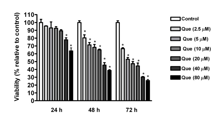
To determine the effect of quercetin on TNBC cells, we treated MDA-MB-231 cells with various quercetin concentrations (2.5~80 µM) for 24 h, 48 h and 72 h and cell viability was determined by MTT assay. As shown in Fig. 1, quercetin demonstrated dose- and time-dependent effects on reduction of the cell viability in MDA-MB-231 cells. Treatment with either 40 µM or 80 µM of quercetin for 24 h reduced cell viability about 20% and 35%, respectively, while treatment with lower concentrations (2.5~20 µM) just caused a minor reduction in cell viability (about 5~10%) in comparison with control. However, the reduction in cell viability increased steadily from 24 h to 72 h in quercetin treatment with all doses.

Quercetin induced apoptosis and cell cycle arrest in MDA-MB-231 breast cancer cells

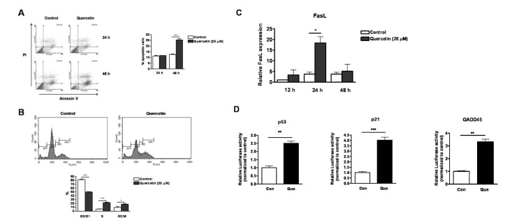
Further, to study whether the reduced viability was due to cell apoptosis triggered by quercetin treatment, FACS analysis was used to detect apoptotic cells followed by treatment of 20 µM quercetin at 24 h and 48 h. Results showed an increase of 15% apoptotic cells (cells with Annexin V positive) after 48 h of quercetin treatment. However, no significant difference for cell death was observed after 24 h of quercetin treatment, compared with control (Fig. 2A). Obtained results are consistent with MTT results as treatment with 20 µM of quercetin for 24 h did not affect cell viability and apoptosis significantly. However, after 48 h of quercetin treatment, cell viability was reduced and population of apoptotic cells was increased significantly. The increase of apoptotic cell death (about 15%) was less than the decrease of cell viability observed by MTT assay (about 30%). It may be further explained by growth arrest. Therefore, we analyzed changes in cell cycle after 48 h of stimulation with quercetin (20 µM) using flow cytometry. As shown in Fig. 2B, quercetin caused cell cycle arrest at S and G2/M phase. Namely, treatment with quercetin resulted in 3 fold increase in cell population of S phase and 2 fold increase in cell population of G2/M phase, along with 30% reduction in cell population of G0/G1 phase. Fas ligand (FasL) is a pro-apoptotic factor that can activate Fas receptor leading to apoptosis. Here we observed that expression of FasL mRNA was enhanced by quercetin after 24 h treatment (Fig. 2C). p53 is known as a cell cycle regulator that activates and inhibit genes involved in cell cycle. GADD45, a p53-regulated and DNA damage-inducible protein have been shown to induce G2-M cell cycle arrest [2829]. p21 (p21WAF1/Cip1), a protein transcriptionally regulated by p53 has been reported to inhibit CDK2 activity, leading to G1/S arrest. [30]. Therefore, signaling activities of p53, p21 and GADD45 were analyzed after quercetin treatment. Fig. 2D shows that quercetin treatment induced signaling activity of p53 (about 3 folds) and promoter activities of p21 (4 folds) and GADD45 (3 folds), suggesting a possibility that increased activities of p53, p21 and GADD45 might lead to cell cycle arrest triggered by quercetin.

Effect of quercetin on Foxo3a activity

Above data showed that quercetin significantly induced apoptosis and cell cycle arrest in breast cancer cells. Identification of signaling molecules that are regulated by quercetin and can contribute to apoptosis and cell cycle arrest was required. It is known that Foxo3a signaling pathway is involved in cell apoptosis and cell cycle arrest by regulating transcription of genes involved in triggering apoptosis [31] and inhibiting cell cycle [32]. Therefore, any involvement of Foxo3a after quercetin treatment in MDA-MB-231 cells was investigated. Data showed that quercetin treatment for 24 h induced about 3 folds of Foxo3a activity in breast cancer cells, as detected by Luciferase assay (Fig. 3A). Further, to elucidate whether quercetin regulated Foxo3a activity was affecting at gene or protein level, we measured mRNA and protein levels of Foxo3a in quercetin-treated cancer cells. The result displayed that quercetin did not affect gene expression of Foxo3a after 12 h, 24 h and 48 h of treatment (Fig. 3B), however, quercetin triggered a steady increase in protein level of Foxo3a after 3~12 h of treatment, maximal increment being 5 folds after 12 h of treatment (Fig. 3C).
Since transcriptional activity of Foxo3a requires its nuclear translocation, we tried to examine the subcellular localization of Foxo3a in quercetin-treated MDA-MB-231 cells. Our immunofluorescence analysis shows increased nuclear Foxo3a of quercetin-treated cells compared to control (Fig. 3D). The result obtained was further confirmed by western blot with nuclear and cytoplasmic fractions. The data shows that quercetin-treated MDA-MB-231 cells exhibited 6~7 folds increases in nuclear Foxo3a protein levels at 6~24 h of treatment, in comparison with control at 0 h. However, no change of cytoplasmic protein levels of Foxo3a by quercetin treatment was observed (Fig. 3E). The results indicate that overall elevated Foxo3a protein in quercetin-treated MDA-MB-231 is associated with its increased levels in nuclei.

Quercetin induced Foxo3a activity through activation of JNK signaling pathway

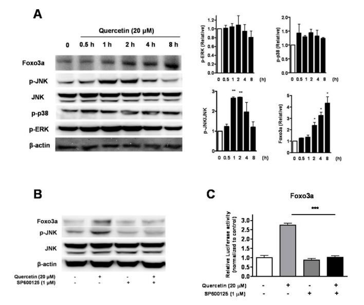
Since quercetin regulated Foxo3a activity, it may be essential to clarify upstream signaling pathways responsible for regulation of Foxo3a activity in quercetin treatment. Therefore, we investigated whether activation of several signaling pathways such as ERK, p38, JNK was induced by quercetin. Western blot analysis using antibodies against phosphorylated forms of ERK, p38 and JNK was carried out to determine activation of respective signaling pathways. As presented in Fig. 4A, a significant increase of JNK activation (p-JNK/JNK) was observed after 1~2 h of quercetin treatment. This activation of JNK might be responsible for increased Foxo3a protein level as observed after 3~12 h of quercetin treatment. However, the other signaling pathway activation (p-ERK and p-p38) showed no significant alteration by quercetin.
To confirm that JNK is one of main signaling pathway participating Foxo3a activity regulation, a JNK specific inhibitor (SP600125) was used. Results showed that pre-treatment with 1 µM of JNK inhibitor (SP600125) completely abolished the effect of quercetin on elevation of protein level and transcriptional activity of Foxo3a (Fig. 4A and 4B). Taken together, the data suggests that JNK signaling pathway participated in regulation of Foxo3a activity in quercetin-treated breast cancer cells.
Moreover, involvement of p38 and ERK signaling pathway in increased Foxo3a activity in response to quercetin treatment was further confirmed. The result shows that treatment with 20 µM of p38 inhibitor SB203580 or 50 µM of ERK inhibitor PD98059 [33] did not affect on quercetin-induced Foxo3a activity (data not shown), suggesting that p38 and ERK signaling pathways may be not necessary for the induction of Foxo3a in response to quercetin treatment.

Quercetin regulated apoptosis and cell cycle arrest through JNK-Foxo3a signaling pathway

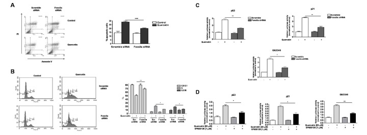
To verify whether Foxo3a was involved in apoptosis and cell cycle arrest triggered by quercetin treatment, MDA-MB-231 cells were transiently transfected with Foxo3a-specific siRNA to deplete the endogenous expression of Foxo3a and the cells transfected with scramble siRNA was used as control. As shown in Fig. 5A, quercetin-induced apoptosis was reduced significantly in Foxo3a siRNA-transfected cells, compared to control. Moreover, Foxo3a knockdown reduced the effect of quercetin on increasing cell population in S and G2/M phases significantly (Fig. 5B). The result suggests that Foxo3a may regulate apoptosis and cell cycle arrest induced by quercetin.
Moreover, effect of Foxo3a knockdown on increased activities of p53, p21 and GADD45 signaling in quercetin treatment was analyzed. MDA-MB-231 cells were co-transfected with Foxo3a shRNA (or scramble shRNA for control) and a plasmid containing p21, p53 or GADD45 luciferase reporter gene prior to quercetin treatment for 24 h. As shown in Fig. 5C, the increase in the reporter activities of p53, p21 and GADD45 were abolished in Foxo3a knockdown cells compared with control. This data suggests that Foxo3a was possibly involved in regulation of p53, p21, and GADD45 signaling activities enhanced by quercetin.
As JNK signaling pathway was involved in regulation of Foxo3a in quercetin treatment, we also examined the involvement of JNK in induction of p21, p53 and GADD45 signaling activities. Similarly to Foxo3a knockdown treatment, JNK inhibitor also abolished the effect of quercetin on increasing reporter activities of p21, p53 and GADD45 in MDA-MB-231 cells (Fig. 5D).

DISCUSSION

In addition to pro-apoptotic activity, quercetin possesses anti-apoptotic activity in several non-tumorigenic cells. As reported, quercetin inhibits hydrogen peroxide-induced apoptosis in mesangial cells [34] and epithelial cells [35]. Quercetin was also known to inhibit apoptosis of neuronal cells caused by trauma brain injury [36]. However, quercetin frequently activates apoptosis in cancer cells. Quercetin-mediated apoptosis may result from mitochondria disruption [37], ROS generation [1438], quercetin intercalation into DNA [39] or downregulation of survival signaling pathways such as PI3K/Akt, NFkB [40]. Besides, quercetin causes cell cycle arrest in cancer cells by modulating several molecules, for examples, p21, p27, cyclin B1 and cyclin-dependent kinases [414243].
Till now, there are only few studies on the anti-cancer effect of quercetin in TNBC. This study revealed that anti-cancer effect of quercetin in TNBC cells was via induction of apoptosis and cell cycle arrest. Our primary observation shows that quercetin reduced cell viability in a time- and dose-dependent manner (Fig. 1). And treatment with quercetin (20 µM) for 48 h induced both cell apoptosis and cell cycle arrest at S and G2/M phases (Fig. 2A and 2B). Moreover, the apoptosis-inducing effect of quercetin was confirmed by the observation that quercetin treatment stimulated FasL mRNA expression at 24 h (Fig. 2C). Besides, S and G2/M cell cycle arrest triggered by quercetin was further confirmed by effect of quercetin on increased signaling activities of p53, p21 and GADD45 (Fig. 2D).
Previous studies showed quercetin induced apoptosis and inhibited cell cycle in TNBC cells [4445], however, in these papers they used high doses of quercetin to treat breast cancer cells. According to study of Chien et al, quercetin treatment of concentration of 250 µM caused ER stress, reduced mitochondrial membrane potential (MMP), activation of capase-3 and reduction of X-linked inhibitor of apoptosis protein (XIAP) which may result in quercetin-induced apoptosis in MDA-MB-231 cells [44]. The other study shows that treatment of 80 µM quercetin to MDA-MB-231 caused an increase in miR-146a, in turn, miR-146a increased Bax and activated capase-3, leading to apoptosis [45]. However, miR-146a may not be mechanism for quercetin-induced apoptosis in quercetin treatment with lower doses as it was shown that effect of a low dose of quercetin (25 µM) on miR-146a was less noticeable. Besides, our study is in agreement with previous studies showing that quercetin increases cell accumulation at G2/M and S phases [4446]. Activation of p53 and increased p21 was also observed by quercetin treatment to TNBC cells [46].
FOXOs transcription factors have been known to regulate proliferation, apoptosis, differentiation and metabolic processes [47]. Till now, suppression of FOXOs in cancer cells was thought to be mainly due to activation of multiple oncokinases such as Akt, serum- and glucocorticoid-induced kinase (SGK), by a phosphorylation-ubiquitylation-mediated cascade [48]. On the other hand, JNK increases nuclear localization of FOXOs allowing transcriptional activity of FOXOs [4950]. FOXOs are known to modulate expression of genes involved in apoptosis and cell cycle [3151].
This study for the first time elucidated that quercetin was able to regulate Foxo3a activity, resulting in apoptosis and cell cycle arrest. Data shows that quercetin increased Foxo3a protein level at about 3~12 h with maximal increment at 12 h (Fig. 3C), while did not alter its mRNA level (Fig. 3B), suggesting that quercetin may affect on stability of Foxo3a protein. Moreover, to enable a transcriptional activity of Foxo3a, Foxo3a translocation to nucleus is required. Our data from immunostaining and subcellular fractionation experiments show elevated nuclear Foxo3a levels in quercetin-treated breast cancer cells compared to control (Fig. 3D and 3E). Moreover, FasL, a pro-apoptotic gene, is also known as a target gene of Foxo3a. The data showed the increase of Foxo3a nuclear translocation at 6~24 h, followed by the increase of FasL mRNA level observed at 24 h after quercetin treatment (Fig. 2C). Therefore, Foxo3a nuclear translocation may be related to its function on activation of target genes.
Investigating the upstream signaling pathway that regulates Foxo3a activity in quercetin treatment, our study found that JNK was a possible signaling pathway involved in induction of Foxo3a activity in quercetin-treated breast cancer cells. The evidence is that quercetin treatment increased JNK activation (pJNK/JNK) at 1~2 h (Fig. 4A). The other evidence is that treatment with JNK inhibitor (SP600125) inhibited JNK activation and decreased Foxo3a protein level and activity (Fig. 4B and 4C). However, ERK and p38 signaling pathways may not be related to regulation of Foxo3a in quercetin-treated breast cancer cells.
Moreover, our study provided evidences suggesting for role of Foxo3a in apoptosis and cell cycle arrest in quercetin-treated breast cancer cells. The data revealed that silencing of the endogenous Foxo3a expression rescued breast cancer cells from apoptosis and cell cycle arrest and abolished the increment of p53, p21 and GADD45 signaling activities in response to quercetin treatment (Fig. 5A, 5B and 5C). In addition, the possible involvement of JNK in effect of quercetin on breast cancer cells was confirmed by effect of JNK inhibitor on signaling activities of p53, p21 and GADD45 in quercetin-treated breast cancer cells (Fig. 5D).
In summary, for the first time our study demonstrated that Foxo3a signaling pathway is one of the main contributor to the apoptotic process and cell cycle arrest by quercetin treatment. Moreover, quercetin effectively induced apoptosis and growth arrest in TNBC cells, suggesting that quercetin may be a potential therapeutic against TNBC.

ACKNOWLEDGEMENTS

This research was supported by Basic Science Research Program through the National Research Foundation of Korea (NRF) funded by the Ministry of Education (2014R1A1A4A03009388 & 2014R1A1A2055560) and by Hallym University Research Fund.

Notes

Author contributions: L.T.N., A.R.S., and S.J. performed Luciferase assay, MTT assay, western blot analysis and real-time RT-PCR. Y.H.L., J.B.P. and G.S. performed immunostaining and FACS analysis. L.T.N. and J.S.N. wrote the manuscript. S.S.L. and J.S.N. supervised and coordinated the study.

CONFLICTS OF INTEREST: The authors declare no conflicts of interest.

References

1. Brenton JD, Carey LA, Ahmed AA, Caldas C. Molecular classification and molecular forecasting of breast cancer: ready for clinical application? J Clin Oncol. 2005; 23:7350–7360. PMID: 16145060.
crossref
2. Eroles P, Bosch A, Pérez-Fidalgo JA, Lluch A. Molecular biology in breast cancer: intrinsic subtypes and signaling pathways. Cancer Treat Rev. 2012; 38:698–707. PMID: 22178455.
crossref
3. Badve S, Dabbs DJ, Schnitt SJ, Baehner FL, Decker T, Eusebi V, Fox SB, Ichihara S, Jacquemier J, Lakhani SR, Palacios J, Rakha EA, Richardson AL, Schmitt FC, Tan PH, Tse GM, Weigelt B, Ellis IO, Reis-Filho JS. Basal-like and triple-negative breast cancers: a critical review with an emphasis on the implications for pathologists and oncologists. Mod Pathol. 2011; 24:157–167. PMID: 21076464.
crossref
4. Cleator S, Heller W, Coombes RC. Triple-negative breast cancer: therapeutic options. Lancet Oncol. 2007; 8:235–244. PMID: 17329194.
crossref
5. Rakha EA, Reis-Filho JS, Ellis IO. Basal-like breast cancer: a critical review. J Clin Oncol. 2008; 26:2568–2581. PMID: 18487574.
crossref
6. Dent R, Trudeau M, Pritchard KI, Hanna WM, Kahn HK, Sawka CA, Lickley LA, Rawlinson E, Sun P, Narod SA. Triple-negative breast cancer: clinical features and patterns of recurrence. Clin Cancer Res. 2007; 13:4429–4434. PMID: 17671126.
crossref
7. Kennecke H, Yerushalmi R, Woods R, Cheang MC, Voduc D, Speers CH, Nielsen TO, Gelmon K. Metastatic behavior of breast cancer subtypes. J Clin Oncol. 2010; 28:3271–3277. PMID: 20498394.
crossref
8. Morel I, Lescoat G, Cogrel P, Sergent O, Pasdeloup N, Brissot P, Cillard P, Cillard J. Antioxidant and iron-chelating activities of the flavonoids catechin, quercetin and diosmetin on iron-loaded rat hepatocyte cultures. Biochem Pharmacol. 1993; 45:13–19. PMID: 8424806.
crossref
9. Guardia T, Rotelli AE, Juarez AO, Pelzer LE. Anti-inflammatory properties of plant flavonoids Effects of rutin, quercetin and hesperidin on adjuvant arthritis in rat. Farmaco. 2001; 56:683–687. PMID: 11680812.
crossref
10. Gupta SC, Kim JH, Prasad S, Aggarwal BB. Regulation of survival, proliferation, invasion, angiogenesis, and metastasis of tumor cells through modulation of inflammatory pathways by nutraceuticals. Cancer Metastasis Rev. 2010; 29:405–434. PMID: 20737283.
crossref
11. van Rijn J, van den Berg J. Flavonoids as enhancers of x-ray-induced cell damage in hepatoma cells. Clin Cancer Res. 1997; 3:1775–1779. PMID: 9815563.
12. Hofmann J, Fiebig HH, Winterhalter BR, Berger DP, Grunicke H. Enhancement of the antiproliferative activity of cis-diamminedichloroplatinum(II) by quercetin. Int J Cancer. 1990; 45:536–539. PMID: 2155185.
crossref
13. Sharmila G, Bhat FA, Arunkumar R, Elumalai P, Raja Singh P, Senthilkumar K, Arunakaran J. Chemopreventive effect of quercetin, a natural dietary flavonoid on prostate cancer in in vivo model. Clin Nutr. 2014; 33:718–726. PMID: 24080313.
14. Kim GT, Lee SH, Kim JI, Kim YM. Quercetin regulates the sestrin 2-AMPK-p38 MAPK signaling pathway and induces apoptosis by increasing the generation of intracellular ROS in a p53-independent manner. Int J Mol Med. 2014; 33:863–869. PMID: 24535669.
crossref
15. Maurya AK, Vinayak M. Anticarcinogenic action of quercetin by downregulation of phosphatidylinositol 3-kinase (PI3K) and protein kinase C (PKC) via induction of p53 in hepatocellular carcinoma (HepG2) cell line. Mol Biol Rep. 2015; 42:1419–1429. PMID: 26311153.
crossref
16. Vidya Priyadarsini R, Senthil Murugan R, Maitreyi S, Ramalingam K, Karunagaran D, Nagini S. The flavonoid quercetin induces cell cycle arrest and mitochondria-mediated apoptosis in human cervical cancer (HeLa) cells through p53 induction and NF-κB inhibition. Eur J Pharmacol. 2010; 649:84–91. PMID: 20858478.
crossref
17. Bishayee K, Ghosh S, Mukherjee A, Sadhukhan R, Mondal J, Khuda-Bukhsh AR. Quercetin induces cytochrome-c release and ROS accumulation to promote apoptosis and arrest the cell cycle in G2/M, in cervical carcinoma: signal cascade and drug-DNA interaction. Cell Prolif. 2013; 46:153–163. PMID: 23510470.
18. Jeong JH, An JY, Kwon YT, Li LY, Lee YJ. Quercetin-induced ubiquitination and down-regulation of Her-2/neu. J Cell Biochem. 2008; 105:585–595. PMID: 18655187.
crossref
19. Huang CY, Chan CY, Chou IT, Lien CH, Hung HC, Lee MF. Quercetin induces growth arrest through activation of FOXO1 transcription factor in EGFR-overexpressing oral cancer cells. J Nutr Biochem. 2013; 24:1596–1603. PMID: 23618529.
crossref
20. Gulati N, Laudet B, Zohrabian VM, Murali R, Jhanwar-Uniyal M. The antiproliferative effect of Quercetin in cancer cells is mediated via inhibition of the PI3K-Akt/PKB pathway. Anticancer Res. 2006; 26:1177–1181. PMID: 16619521.
21. Sun ZJ, Chen G, Hu X, Zhang W, Liu Y, Zhu LX, Zhou Q, Zhao YF. Activation of PI3K/Akt/IKK-alpha/NF-kappaB signaling pathway is required for the apoptosis-evasion in human salivary adenoid cystic carcinoma: its inhibition by quercetin. Apoptosis. 2010; 15:850–863. PMID: 20386985.
22. Hu MC, Lee DF, Xia W, Golfman LS, Ou-Yang F, Yang JY, Zou Y, Bao S, Hanada N, Saso H, Kobayashi R, Hung MC. IkappaB kinase promotes tumorigenesis through inhibition of forkhead FOXO3a. Cell. 2004; 117:225–237. PMID: 15084260.
23. Zou Y, Tsai WB, Cheng CJ, Hsu C, Chung YM, Li PC, Lin SH, Hu MC. Forkhead box transcription factor FOXO3a suppresses estrogen-dependent breast cancer cell proliferation and tumorigenesis. Breast Cancer Res. 2008; 10:R21. PMID: 18312651.
crossref
24. Lin CH, Chang CY, Lee KR, Lin HJ, Chen TH, Wan L. Flavones inhibit breast cancer proliferation through the Akt/FOXO3a signaling pathway. BMC Cancer. 2015; 15:958. PMID: 26675309.
crossref
25. Sunters A, Madureira PA, Pomeranz KM, Aubert M, Brosens JJ, Cook SJ, Burgering BM, Coombes RC, Lam EW. Paclitaxel-induced nuclear translocation of FOXO3a in breast cancer cells is mediated by c-Jun NH2-terminal kinase and Akt. Cancer Res. 2006; 66:212–220. PMID: 16397234.
crossref
26. Krol J, Francis RE, Albergaria A, Sunters A, Polychronis A, Coombes RC, Lam EW. The transcription factor FOXO3a is a crucial cellular target of gefitinib (Iressa) in breast cancer cells. Mol Cancer Ther. 2007; 6:3169–3179. PMID: 18089711.
crossref
27. Sunters A, Fernández de Mattos S, Stahl M, Brosens JJ, Zoumpoulidou G, Saunders CA, Coffer PJ, Medema RH, Coombes RC, Lam EW. FoxO3a transcriptional regulation of Bim controls apoptosis in paclitaxel-treated breast cancer cell lines. J Biol Chem. 2003; 278:49795–49805. PMID: 14527951.
crossref
28. Jin S, Tong T, Fan W, Fan F, Antinore MJ, Zhu X, Mazzacurati L, Li X, Petrik KL, Rajasekaran B, Wu M, Zhan Q. GADD45-induced cell cycle G2-M arrest associates with altered subcellular distribution of cyclin B1 and is independent of p38 kinase activity. Oncogene. 2002; 21:8696–8704. PMID: 12483522.
crossref
29. Maeda T, Hanna AN, Sim AB, Chua PP, Chong MT, Tron VA. GADD45 regulates G2/M arrest, DNA repair, and cell death in keratinocytes following ultraviolet exposure. J Invest Dermatol. 2002; 119:22–26. PMID: 12164919.
crossref
30. Abbas T, Dutta A. p21 in cancer: intricate networks and multiple activities. Nat Rev Cancer. 2009; 9:400–414. PMID: 19440234.
crossref
31. Fu Z, Tindall DJ. FOXOs, cancer and regulation of apoptosis. Oncogene. 2008; 27:2312–2319. PMID: 18391973.
crossref
32. Schmidt M, Fernandez de, van der Horst A, Klompmaker R, Kops GJ, Lam EW, Burgering BM, Medema RH. Cell cycle inhibition by FoxO forkhead transcription factors involves downregulation of cyclin D. Mol Cell Biol. 2002; 22:7842–7852. PMID: 12391153.
crossref
33. Zhang K, Corsa CA, Ponik SM, Prior JL, Piwnica-Worms D, Eliceiri KW, Keely PJ, Longmore GD. The collagen receptor discoidin domain receptor 2 stabilizes SNAIL1 to facilitate breast cancer metastasis. Nat Cell Biol. 2013; 15:677–687. PMID: 23644467.
crossref
34. Ishikawa Y, Kitamura M. Anti-apoptotic effect of quercetin: intervention in the JNK- and ERK-mediated apoptotic pathways. Kidney Int. 2000; 58:1078–1087. PMID: 10972672.
crossref
35. Hu XT, Ding C, Zhou N, Xu C. Quercetin protects gastric epithelial cell from oxidative damage in vitro and in vivo. Eur J Pharmacol. 2015; 754:115–124. PMID: 25701726.
crossref
36. Yang T, Kong B, Gu JW, Kuang YQ, Cheng L, Yang WT, Xia X, Shu HF. Anti-apoptotic and anti-oxidative roles of quercetin after traumatic brain injury. Cell Mol Neurobiol. 2014; 34:797–804. PMID: 24846663.
crossref
37. Ferraresi R, Troiano L, Roat E, Lugli E, Nemes E, Nasi M, Pinti M, Fernandez MI, Cooper EL, Cossarizza A. Essential requirement of reduced glutathione (GSH) for the anti-oxidant effect of the flavonoid quercetin. Free Radic Res. 2005; 39:1249–1258. PMID: 16298752.
crossref
38. Lee YK, Hwang JT, Kwon DY, Surh YJ, Park OJ. Induction of apoptosis by quercetin is mediated through AMPKalpha1/ASK1/p38 pathway. Cancer Lett. 2010; 292:228–236. PMID: 20083342.
39. Srivastava S, Somasagara RR, Hegde M, Nishana M, Tadi SK, Srivastava M, Choudhary B, Raghavan SC. Quercetin, a natural flavonoid interacts with DNA, arrests cell cycle and causes tumor regression by activating mitochondrial pathway of apoptosis. Sci Rep. 2016; 6:24049. PMID: 27068577.
crossref
40. Youn H, Jeong JC, Jeong YS, Kim EJ, Um SJ. Quercetin potentiates apoptosis by inhibiting nuclear factor-kappaB signaling in H460 lung cancer cells. Biol Pharm Bull. 2013; 36:944–951. PMID: 23727915.
crossref
41. Bach A, Bender-Sigel J, Schrenk D, Flügel D, Kietzmann T. The antioxidant quercetin inhibits cellular proliferation via HIF-1-dependent induction of p21WAF. Antioxid Redox Signal. 2010; 13:437–448. PMID: 19958256.
42. Mu C, Jia P, Yan Z, Liu X, Li X, Liu H. Quercetin induces cell cycle G1 arrest through elevating Cdk inhibitors p21 and p27 in human hepatoma cell line (HepG2). Methods Find Exp Clin Pharmacol. 2007; 29:179–183. PMID: 17520098.
43. Jeong JH, An JY, Kwon YT, Rhee JG, Lee YJ. Effects of low dose quercetin: cancer cell-specific inhibition of cell cycle progression. J Cell Biochem. 2009; 106:73–82. PMID: 19009557.
crossref
44. Chien SY, Wu YC, Chung JG, Yang JS, Lu HF, Tsou MF, Wood WG, Kuo SJ, Chen DR. Quercetin-induced apoptosis acts through mitochondrial- and caspase-3-dependent pathways in human breast cancer MDA-MB-231 cells. Hum Exp Toxicol. 2009; 28:493–503. PMID: 19755441.
crossref
45. Tao SF, He HF, Chen Q. Quercetin inhibits proliferation and invasion acts by up-regulating miR-146a in human breast cancer cells. Mol Cell Biochem. 2015; 402:93–100. PMID: 25596948.
crossref
46. Seo HS, Ju JH, Jang K, Shin I. Induction of apoptotic cell death by phytoestrogens by up-regulating the levels of phospho-p53 and p21 in normal and malignant estrogen receptor α-negative breast cells. Nutr Res. 2011; 31:139–146. PMID: 21419318.
crossref
47. Accili D, Arden KC. FoxOs at the crossroads of cellular metabolism, differentiation, and transformation. Cell. 2004; 117:421–426. PMID: 15137936.
crossref
48. Yang JY, Hung MC. A new fork for clinical application: targeting forkhead transcription factors in cancer. Clin Cancer Res. 2009; 15:752–757. PMID: 19188143.
crossref
49. Sunayama J, Tsuruta F, Masuyama N, Gotoh Y. JNK antagonizes Akt-mediated survival signals by phosphorylating 14-3-3. J Cell Biol. 2005; 170:295–304. PMID: 16009721.
crossref
50. Lam EW, Francis RE, Petkovic M. FOXO transcription factors: key regulators of cell fate. Biochem Soc Trans. 2006; 34:722–726. PMID: 17052182.
crossref
51. Huang H, Tindall DJ. Dynamic FoxO transcription factors. J Cell Sci. 2007; 120:2479–2487. PMID: 17646672.
crossref
Fig. 1

Quercetin reduced viability of MDA-MB-231 breast cancer cells.

MDA-MB-231 cells were treated with various concentration of quercetin (2.5~80 µM) for 24 h, 48 h and 72 h. Cell viability was assessed by MTT assay. *p<0.001, significant difference compared to control.
kjpp-21-205-g001
Fig. 2

Quercetin induced apoptosis and cell cycle arrest in MDA-MB-231 breast cancer cells.

(A) FACS analysis via Annexin V-FITC/PI staining was used to observe the induction of apoptosis in MDA-MB-231 by quercetin treatment. Representative images of the flow cytometry analysis are shown (left). Cells in the lower and upper right quadrant indicate cells with early and late apoptosis, respectively. Statistic graph presents apoptotic levels of quercetin-treated samples and untreated controls at 24 h and 48 h (right). (B) MDA-MB-231 cells were treated with quercetin for 48 h and stained with PI. Cell cycle was analyzed by FACS. M1, M2, M3 and M4 indicate sub G1, G0/G1, S and G2/M phases of cell cycle, respectively (up). The graph shows percentage of cells in each phase (down). (C) RT real-time PCR analysis for expression of FasL mRNA in MDA-MB-231 cells treated with 20 µM quercetin for 12 h, 24 h and 48 h. (D) MDA-MB-231 cells were transiently transfected with p53, p21 or GADD45-luciferase reporter for 24 h prior to treatment of 20 µM quercetin for another 24 h. Cells lysate were collected for Luciferase assay. Relative luciferase activity after normalized with Renilla luciferase reporter is shown as fold change from control. *p<0.05, **p<0.01, ***p<0.001.
kjpp-21-205-g002
Fig. 3

Quercetin induced Foxo3a activity.

(A) MDA-MB-231 cells were transiently transfected with Foxo3a luciferase reporter for 24 h prior to treatment of 20 µM quercetin for another 24 h. Thereafter, cell lysates were collected for Luciferase assay. The graph shows relative luciferase activity. (B) RT real-time PCR analysis for expression of Foxo3a mRNA in MDA-MB-231 cells treated with 20 µM quercetin for 12 h, 24 h and 48 h. (C) MDA-MB-231 cells were treated with 20 µM quercetin for various time points from 0 to 48 h and total Foxo3a was detected by western blot. β-actin was used as loading control (left). Densitometric quantification of Foxo3a protein levels from western blot analysis (right). Foxo3a levels were compared to basal level at 0 h. (D) After treatment with quercetin for 24 h, cells were fixed and stained for Foxo3a, and DNA was counterstained with DAPI. Representative microcopy images are shown (E) Breast cancer cells were treated with quercetin for 6 h, 12 h and 24 h, and subcellular fractions were isolated and immunoblotted for Foxo3a. Lamin B1 and β-actin were used as loading controls of nucleus and cytoplasm respectively. Western blot analysis image (up) and densitometric quantification of Foxo3a protein expression from western blot analysis (down). *p<0.05, **p<0.01, ***p<0.001.
kjpp-21-205-g003
Fig. 4

Quercetin increased Foxo3a activity via activation of JNK signaling pathway.

(A) MDA-MB-231 was treated with quercetin and levels of p-ERK, p-38, JNK, p-JNK and Foxo3a at 0~8 h were detected by western blot (left). Densitometric quantification of protein expression from western blot analysis (right). The protein levels were compared to basal level at 0 h (B) MDA-MB-231 cells were pre-treated for 30 mins with SP600125 prior to treatment of quercetin for 3 h. Samples were collected and levels of Foxo3a, JNK and p-JNK were determined by western blot. β-actin was used as loading control. (C) MDA-MB-231 cells were transiently transfected with Foxo3a luciferase reporter for 24 h prior to pre-treatment of SP600125 for 1 h and treatment of 20 µM quercetin for another 24 h. Luciferase assay was used to measure activity of Foxo3a. Relative luciferase activity after normalized with Renilla luciferase reporter is shown as fold change from control. *p<0.05, **p<0.01, ***p<0.001.
kjpp-21-205-g004
Fig. 5

Quercetin regulates apoptosis and cell cycle arrest through the JNK-Foxo3a axis.

(A) Cancer cells were transfected with Foxo3a siRNA or scramble siRNA for control prior to treatment with 20 µM quercetin for 48 h. FACS analysis was applied to measure population of apoptotic cells. Representative images of the flow cytometry analysis are shown (left). Statistic graph presents apoptotic levels (right). (B) Cancer cells were transfected with Foxo3a siRNA or scramble siRNA for control prior to treatment with 20 µM quercetin for 48 h. Then, cells were collected and stained with PI, cell cycle was analyzed by FACS. M1, M2, M3, M4 indicate sub G1, G0/G1, S and G2/M phase, respectively (up). The graph shows the percentage of cells in each phase (down). (C) MDA-MB-231 cells were transiently co-transfected with Foxo3a shRNA and a plasmid containing p53, p21 or GADD45 luciferase reporter for 24 h prior to treatment of quercetin. Luciferase activity was measured after 24 h. (D) MDA-MB-231 cells were transfected with p53, p21 or GADD45 luciferase reporter for 24 h prior to pre-treatment of 1 µM SP600125 and treatment of 20 µM quercetin. Luciferase activity was measured after 24 h. *p<0.05, **p<0.01, ***p<0.001.
kjpp-21-205-g005
TOOLS
Similar articles