Journal List > Korean J Physiol Pharmacol > v.20(6) > 1026133

Kim, Kim, Bae, Wang, Park, Min, Je, and Sohn: The p90rsk-mediated signaling of ethanol-induced cell proliferation in HepG2 cell line

Abstract

Ribosomal S6 kinase is a family of serine/threonine protein kinases involved in the regulation of cell viability. There are two subfamilies of ribosomal s6 kinase, (p90rsk, p70rsk). Especially, p90rsk is known to be an important downstream kinase of p44/42 MAPK. We investigated the role of p90rsk on ethanol-induced cell proliferation of HepG2 cells. HepG2 cells were treated with 10~50 mM of ethanol with or without ERK and p90rsk inhibitors. Cell viability was measured by MTT assay. The expression of pERK1, NHE1 was measured by Western blots. The phosphorylation of p90rsk was measured by ELISA kits. The expression of Bcl-2 was measured by qRT-PCR. When the cells were treated with 10~30 mM of ethanol for 24 hour, it showed significant increase in cell viability versus control group. Besides, 10~30 mM of ethanol induced increased expression of pERK1, p-p90rsk, NHE1 and Bcl-2. Moreover treatment of p90rsk inhibitor attenuated the ethanol-induced increase in cell viability and NHE1 and Bcl-2 expression. In summary, these results suggest that p90rsk, a downstream kinase of ERK, plays a stimulatory role on ethanol-induced hepatocellular carcinoma progression by activating anti-apoptotic factor Bcl-2 and NHE1 known to regulate cell survival.

INTRODUCTION

The p90 ribosomal s6 kinase (p90rsk) is a family of serine/threonine kinases that is located downstream of MAPK cascade [1]. In humans, three isoforms of p90rsk have been identified which show similar overall structures with two kinase domains, amino- and carboxy-terminal domains. The amino-terminal is similar to p70 ribosomal s6 kinase showing ~60% of sequence identity, whereas carboxy-terminal kinase is most closely related to calcium/calmodulin-dependent kinase group of kinases (35% sequence identity) [2]. Activation of the amino-terminal domain leads to phosphorylation of all known targets of p90rsk, whereas activation of carboxy-terminal is involved in autophosphorylation [3]. MAPK-catalyzed phosphorylation of Ser364 and Thr574 is essential for activation of amino-terminal domain and carboxy-terminal domain, respectively [4].
P90rsk is revealed to play an essential role in the cell survival and cell cycle regulation with the ability to phosphorylate and regulate the activity of several substrates, including many transcription factors and kinases, the cyclin-dependent kinase inhibitor, tumor suppressor, several cell survival factors [5]. Activation of p90rsk accompanies oncogenic transformation, stimulation of G0/G1 transition and differentiation of many types of cells [678]. Besides, increased activation of p90rsk is reported during meiotic maturation [9]. During maturation of Xenopus oocytes, activation of p90rsk is needed at the start of meiosis to suppress entry into S phase and to facilitate cyclin accumulation [10]. P90rsk is also related to anti-apoptotic effect. In various cancer cells, p90rsk is generally overexpressed for anti-apoptotic regulation [151112]. Moreover, a recent study reported that p90rsk directly promotes cancer cell survival by interacting with heat shock protein 27 [13]. However, the detailed mechanism related to p90rsk is still elusive.
Among targets downstream of p90rsk is NHE1 (Na+/H+ exchanger isoform-1) [14]. NHE1 is expressed ubiquitously both in the plasma membrane and mitochondrial inner membrane of mammalian cells and extrude intracellular H+ in exchange for extracellular Na+ to regulate intracellular pH and the concentration of intracellular Na+ [15]. In normal cells, the activity of NHE1 remains inactive at the resting pH, while in malignancy cells, it is usually hyperactive [16]. NHE1-dependent H+ efflux leads to intracellular alkalinization, which prevents apoptotic event [1718] and promotes cell proliferation and mitogenic stimulation [19]. For instance, NHE1 defends against apoptotic stress by inhibiting of caspase activity and is reported to be involved in the survival of several cell lines [2021]. Furthermore, Inhibition of NHE1 has been reported in an early signal transduction that may participate in the regulation of apoptotic response by many drugs [22232425]. Many data also suggest that anti-apoptotic event of Bcl-2 family is shown to be dependent on NHE1-assocaited cellular alkalinization [262728].
A series of studies have demonstrated that bcl-2 family members are associated with ERK-p90rsk pathway [2930]. Bcl-2 family members can induce or inhibit cell death, with the ratios of proapoptotic protein family members to anti-apoptotic protein family members representing a critical indicator of sensitivity of mammalian cells to many kinds of apoptotic stress [31]. Several members of anti-apoptotic Bcl-2 family proteins can physically interact with each other, forming hetero- or homo-dimers [32]. Bcl-2 family members contain up to four Bcl-2 homology (BH) domains: BH1, BH2, BH3, and BH4. Some Bcl-2 family members contain only a BH3 domain. BH3-only subfamily of Bcl-2 family proteins heterodimerizes and antagonizes the activity of prosurvival proteins (Bcl-2, Bcl-xL) and promotes apoptosis [33].
Many studies have established that the increased activity of ERK is critical to progression of hepatocellular carcinoma [34]. Furthermore, recent studies have established that ethanol-induced growth of hepatocellular carcinoma involve increases in ERK-MAPK signaling [35]. Among the substrates downstream of ERK is the p90rsk. Importance of p90rsk in many diseases such as cancer is being appreciated. In prostate cancer, overexpression of p90rsk has been reported [5]. In HepG2 cells, among p90rsk subtype, rsk1 is known to be expressed [36]. Therefore, we questioned whether p90rsk, especially rsk1, may play an important role in ethanol-induced growth of human hepatocellular carcinoma in association with several substrates, which are known to regulate cell survival and apoptosis, downstream of p90rsk. In this study, we explored molecular changes within HepG2 cells treated with ethanol (10~50 mM) with or without various kinds of inhibitors associated with ERK-p90rsk signaling pathway.

METHODS

Reagents

Aprotinin, leupeptin, bovine serum albumin (BSA), β-mercaptoethanol, ethylene glycol-bis-(β-aminoethylether)-N,N,N',N'-tetraacetic acid (EGTA), phenylmethyl-sulfonylfluoride (PMSF), thiazolyl blue tetrazolium bromide, ethylenediamine tetra acetic acid (EDTA), Hank's Balanced Salt Solution-Modified (HBSS), Phosphatase Inhibitor Cocktail 3 and PD98059 were purchased from Sigma Chemical Co. (St. Louis, MO, USA); Fetal bovine serum (FBS), Antibiotic-Antimycotic (penicillin, streptomycin, amphotericin B), TRIzol reagent and trypsin-EDTA from Invitrogen (Grand Island, NY, USA); Ethanol from Merck (Darmstadt, Germany) Tris-buffered Saline(TBS), Dulbecco's modified Eagle's medium (DMEM) and phosphatebuffered saline (PBS) from Welgene Inc. (Daegu, South Korea); p90rsk ELISA kit from Cell Signaling Technology (Beverly, MA, USA); Actin antibody SL-0101 and cariporide and ERK1 antibody from Santa Cruz Biotechnology (Santa Cruz, CA, USA) goat anti-rabbit IgG-HRP and goat anti-mouse IgG-HRP from Bethyl (Montgomery, TX, USA); NHE1 antibody from Abcam (Cambridge, UK); Raddinbow prestained molecular weight marker from Amersham (Arlington Heights, IL, USA); Ammonium persulfate from PerkinElmer Life Sciences (Boston, MA, USA); nitrocellulose (NC) membrane, Tris/Glycine/SDS buffer, Tris/Glycine buffer, Restore TM Western Blot Stripping Buffer from Thermo (Rockford, IL, USA); DEPC-DW from BIONEER (Daejeon, South Korea); TOPscript™ cDNA Synthesis kit and TOPreal™ qPCR 2X PreMIX (SYBR Green with low ROX) from Enzynomics (Daejeon, South Korea); 30% acrylamide/bis solution from BioRad (Richmond, CA, USA).

Cultures of HepG2 cells

Human hepatoma HepG2 cells were obtained from Korean Cell Line Bank (KCLB, Seoul, Korea). Cells were cultured in DMEM supplemented with 10% FBS containing 100 U/ml penicillin, 0.1 mg/ml streptomycin, and 0.25 µg/ml amphotericin B and incubated in a humidified atmosphere of 5% CO2 and 95% air at 37℃. After reaching confluence, the cells were detached using 1% trypsin-EDTA in HBSS with bicarbonate. The cells were then counted, seeded at 2×10⁵ cells/mL on 100 mm culture dishes, and maintained in DMEM containing 10% FBS. The medium was changed every 48 hours until the cells reached confluence.

Measurement of cell viability

The cell viability was measured using the conventional 3-(4,5-dimethylthiazol-2-yl)-2,5-diphenyl-tetrazolium bromide (MTT) reduction assay. In this assay, viable cells convert MTT to insoluble blue formazan crystals via the mitochondrial respiratory chain enzyme succinate dehydrogenase. The cells were plated at a density of 2.5×10⁵/well in 6-well plates and grown in DMEM with 10% FBS and Antibiotic-Antimycotic. When the cells were made quiescent at confluence by incubation, the cells were synchronized in serum-free medium for 24 hours, followed by treatment with each indicated agent for the indicated time periods. After incubation, the cells were rapidly washed twice with PBS and incubated with MTT solution (final concentration, 5 mg/mL) for 4 hours at 37℃. Then, the supernatant was removed and the formazan crystals were dissolved in DMSO with gently shaking for 15 min. Absorbance was monitored at 570 nm with a microplate reader (Molecular Devices, Sunnyvale, CA, USA).

Preparation of cell extracts

The cells were plated at a density of 2.5×10⁵ cells/well in 6-well plates. After the incubation of 48 hours they were serum starved by incubation in serum-free DMEM for 24 hours. The cells were then stimulated with each compound for the indicated time periods or at the indicated concentrations. After incubation, the cells were rapidly washed twice with PBS and lysed on ice for 5 min in 200 µl of lysis buffer (20 mM Tris-HCl (pH7.4), 0.5 mM EDTA, 0.5 mM EGTA, 1% (w/v) Triton X-100, 0.01% (w/v) SDS, 10 µg/mL leupeptin, 10 µg/mL aprotinin, 1 mM PMSF, and 0.7 µg/mL β-mercaptoethanol, Phosphatase inhibitor cocktail-3 10 µl/mL). The lysates were scraped with a cell scraper and collected in Eppendorf tubes. They were then sonicated (6 seconds, 2x) and centrifuged for 15 min at 13,000 rpm at 4℃ to remove cellular debris. After denaturation, the supernatants were collected and stored at –80℃ for protein assay and Western blot analysis.

Protein assays

The protein concentration of the supernatant in each lysate was determined spectrophotometrically by the Bradford reagents according to the instructions of the manufacturer (Bio-Rad Chemical Division, Richmond, CA, USA). Absorption was measured at a wavelength of 595 nm.

Western blot analysis

Equal amounts of proteins from each sample were subjected to electrophoresis on a 7.5% SDS-polyacrylamide gel and transferred to a NC membrane using the Power Pac 1000 (Bio-Rad, Melville, NY, U.S.A.) power supply. After blocking the NC membrane with 5% nonfat dried milk powder/ TBS containing 0.1% Tween 20 for 60 min followed by three rinses in milk-free TBS, the membranes were probed overnight at 4℃ with primary antibodies against p-ERK1, NHE1 and Actin. Primary antibodies were then removed by washing the membranes 3 times in TBS containing 0.1% Tween 20. This was followed by 70 min incubation in horseradish peroxidase-conjugated secondary antibody. Immunoreactive proteins were detected with ECL agent. Molecular masses were estimated by comparison with a prestained molecular mass marker. To confirm the uniformity of protein loading, the same blots were subsequently stripped with Western blot stripping buffer and reprobed with Actin antibodies. The results were analyzed by Quantity One analysis software (Bio-Rad Chemical Division, Richmond, CA, USA). Percent of p-ERK1 or NHE1 activation was calculated as the ratio of p-ERK1 or NHE1 to Actin.

Quantitative real-time PCR

At the end of the each treatment, total RNA was isolated from HepG2 cells using TRIzol reagent. The quality of RNA preparation was verified by NanoDrop ND-1000 Spectrophotometer (NanoDrop Technologies Inc, Rockland, DE) First-strand cDNA was generated by reverse transcription of the isolated RNA using the TOPscript cDNA synthesis kit according to the manufacturer's instruction. Quantitative real-time PCR reactions were performed with TOPreal™ qPCR 2X PreMIX (SYBR Green with low ROX) and CFX Connect Real-Time System (Bio-Rad, Richmond, CA, USA). In brief, each reaction included 1.5 µL of diluted cDNA, 2 µL of primer mix, 10 µL of TOPreal™ qPCR 2X PreMIX and 6.5 µL of distilled water in a final reaction volume of 20µL. Each sample was quantified in duplicate. PCR conditions were initial denaturation of 15 min at 95℃, followed by cycles of 10 s at 95℃ and 30 s at 54℃. Data were analyzed using the Bio-Rad CFX Manager software. The Bcl-2 gene expression was normalized to the β-actin gene expression after the analysis. The primer sequences were as follows: Bcl-2; 5'-GTG AAC TGG GGG AGG ATT GT-3' (forward) and 5'-GGA GAA ATC AAA CAG AGG CC-3' (reverse), β-actin; 5'-CGA GCT GTC TTC CCA TCC A-3' (forward) and 5'-TCA CCA ACG TAG CTG TCT TTC TG-3' (reverse).

Measurements of phosphorylated-p90rsk from HepG2 cells

Cells were cultured in 100-mm culture dishes. HepG2 cells were then stimulated with ethanol in the presence or absence with PD98059 at the indicated concentration. The cell lysates were obtained as the manual and stored at –70℃ until the assays. The levels of phosphorylated p90rsk were quantified using p-p90rsk ELISA kit. Assays were performed according to the manufacturer's instructions.

Data analysis

Differences among the groups were analyzed using one way ANOVA and Student's t-test. Data are expressed as the means±S.E.M. of 3~6 experiments and differences between groups were considered significant at p<0.05.

RESULTS

Ethanol induces cell proliferation of HepG2 cells

To investigate the effect of ethanol on the proliferation of HepG2 cells, MTT assays were performed in HepG2 cells in the absence or presence of ethanol. The cells were incubated with ethanol at the indicated concentration for 24 hours, and then cell viability was measured using the MTT assay (Fig. 1). Addition of 10~30 mM ethanol led to significant increases in cell viability compared to control. The maximal viability of cells was observed when exposed to 20 mM ethanol (150% versus control).

Activity of ERK is increased after ethanol treatment in HepG2 cells

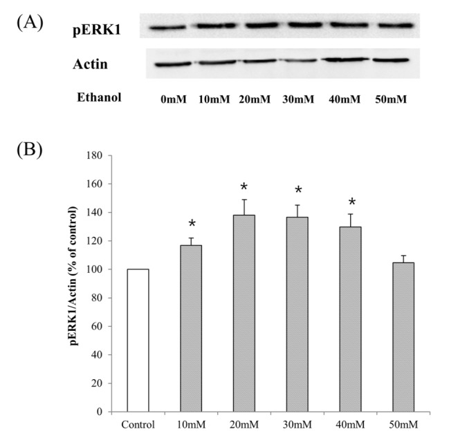
To examine whether ethanol induces activation ERK in HepG2 cells, serum starved cells were exposed to 10~50 mM ethanol for 24 hr, and then phosporylated-ERK1 expression was measured by Western blot (Fig. 2A). Densitometric analysis demonstrated that ethanol treatment stimulated ERK1 activation between 10~40 mM ethanol, and the maximal stimulation was occurred at 20 mM (Fig. 2B), suggesting that ethanol is involved in activation of ERK pathway.

Ethanol treatment activates ERK-p90rsk pathway in HepG2 cells

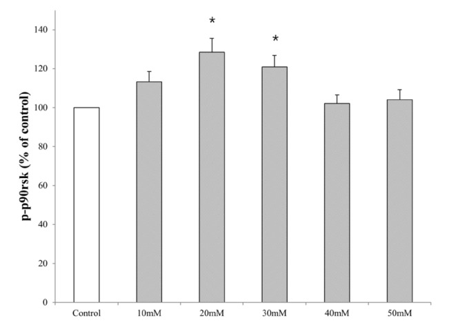
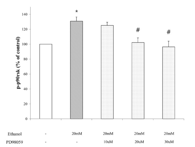
P90rsk, which is the substrate downstream of ERK, regulates cell proliferation and survival [5]. Thus, to investigate whether ethanol induces activation of p90rsk in HepG2 cells, we measured the amount of phosphorylated-p90rsk after the treatment of ethanol (10~50 mM) for 24 hr. As shown in Fig. 3, HepG2 cells maintained high expression of phosphorylated-p90rsk versus control with the treatment of 20~30 mM ethanol. To analyze the effect of ethanol upstream of p90rsk, we tested the changes in phosphorylated-p90rsk expression after the treatment of ethanol in the presence or absence of PD98059 (ERK inhibitor). Fig. 4 shows that PD98059 suppresses increased expression of phosphorylated-p90rsk in ethanol-stimulated HepG2 cells. These results suggest that ethanol induces p90rsk activation in HepG2 cells via activation of MAPK-p90rsk pathway.

Ethanol-induced cell proliferation of HepG2 cells is reduced by inhibition of p90rsk activity

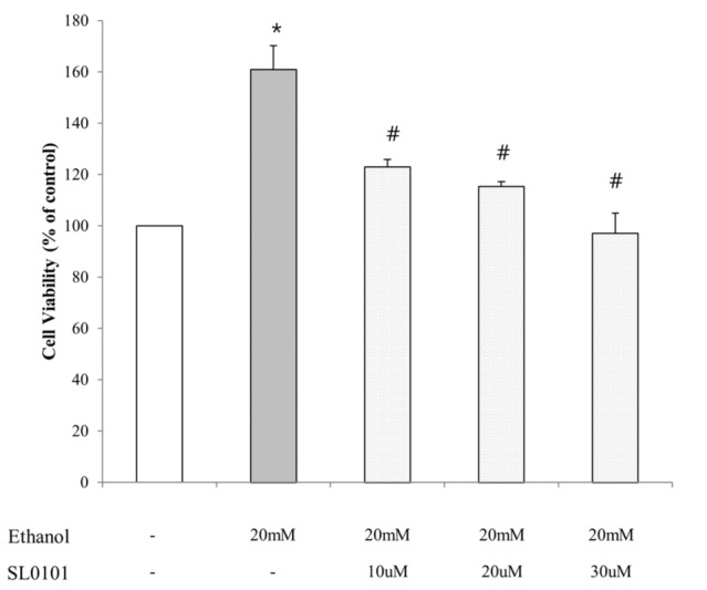
To identify the cell viability after the inhibition of p90rsk, MTT assay had been performed with the treatment of ethanol in the presence or absence of SL0101 (selective inhibitor of p90rsk). Serum-starved HepG2 cells were exposed to ethanol (20 mM) with or without SL0101 (10, 20, 30 µM) for 24 hours. As shown in Fig. 5 treatment with SL0101 diminished ethanol-induced cell proliferation of HepG2 cells in a dose-dependent manner, demonstrating that activation of p90rsk is associated with ethanol-induced cell proliferation of HepG2 cells.

Stimulation of NHE1 activity induced by ethanol treatment in HepG2 cells depends on activation of p90rsk

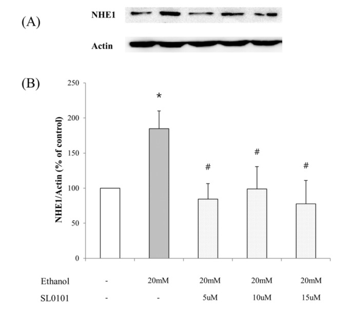
Next, we tested the effect of ethanol on the expression of NHE1, which is stimulated by ERK-p90rsk signaling pathway. In our study, to determine the effect of ethanol on expression of NHE1, the cells were treated with 10~50 mM ethanol. Western blot analysis of lysates from HepG2 cells treated with 10~30 mM ethanol showed upregulation in the expression of NHE1 compared with lysates from naïve cells (Fig. 6A, 6B). To further explore the mechanism by which ethanol influences NHE1 expression, HepG2 cells were exposed to ethanol (20 mM) with or without SL0101 (5, 10, 15 µM). As shown in Fig. 7, inhibition of p90rsk by the treatment of SL0101 diminished the upregulation in the expression of NHE1 induced by ethanol, suggesting that activation of NHE1 in ethanol-stimulated HepG2 cells is mediated by p90rsk activation.

Ethanol-induced increase in expression of Bcl-2 in HepG2 cells is mediated by p90rsk-NHE1 signaling pathway

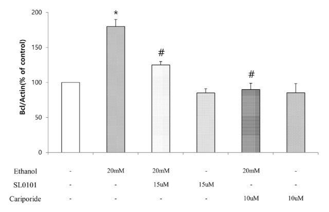
To examine whether inhibition of p90rsk or NHE1 down-regulates Bcl-2 expression in HepG2 cells, cells were treated with or Cariporide (10 µM) SL0101 (15 µM) and exposed to 20 mM ethanol, and then Bcl-2 expression in mRNA level was measured by qRT-PCR (Fig. 8). The expression of Bcl-2 was upregulated when treated with 20 mM ethanol and this effect was diminished with the treatment of SL0101 or Cariporide.

DISCUSSION

It has been previously shown that 10~40 mM of ethanol treatment increases ERK activity, resulting in growth of a series of human hepatocellular carcinoma cells, while it showed no effect in normal liver cells [35]. The detailed mechanism downstream of ERK is not well understood. P90rsk, which is a well-known downstream substrate of ERK and an important regulator of apoptosis, is reported to be associated with cancer progression in various types of cells [1337]. In this study, we investigated the role of p90rsk and its downstream substrates mediating the ethanol-induced cell proliferation.

Ethanol induces cell proliferation of HepG2 cells through ERK-p90rsk pathway

In the present study, ethanol, which is widely known to be hepatotoxic, exhibited stimulatory effect on growth of HepG2 cells. This stimulatory effect of ethanol on carcinoma cell proliferation is related with its ability to activate ERK-p90rsk pathway, suggesting an increase in cell cycle progression and decrease in apoptosis. The results showed that 24 hr of ethanol treatment (10~30 mM) induced increase in cell viability and the maximal increase in cell viability was occurred in the group treated with 20 mM of ethanol, which is consistent to the results of ERK1 and p90rsk activation confirmed by western blot analysis and ELISA, respectively. Ethanol treatment (10~30 mM) enhanced the activities of ERK1 and p90rsk, and the maximal effects were shown at the concentration of 20 mM both. Moreover, the elevated level of phosphorylated p90rsk was diminished with the treatment of ERK inhibitor, PD98059, demonstrating that ethanol increases p90rsk activity by ERK pathway.

Ethanol activates NHE1 and anti-apopotitc Bcl-2 through p90rsk pathway

Herein, SL0101 which inhibits p90rsk specifically was used to further explore the role of p90rsk in ethanol-induced cell proliferation of HepG2 cells. Inhibition of p90rsk has been studied and showed the efficacy in many types of cells. For example, inhibition of p90rsk was effective in radiation-induced cell proliferation of human breast carcinoma cells [38]. In this study, inhibition of p90rsk activity diminished ethanol-induced increase in cell viability dose-dependently implicating that ethanol-induced hepatocellular carcinoma cell proliferation is mediated by p90rsk activation.
Among downstream substrates of p90rsk is NHE1 which can increase cellular pH by extruding intracellular H+ [15]. It is previously studied that activation of NHE1 leads to increases in intracellular pH and anti-apoptotic effect of Bcl-2 family is dependent on its ability to alkalinize the cell [39]. Besides, NHE1-dependent intracellular alkalinization is reported to be critical in malignant transformation [40]. Because activation of p90rsk was also responsible for regulation both of anti-apoptotic Bcl-2 family and NHE1 [2930], we examined whether ethanol can induce increases in NHE1 and Bcl-2 expression, and this ethanol-induced upregulation of Bcl-2 was mediated by ERK-p90rsk pathway and stimulation of NHE1.
When treated with ethanol (10~30 mM), the expression of NHE1 was increased and the maximal increase was shown at the concentration of 20 mM, which is same with previous results about ERK1 and p90rsk activation. Furthermore, this ethanol-induced elevation in expression of NHE1 was abrogated with the treatment of SL0101 (5~15 µM) which can selectively inhibit p90rsk activity.
Bcl-2 is a well-known anti-apoptotic factor. Upstream pathways that affect bcl-2 expression include ERK pathway as well as many other pathways [2930]. Several recent studies have revealed a survival pathway leading to activation of anti-apoptotic Bcl-2 that involves p90rsk pathway [4142]. The studies presented here demonstrate that ethanol can induce increases in Bcl-2 expression and these effects are mediated by p90rsk and NHE1 activation. Treatment of ethanol (20 mM) increased Bcl-2 expression up to 2-fold. Since Bcl-2 is a major anti-apoptotic factor this suggests that 20 mM of ethanol can inhibit apoptosis. Moreover, ethanol-induced increase in expression of Bcl-2 was abrogated with the treatment of cariporide or SL0101. Inhibition of p90rsk with the treatment of SL0101 diminished ethanol-induced Bcl-2 upregulation, demonstrating that ethanol induces anti-apoptotic Bcl-2 activation through p90rsk pathway, while the results when treated with ethanol in the presence of cariporide implicate that increased intracellular pH may contribute to activation of anti-apoptotic Bcl-2, since NHE1 extrudes intracellular H+ out and increases intracellular pH [39].
The present study provides that mechanism of alcohol-induced hepatocellular carcinoma progression may involve increases in ERK-p90rsk pathways and activation of NHE1, resulting in decreased apoptosis. There have been many therapeutic approaches targeting p90rsk and many researches demonstrate that elevated levels of p90rsk are reported during stimulation of cell proliferation in many types of carcinoma cell lines [4344]. Besides p90rsk, hyperactive NHE1 even at the resting pH and the resulting cellular alkalinization have been reported to be directly related to uncontrolled proliferation in malignancy cells [39]. Thus, p90rsk that regulates cellular proliferation, as well as NHE1, may be an important molecule for therapeutic targeting in ethanol-induced hepatocellular carcinoma progression.

Notes

Author contributions: H.S.K and S.J.K performed the model design, cell-based assay experiments. J.H.B and Y.Y.W performed experiments for data. S.Y.P and Y.S.M and H.D.J assisted modeling and simulation design and writing of the manuscript. U.D.S supervised and coordinated the study and reviewed the manuscript.

CONFLICTS OF INTEREST: The authors declare no conflicts of interest.

References

1. Smith JA, Poteet-Smith CE, Xu Y, Errington TM, Hecht SM, Lannigan DA. Identification of the first specific inhibitor of p90 ribosomal S6 kinase (RSK) reveals an unexpected role for RSK in cancer cell proliferation. Cancer Res. 2005; 65:1027–1034. PMID: 15705904.
2. Frödin M, Gammeltoft S. Role and regulation of 90 kDa ribosomal S6 kinase (RSK) in signal transduction. Mol Cell Endocrinol. 1999; 151:65–77. PMID: 10411321.
crossref
3. Roux PP, Richards SA, Blenis J. Phosphorylation of p90 ribosomal S6 kinase (RSK) regulates extracellular signal-regulated kinase docking and RSK activity. Mol Cell Biol. 2003; 23:4796–4804. PMID: 12832467.
crossref
4. Zhang Y, Zhong S, Dong Z, Chen N, Bode AM, Ma W, Dong Z. UVA induces Ser381 phosphorylation of p90RSK/MAPKAP-K1 via ERK and JNK pathways. J Biol Chem. 2001; 276:14572–14580. PMID: 11278279.
crossref
5. Clark DE, Errington TM, Smith JA, Frierson HF Jr, Weber MJ, Lannigan DA. The serine/threonine protein kinase, p90 ribosomal S6 kinase, is an important regulator of prostate cancer cell proliferation. Cancer Res. 2005; 65:3108–3116. PMID: 15833840.
crossref
6. Nguyen TT, Scimeca JC, Filloux C, Peraldi P, Carpentier JL, Van Obberghen E. Co-regulation of the mitogen-activated protein kinase, extracellular signal-regulated kinase 1, and the 90-kDa ribosomal S6 kinase in PC12 cells. Distinct effects of the neurotrophic factor, nerve growth factor, and the mitogenic factor, epidermal growth factor. J Biol Chem. 1993; 268:9803–9810. PMID: 8387505.
crossref
7. Chen RH, Blenis J. Identification of Xenopus S6 protein kinase homologs (pp90rsk) in somatic cells: phosphorylation and activation during initiation of cell proliferation. Mol Cell Biol. 1990; 10:3204–3215. PMID: 2342472.
crossref
8. Blenis J. Signal transduction via the MAP kinases: proceed at your own RSK. Proc Natl Acad Sci U S A. 1993; 90:5889–5892. PMID: 8392180.
crossref
9. Kalab P, Kubiak JZ, Verlhac MH, Colledge WH, Maro B. Activation of p90rsk during meiotic maturation and first mitosis in mouse oocytes and eggs: MAP kinase-independent and -dependent activation. Development. 1996; 122:1957–1964. PMID: 8674434.
crossref
10. Gross SD, Schwab MS, Taieb FE, Lewellyn AL, Qian YW, Maller JL. The critical role of the MAP kinase pathway in meiosis II in Xenopus oocytes is mediated by p90(Rsk). Curr Biol. 2000; 10:430–438. PMID: 10801413.
crossref
11. Tan Y, Ruan H, Demeter MR, Comb MJ. p90(RSK) blocks bad-mediated cell death via a protein kinase C-dependent pathway. J Biol Chem. 1999; 274:34859–34867. PMID: 10574959.
crossref
12. Law JH, Habibi G, Hu K, Masoudi H, Wang MY, Stratford AL, Park E, Gee JM, Finlay P, Jones HE, Nicholson RI, Carboni J, Gottardis M, Pollak M, Dunn SE. Phosphorylated insulin-like growth factor-i/insulin receptor is present in all breast cancer subtypes and is related to poor survival. Cancer Res. 2008; 68:10238–10246. PMID: 19074892.
crossref
13. Zoubeidi A, Zardan A, Wiedmann RM, Locke J, Beraldi E, Fazli L, Gleave ME. Hsp27 promotes insulin-like growth factor-I survival signaling in prostate cancer via p90Rsk-dependent phosphorylation and inactivation of BAD. Cancer Res. 2010; 70:2307–2317. PMID: 20197463.
crossref
14. Lucien F, Brochu-Gaudreau K, Arsenault D, Harper K, Dubois CM. Hypoxia-induced invadopodia formation involves activation of NHE-1 by the p90 ribosomal S6 kinase (p90RSK). PLoS One. 2011; 6:e28851. PMID: 22216126.
crossref
15. Liu Z, Wang S, Zhou H, Yang Y, Zhang M. Na+/H+ exchanger mediates TNF-alpha-induced hepatocyte apoptosis via the calpain-dependent degradation of Bcl-xL. J Gastroenterol Hepatol. 2009; 24:879–885. PMID: 19220664.
16. Gillies RJ. The tumour microenvironment: causes and consequences of hypoxia and acidity. Introduction. Novartis Found Symp. 2001; 240:1–6. PMID: 11727923.
17. Segal MS, Beem E. Effect of pH, ionic charge, and osmolality on cytochrome c-mediated caspase-3 activity. Am J Physiol Cell Physiol. 2001; 281:C1196–C1204. PMID: 11546656.
18. Matsuyama S, Llopis J, Deveraux QL, Tsien RY, Reed JC. Changes in intramitochondrial and cytosolic pH: early events that modulate caspase activation during apoptosis. Nat Cell Biol. 2000; 2:318–325. PMID: 10854321.
crossref
19. Putney LK, Denker SP, Barber DL. The changing face of the Na+/H+ exchanger, NHE1: structure, regulation, and cellular actions. Annu Rev Pharmacol Toxicol. 2002; 42:527–552. PMID: 11807182.
20. Wu KL, Khan S, Lakhe-Reddy S, Wang L, Jarad G, Miller RT, Konieczkowski M, Brown AM, Sedor JR, Schelling JR. Renal tubular epithelial cell apoptosis is associated with caspase cleavage of the NHE1 Na+/H+ exchanger. Am J Physiol Renal Physiol. 2003; 284:F829–F839. PMID: 12453872.
21. Li J, Eastman A. Apoptosis in an interleukin-2-dependent cytotoxic T lymphocyte cell line is associated with intracellular acidification. Role of the Na+/H+-antiport. J Biol Chem. 1995; 270:3203–3211. PMID: 7852405.
22. Luo J, Tannock IF. Inhibition of the regulation of intracellular pH: potential of 5-(N,N-hexamethylene) amiloride in tumour-selective therapy. Br J Cancer. 1994; 70:617–624. PMID: 7917906.
crossref
23. Maidorn RP, Cragoe EJ Jr, Tannock IF. Therapeutic potential of analogues of amiloride: inhibition of the regulation of intracellular pH as a possible mechanism of tumour selective therapy. Br J Cancer. 1993; 67:297–303. PMID: 8381657.
crossref
24. Thangaraju M, Sharma K, Liu D, Shen SH, Srikant CB. Interdependent regulation of intracellular acidification and SHP-1 in apoptosis. Cancer Res. 1999; 59:1649–1654. PMID: 10197642.
25. Shrode LD, Tapper H, Grinstein S. Role of intracellular pH in proliferation, transformation, and apoptosis. J Bioenerg Biomembr. 1997; 29:393–399. PMID: 9387100.
26. Packham G. Mutation of BCL-2 family proteins in cancer. Apoptosis. 1998; 3:75–82. PMID: 14646504.
27. Reynolds JE, Li J, Craig RW, Eastman A. BCL-2 and MCL-1 expression in Chinese hamster ovary cells inhibits intracellular acidification and apoptosis induced by staurosporine. Exp Cell Res. 1996; 225:430–436. PMID: 8660932.
crossref
28. Harguindey S, Pedraz JL, Cañero RG, Katin M. Edelfosine, apoptosis, MDR and Na+/H+ exchanger: induction mechanisms and treatment implications. Apoptosis. 2000; 5:87–89. PMID: 11227496.
29. Lee KW, Kim SG, Kim HP, Kwon E, You J, Choi HJ, Park JH, Kang BC, Im SA, Kim TY, Kim WH, Bang YJ. Enzastaurin, a protein kinase C beta inhibitor, suppresses signaling through the ribosomal S6 kinase and bad pathways and induces apoptosis in human gastric cancer cells. Cancer Res. 2008; 68:1916–1926. PMID: 18339873.
30. Meng XB, Sun GB, Wang M, Sun J, Qin M, Sun XB. P90RSK and Nrf2 activation via MEK1/2-ERK1/2 pathways mediated by Notoginsenoside R2 to prevent 6-hydroxydopamine-induced apoptotic death in SH-SY5Y cells. Evid Based Complement Alternat Med. 2013; DOI: 10.1155/2013/971712.
crossref
31. Oltvai ZN, Milliman CL, Korsmeyer SJ. Bcl-2 heterodimerizes in vivo with a conserved homolog, Bax, that accelerates programmed cell death. Cell. 1993; 74:609–619. PMID: 8358790.
32. Gross A, McDonnell JM, Korsmeyer SJ. BCL-2 family members and the mitochondria in apoptosis. Genes Dev. 1999; 13:1899–1911. PMID: 10444588.
crossref
33. Kubasiak LA, Hernandez OM, Bishopric NH, Webster KA. Hypoxia and acidosis activate cardiac myocyte death through the Bcl-2 family protein BNIP3. Proc Natl Acad Sci U S A. 2002; 99:12825–12830. PMID: 12226479.
crossref
34. Anjum R, Blenis J. The RSK family of kinases: emerging roles in cellular signalling. Nat Rev Mol Cell Biol. 2008; 9:747–758. PMID: 18813292.
crossref
35. Hennig M, Yip-Schneider MT, Klein P, Wentz S, Matos JM, Doyle C, Choi J, Wu H, O'Mara A, Menze A, Noble S, McKillop IH, Schmidt CM. Ethanol-TGFalpha-MEK signaling promotes growth of human hepatocellular carcinoma. J Surg Res. 2009; 154:187–195. PMID: 19321179.
36. Smadja-Lamère N, Shum M, Déléris P, Roux PP, Abe J, Marette A. Insulin activates RSK (p90 ribosomal S6 kinase) to trigger a new negative feedback loop that regulates insulin signaling for glucose metabolism. J Biol Chem. 2013; 288:31165–31176. PMID: 24036112.
37. Chen SJ, Karan D, Johansson SL, Lin FF, Zeckser J, Singh AP, Batra SK, Lin MF. Prostate-derived factor as a paracrine and autocrine factor for the proliferation of androgen receptor-positive human prostate cancer cells. Prostate. 2007; 67:557–571. PMID: 17221842.
crossref
38. Amorino GP, Hamilton VM, Valerie K, Dent P, Lammering G, Schmidt-Ullrich RK. Epidermal growth factor receptor dependence of radiation-induced transcription factor activation in human breast carcinoma cells. Mol Biol Cell. 2002; 13:2233–2244. PMID: 12134064.
crossref
39. Harguindey S, Orive G, Luis Pedraz J, Paradiso A, Reshkin SJ. The role of pH dynamics and the Na+/H+ antiporter in the etiopathogenesis and treatment of cancer. Two faces of the same coin--one single nature. Biochim Biophys Acta. 2005; 1756:1–24. PMID: 16099110.
40. Reshkin SJ, Bellizzi A, Caldeira S, Albarani V, Malanchi I, Poignee M, Alunni-Fabbroni M, Casavola V, Tommasino M. Na+/H+ exchanger-dependent intracellular alkalinization is an early event in malignant transformation and plays an essential role in the development of subsequent transformation-associated phenotypes. FASEB J. 2000; 14:2185–2197. PMID: 11053239.
41. Tan N, Wong M, Nannini MA, Hong R, Lee LB, Price S, Williams K, Savy PP, Yue P, Sampath D, Settleman J, Fairbrother WJ, Belmont LD. Bcl-2/Bcl-xL inhibition increases the efficacy of MEK inhibition alone and in combination with PI3 kinase inhibition in lung and pancreatic tumor models. Mol Cancer Ther. 2013; 12:853–864. PMID: 23475955.
crossref
42. Rajah TT, Peine KJ, Du N, Serret CA, Drews NR. Physiological concentrations of genistein and 17β-estradiol inhibit MDA-MB-231 breast cancer cell growth by increasing BAX/BCL-2 and reducing pERK1/2. Anticancer Res. 2012; 32:1181–1191. PMID: 22493348.
43. Zhou J, Wulfkuhle J, Zhang H, Gu P, Yang Y, Deng J, Margolick JB, Liotta LA, Petricoin E 3rd, Zhang Y. Activation of the PTEN/mTOR/STAT3 pathway in breast cancer stem-like cells is required for viability and maintenance. Proc Natl Acad Sci U S A. 2007; 104:16158–16163. PMID: 17911267.
crossref
44. Gwin J, Drews N, Ali S, Stamschror J, Sorenson M, Rajah TT. Effect of genistein on p90RSK phosphorylation and cell proliferation in T47D breast cancer cells. Anticancer Res. 2011; 31:209–214. PMID: 21273600.
Fig. 1

Effect of ethanol on the growth of HepG2 cell line.

Serum-starved HepG2 cells were incubated with ethanol for 24 hours at the indicated concentration. The cell viability was estimated using MTT assay. Data are expressed as means±S.E of three experiments (student's t-test; *p<0.05 vs. control).
kjpp-20-595-g001
Fig. 2

Activation of ERK in HepG2 cell lines after the treatment with ethanol (10~50 mM) for 24 hours.

(A) Cell lysates were analyzed by Western blot to detect pERK1. (B) The graph represents fold expression of pERK1 relative to β-actin averaged from four independent experiments. Representative Western immunoblots are presented. Actin expression is shown for a control loading. Data are expressed as means±S.E of four experiments (student's t-test; *p<0.05 vs. control).
kjpp-20-595-g002
Fig. 3

Activation of p90rsk in HepG2 cell lines after the treatment with ethanol (10~50 mM) for 24 hours.

Serum-starved HepG2 cells were incubated with ethanol for 24 hours at the indicated concentration. The concentration of phosphorylated-p90rsk in the cell lysate was determined by ELISA. Data are expressed as means±S.E of four experiments (student's t-test; *p<0.05 vs. control).
kjpp-20-595-g003
Fig. 4

The effect of inhibitor of ERK on the expression of p90rsk in HepG2 cell lines after the treatment with ethanol (20 mM) for 24 hours.

Serum-starved HepG2 cells were incubated with ethanol for 24 hours with or without PD98059 (ERK inhibitor) at the indicated concentration. The concentration of phosphorylated-p90rsk in the cell lysate was determined by ELISA. Data are expressed as means±S. E of four experiments (student's t-test; *p<0.05 vs. control, #p<0.05 vs. ethanol alone).
kjpp-20-595-g004
Fig. 5

The effect of inhibitor of p90rsk on the ethanol-induced growth of HepG2 cells.

Serum-starved HepG2 cells were treated for 24 hours with ethanol (20 mM) alone or in combination with SL0101 (p90rsk inhibitor) at each concentration. Cell viability was assessed with MTT assay. Data are expressed as means±S.E of three experiments (student's t-test; *p<0.05 vs. normal group, #p<0.05 vs. ethanol alone).
kjpp-20-595-g005
Fig. 6

Activation of NHE1 in HepG2 cell lines after the treatment with ethanol (10~50 mM) for 24 hours.

A, serum-starved HepG2 cells were treated with ethanol for 24 hours at the indicated concentration. Identical amounts of lysate proteins were subjected to 7.5% SDS-PAGE and immunoblotted (IB) with anti-NHE1 antibody. β–actin content within lystates is shown as an loading control. B, the graph represents fold expression of NHE1 relative to β–actin averaged from four independent experiments. Data are expressed as means±S.E of four experiments (student's t-test; *p<0.05 vs. control).
kjpp-20-595-g006
Fig. 7

The effect of inhibitor of p90rsk on the expression of NHE1 in HepG2 cell lines after the treatment with ethanol (20 mM) for 24 hours.

A, Serum-starved HepG2 cells were treated for 24 hours with ethanol (20 mM) alone or in combination with SL0101 (p90rsk inhibitor) at each concentration. Identical amounts of lysate proteins were subjected to 7.5% SDS-PAGE and immunoblotted (IB) with anti-NHE1 antibody. β–actin content within lystates is shown as an loading control. B, the graph represents fold expression of NHE1 relative to β–actin averaged from three independent experiments. Data are expressed as means±S.E of four experiments (student's t-test; *p<0.05 vs. control, #p<0.05 vs. ethanol alone).
kjpp-20-595-g007
Fig. 8

The effect of inhibitor of p90rsk and NHE1 on the expression of Bcl-2 in HepG2 cell lines after the treatment with ethanol (20 mM) for 24 hours.

HepG2 cells were treated with 20 mM of ethanol alone or in combination of p90rsk or NHE1 inhibitor for 24 hours. Total RNA was extracted and subjected to quantitative RT-PCR (qRT-PCR) for Bcl-2 mRNA. Data are expressed as means±S.E of three experiments (student's t-test; *p<0.05 vs. control, #p<0.05 vs. ethanol alone).
kjpp-20-595-g008
TOOLS
Similar articles