Abstract
Adhesion events of monocytes represent an important step in inflammatory responses induced by chemokines. The β1-integrin CD29 is a major adhesion molecule regulating leukocyte migration and extravasation. Although several adhesion molecules have been known as regulators of CD29, the molecular interactions between CD29 and its regulatory adhesion molecules (such as CD98 and CD147) have not been fully elucidated. Therefore, in this study, we examined whether these molecules are functionally, biochemically, and cell-biologically associated using monocytic U937 cells treated with aggregation-stimulating and blocking antibodies, as well as enzyme inhibitors. The surface levels of CD29, CD98, and CD147 (but not CD43, CD44, and CD82) were increased. The activation of CD29, CD98, and CD147 by ligation of them with aggregation-activating antibodies triggered the induction of cell-cell adhesion, and sensitivity to various enzyme inhibitors and aggregation-blocking antibodies was similar for CD29-, CD98-, and CD147-induced U937 cell aggregation. Molecular association between these molecules and the actin cytoskeleton was confirmed by confocal microscopy and immunoprecipitation. These results strongly suggest that CD29 might be modulated by its biochemical and cellular regulators, including CD98 and CD147, via the actin cytoskeleton.
Inflammation is one of the innate immunity barriers involved in the process of removing pathogens [1]. This response includes numerous molecular and cellular reactions such as the production of cytokines, chemokines, inflammatory mediators, and complement proteins by modulating NF-κB- or AP-1-mediated transcriptional activation pathways [23]. The activation of inflammatory responses also increases the movement of blood leukocytes into inflamed areas by adhesion and chemotaxis processes [4]. These events require functional activation of adhesion molecules triggered by chemokines and ligation with counter-ligands when cells homotypically and heterotypically interact with leukocytes and endothelial cells [5].
Monocytes are important cells in inflammatory responses because blood monocytes are needed to supply fresh macrophages after full differentiation [6]. Macrophages are phagocytes which play a critical role in removing microorganisms [7]. Therefore, stimulation of monocyte migration could help to boost inflammatory responses by increasing numbers of tissue-resident macrophages. The migration activity of monocytes is managed by adhesion molecule activity. So far, variety of adhesion molecules are known to participate in the activation of monocytes. Previously, we and others have identified that β1-integrin (CD29), CD98, CD147, and CD43 are major functional adhesion molecules expressing in monocytes. The activation of these molecules by ligation with homotypic or heterotypic ligands is reported to induce intracellular signaling pathways leading to functional activation of monocytes playing a critical roles in inflammation and virus-derived fusion events [8]. Activation signals of monocytes induced by these adhesion molecules include small GTPase Rho, tyrosine kinases (Syk and Lyn), and the phosphatidylinositol-3-kinase during CD29 activation [910], ERK, Syk, and protein kinase Cθ in CD43 activation [1112], conventional PKC isoforms (α, β, γand δ), ERK, and p38 in case of CD98 stimulation [111314], and VEGFR-2 tyrosine kinase receptor, PI3K, AKT, and ERK1/2 under CD147 activation conditions [151617].
Of them, CD29 is regarded as a significant adhesion molecule that is critically important in allowing strong interactions between leukocytes and endothelial cells in the process known as extravasation [18]. Previously, we and other groups have found that CD29 is functionally associated with CD98 [1920]. Moreover, it was found that CD98 is regulated by CD147 [2122]. Although several reports have suggested cross-regulation between CD98 and CD147 [23], detailed mechanisms of their molecular interactions have not been fully elucidated yet. These findings raise the hypothesis that these molecules might be important for the functional activation of CD29. In this study, we aimed to further clarify the regulatory mechanisms between these three adhesion molecules at the molecular level in order to understand how CD29 is regulated by other adhesion molecules.
Enzyme inhibitors [U0126, an MEK1 inhibitor, rottlerin, a protein kinase Cd inhibitor, and cytochalasin B (Cyto B), an actin cytoskeleton disruptor] were purchased from Calbiochem (La Jolla, CA, USA). U937 cells, a human pleura/pleural effusion promonocyte-like cell line (no. CRL-1593.2), were obtained from the American Type Culture Collection (ATCC, Rockville, MD, USA). All other chemicals were of reagent grade. The following antibodies were used in this study for cell-cell adhesion assays: CD29 (MEM 101A, IgG1, ascites, kindly provided by V. Horejsi); CD43 (161-46, ascites, IgG1, kindly provided by R. Villela); CD98 (ANH-18, purified IgG1, kindly provided by K. Skubiz); and CD147 (MEM M6/1, IgG1, ascites, V. Horejsi). Rhodamine phalloidin was purchased from Molecular Probes (Carlsbad, CA, USA). Antibodies to CD98 (mouse, 4F2) and CD147 (rabbit, EMMPRIN) for immunoprecipitation and immunoblotting were from Santa Cruz Biotechnology (Santa Cruz, CA, USA) and Sino Biological Inc. (Beijing, China), respectively. The following antibodies were used for flow cytometic analysis: CD18 (BU86, IgG1, ascites); CD29 (MAR4, IgG1, ascites); CD43 (161-46, IgG1, ascites); CD44 (E1/2, purified IgG1); CD98 (BK19.9, IgG1, purified antibody); and CD147 (MEM M6/1, IgG1, ascites), as reported previously [21].
U937 cells were cultured in RPMI1640 supplemented with antibiotics (100 U/ml of penicillin and 100 µg/ml of streptomycin) and 10% FCS, and were maintained at 37℃ and 5% CO2 under humidified air conditions.
Expression of U937 surface adhesion molecules was determined by flow cytometric analysis as reported previously [2124]. Cells (105) were washed with staining buffer (PBS containing 2% FBS and 1% sodium azide) and incubated in 50 ml staining buffer containing 10% rabbit serum for 10 minutes on ice and then with the primary antibodies to CD18, CD29, CD43, CD44, CD82, CD98, and CD147 for a further 45 minutes. After washing 3 times with staining buffer, cells were treated with 1/20 dilution of fluorescein isothiocyanate (FITC)–conjugated rabbit anti–mouse IgG secondary antibody (Dako, Dakopatts, High Wycombe, United Kingdom). Cells were then washed 3 times with staining buffer and analyzed on a FACScan (Becton Dickinson, San Jose, CA).
A quantitative homotypic aggregation assay was carried out as reported previously [21]. Briefly, U937 cells were preincubated with enzyme inhibitors or aggregation-blocking antibodies at 37℃ for 3 h or 6 h and incubated further with aggregative antibodies (1 µg/ml) for 3 to 6 h in round-bottom microwell plates. The numbers of non-aggregated and total cells were counted in a hemocytometer and the percentage of cells in aggregates determined as 100 × (total cells–non-aggregated cells)/total cells, and referred to a control (as a percentage) calculated from only the aggregation-activating antibody treatment group. Inhibitory effects of aggregation-blocking antibodies or enzyme inhibitors were calculated as % of control by setting the control group as 100%. For the cell–fibronectin (FN) adhesion assay, drug (U0126 or Cyto B) or blocking antibody (P5D2)-treated U937 cells (5×10⁵ cells/well) were seeded on a fibronectin (50 µg/ml)-coated plate and incubated for 3 h, as reported previously. After removing unbound cells with PBS, the attached cells were treated with 0.1% crystal violet for 15 min. The OD value at 540 nm was measured by a Spectramax 250 microplate reader.
Images of cells in culture were observed using an inverted phase-contrast microscope attached to a video camera, and captured using NIH image software as reported previously [25].
Confocal analysis of actin and adhesion molecules (CD29, CD98, and CD147) on the membrane of U937 cells (1×10⁵ cells/ml) treated with aggregation-activating antibodies was carried out on glass-bottom culture dishes (MatTek Corp., Ashland, MA, USA) using FITC- or PE-labeled antibodies to CD29, CD147, or CD98, as well as the actin-staining chemical rhodamine phalloidin. Intensity changes in CD29, CD147, CD98, or actin cytoskeleton were imaged by LSCM on a Zeiss LSM 510 META confocal microscope equipped with a Zeiss 37℃ incubation system. Images were analyzed using the Zeiss LSM Image Examiner.
Cells (5×10⁶ cells/ml) were lysed in lysis buffer (in mM: 20 TRIS-HCl, pH 7.4, 2 EDTA, 2 EGTA, 50 β-glycerophosphate, 1 sodium orthovanadate, 1 dithiothreitol, 1% Triton X-100, 10% glycerol, 10 µg/ml leupeptin, 10 µg/ml aprotinin, 10 µg/ml pepstatin, 1 benzimide, and 2 hydrogen peroxide) for 30 min under rotation in a cold room. The lysates were clarified by centrifugation at 16,000 g for 10 min at 4℃. Soluble cell lysates were immunoblotted and CD98 and CD147 were visualized as reported previously [26]. For immunoprecipitation, cell lysates containing equal amounts of protein (500 µg) from ANH-18-treated U937 cells (1×10⁷ cells/ml) were pre-cleared with 10 µl of protein A-coupled Sepharose beads (50% v/v) (Amersham, UK) for 1 h at 4℃. Pre-cleared samples were then incubated with 5 µl of antibody to CD147 or CD98 overnight at 4℃. Immune complexes were mixed with 10 ml of protein A-coupled Sepharose beads (50% v/v) and incubated for 3 h at 4℃.
All values are presented as means±standard deviations (SDs) of three (Fig. 1, 4C, and 5B) or six (Fig. 2A, 3A, and 4A) experiments. For statistical comparisons, results were analyzed using ANOVA/Scheffe's post hoc test and the Kruskal–Wallis/Mann–Whitney tests. A p-value<0.05 was considered statistically significant. All statistical tests were performed using SPSS software (SPSS Inc., Chicago, IL, USA). Similar results were obtained in a second experiment.
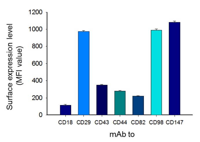
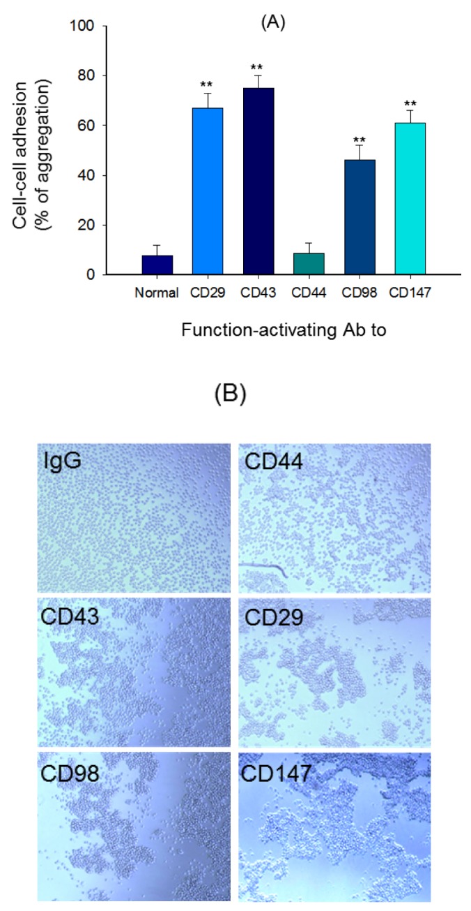
To check expression levels of surface adhesion molecules in U937 cells, flow cytometric analysis was employed. As Fig. 1 shows, the MFI values of CD147, CD98, CD29, and CD43 were 1140, 1013, 992, and 386, respectively. The lowest expression level was seen in CD18 (Fig. 1). Similar to expression levels, aggregation-inducing activity of U937 cells was also exhibited in the ligation of CD43, CD29, CD147, and CD98 up to 76, 68, 61, and 48% compared to 9% of normal cells (Fig. 2A and 2B).
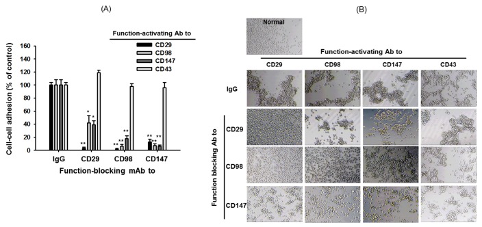
Whether the function-blocking antibodies to CD29, CD98, and CD147 are able to interrupt the cell-cell adhesion induced by the activation of CD29, CD43, CD98, and CD147 was examined. Interestingly, P5D2, a blocking antibody to CD29, blocked the aggregation events induced by CD29, CD98, and CD147 up to 92, 57, and 62%, respectively, while MEM 108 (a CD98 blocking antibody) also suppressed the aggregation levels up to 94, 89, and 78%, respectively (Fig. 3A and 3B). Similarly, the adhesion events induced by CD29, CD98, and CD147 were inhibited by CD147 blocking antibody MEM M6/1 up to 82, 86, and 90%, respectively (Fig. 3A and 3B). However, there was no significant inhibition of blocking antibodies in CD43-triggered cell aggregation (Fig. 3A and 3B).
In addition, sensitivity of cell-cell adhesion events to several enzyme inhibitors was also tested under the aggregation-inducing conditions. Thus, U0126, a MEK1 inhibitor, displayed strong suppressive activity toward U937 cell-cell adhesion events boosted by CD29, CD98, and CD147 up to 92, 84, and 82%, respectively (Fig. 4A). Rottlerin, a PKCδ inhibitor, also diminished the aggregation events by 90, 86, and 72%, whereas Cyto B, an actin cytoskeleton disruptor with blocking activity on actin polymerization, almost completely inhibited the cell-cell adhesion processes managed by CD29, CD98, and CD147 up to 98%. Like blocking antibody effects, these three inhibitors failed to block CD43-induced U937 cell aggregation (Fig. 4A). Interestingly, the inhibitory levels of U0126 and Cyto B in FN-mediated U937 cell adhesion events were dramatically changed exhibiting not only marginal suppression (30%) in U0126 treatment, but also strong stimulation (170%) in Cyto B exposure (Fig. 4B). Meanwhile, no dramatic inhibition (8, 3, and 14%) of U937 cell viability by U0126, rottlerin, and Cyto B was also observed (Fig. 4C).
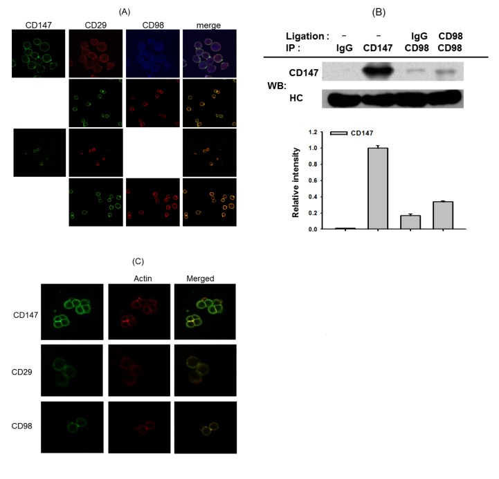
To check whether these molecules are also cellularly and biochemically associated, we employed confocal and immunoprecipitation analyses. According to confocal analysis, there was clear associated between CD147, CD29, and CD98, which are appeared on the membrane of U937 cells (Fig. 5A). In addition, by immunoprecipitation and immunoblotting analyses, binding property of CD147 to CD98 was clearly observed (Fig. 5B). Finally, tight association between actin cytoskeleton and CD147, CD98 or CD29 was also found by confocal microscopy (Fig. 5C).
The adhesion of monocytes is one of the important steps in supplying additional macrophages during the inflammatory response [29]. Among adhesion molecules, β1-integrins are known to be functionally important [30]. In this study, we focused on understanding the regulatory mechanism of CD29 in terms of its associated co-adhesion molecules.
Fig. 1 shows the importance of CD29 and its regulatory proteins in U937 promonocytic cells. The surface levels of CD29, CD98, and CD147 were found to be high, while others (CD43, CD44, and CD82) displayed low levels on the surface. These data suggest that these three molecules might act as major adhesion molecules in monocytes. In fact, ligation of these three molecules with aggregation-activating antibodies strongly induced homotypic U937 cell-cell adhesion events, as assessed by quantitative and qualitative approaches (Fig. 2A and 2B). As reported previously, CD43 activation also led to homotypic cell adhesion patterns in U937 cells.
Aggregation-blocking antibodies to CD29, CD98, and CD147 (but not CD43) almost completely blocked each cell-cell aggregation pattern generated by CD29, CD98, or CD147 activation conditions (Fig. 3A and 3B), indicating that these molecules are closely and functionally associated. The sensitivity of cell-cell adhesion to treatment with inhibitors of signaling enzymes showed very similar patterns for CD29-, CD98-, and CD147-induced conditions, while CD43-induced aggregation was not blocked by these inhibitors (Fig. 4A and 4B). There was no cytotoxicity under treatment with these compounds (Fig. 4C), implying that the inhibitory activity of these compounds was not due to cell death.
Biochemical association between CD29, CD98, and CD147 was examined using confocal and immunoprecipitation analyses. As Fig. 5A shows, these three molecules appeared to be focally associated on the membrane of U937 cells. The molecular interaction between CD147 and CD98 was also confirmed by immunoprecipitation analysis (Fig. 5B). This interaction increased under CD98 ligation conditions, indicating that the active form of CD98 could better associate with CD147. Moreover, molecular association between CD29 and CD98 as well as between CD29 and CD147 has been also previously demonstrated by employing immunoprecipitation analysis [3132]. Since actin is known as an adaptor molecule that allows intracellular signaling molecule (e.g. Src) association [333435], we examined whether actin is important in this complex formation. As Fig. 5C shows, actin stained with rhodamine phalloidin was detected in the focally concentrated area with CD147, CD98, or CD29, implying that these molecules might be associated with the actin cytoskeleton. Indeed, it has been reported that cytochalasin B is capable of completely suppressing the clustering formation induced by CD29 and CD98 [36]. So far, it is unclear whether actin simultaneously binds to these adhesion molecules to manage the formation of molecular complex. Therefore, relevant works regarding the functional role of actin in CD29/CD98/CD147-mediated cell-cell adhesion events will be further continued.
In terms of pathophysiology, modulation of CD29 function is regarded as a therapeutic target to treat various chronic inflammatory and autoimmune diseases [303738]. CD29 was found to be highly expressed in monocytes to interact with vascular endothelial cells and lung fibroblasts and in autoreactive T lymphocytes analyzed from autoimmune patients [39404142]. In addition, engagement of CD29 on monocytes and SLE T cells was reported to trigger focal adhesion kinase- or spleen tyrosine kinase-induced intracellular signaling cascade linked to the proliferation of T cells and secretion of chemokines from monocytes [394344], implying that CD29 may play a critical role in increasing adhesive activity of monocytes and autoreactive T cell activation. The inflammatory processes mediated by neutrophils and macrophages were also found to be critically regulated by CD29 [45]. These findings have raised the possibility that functional regulation of CD29 by small molecules or therapeutic antibodies could become a useful therapeutic strategy in treating autoimmune and chronic inflammatory diseases. We have reported that lancemaside A, 20S-dihydroprotopanaxadiol, cinnamaldehyde, ceramide, chloroquine, and cynaropicrin are regarded as good modulators of CD29-mediated cell-cell adhesion [254647]. Others have found that ibrutinib (an orally active covalent BTK inhibitor) [48], shikonin [49], and biodentine [50] can inhibit the functional activation of integrins. Although there have been numerous attempts to develop CD29-function inhibitors with small molecules, none of these drugs has been released for clinical trials. There is also no therapeutic CD29 antibody (e.g. OS2966) yet prescribed for the clinic [51]. Indirect approaches with other CD29-associated molecules, such as CD98 and CD147, could lead to acceptable methods for regulating CD29 function. Treatment with aggregation-blocking antibodies against CD98 and CD147 strongly suppressed CD29-mediated cell adhesion events (Fig. 3). Further development of therapeutic approaches against autoimmune and chronic inflammatory diseases using CD98 and CD147 will provide additional opportunities for CD29-targeted drug development.
In summary, our study demonstrated that CD29 can be regulated by its associated proteins, CD98 and CD147. Monocytic U937 cells showed similar expression patterns for CD29, CD147, and CD98, and the adhesion events induced by these three molecules exhibited similar inhibitory sensitivity to enzyme inhibitors and blocking antibodies. These molecules were found to be focally associated with the actin cytoskeleton and also co-immunoprecipitated, as shown by confocal and immunoprecipitation analyses, respectively. These results suggest that co-regulation of β1-integrins and the adhesion molecules CD98 and CD147 may significantly contribute to the adhesion of monocytic U937 cells.
ACKNOWLEDGEMENTS
This work was supported by the BK21 plus program through the National Research Foundation (NRF). The authors specially acknowledge Prof. V. Horejsi, Prof. K. Skubitz, Dr. R. Villela, Prof. W. Kasinrerk, and Dr. Joo Young Kim for kindly supplying the valuable antibodies to CD29, CD98, CD43, and CD147 used in this study as well as technical supports.
References
1. Kim CH, Park J, Kim M. Gut microbiota-derived short-chain fatty acids, T cells, and inflammation. Immune Netw. 2014; 14:277–288. PMID: 25550694.
2. Yu T, Li YJ, Bian AH, Zuo HB, Zhu TW, Ji SX, Kong F, Yin de Q, Wang CB, Wang ZF, Wang HQ, Yang Y, Yoo BC, Cho JY. The regulatory role of activating transcription factor 2 in inflammation. Mediators Inflamm. 2014; 2014:950472. PMID: 25049453.
3. Ku SK, Bae JS. Baicalin, baicalein and wogonin inhibits high glucose-induced vascular inflammation in vitro and in vivo. BMB Rep. 2015; 48:519–524. PMID: 25739393.
4. Schnoor M, Alcaide P, Voisin MB, van Buul JD. Crossing the vascular wall: common and unique mechanisms exploited by different leukocyte subsets during extravasation. Mediators Inflamm. 2015; 2015:946509. PMID: 26568666.
5. Haertel B, von Woedtke T, Weltmann KD, Lindequist U. Non-thermal atmospheric-pressure plasma possible application in wound healing. Biomol Ther (Seoul). 2014; 22:477–490. PMID: 25489414.
6. Italiani P, Boraschi D. New insights into tissue macrophages: from their origin to the development of memory. Immune Netw. 2015; 15:167–176. PMID: 26330802.
7. Kim MJ, Rah SY, An JH, Kurokawa K, Kim UH, Lee BL. Human anti-peptidoglycan-IgG-mediated opsonophagocytosis is controlled by calcium mobilization in phorbol myristate acetate-treated U937 cells. BMB Rep. 2015; 48:36–41. PMID: 24856825.
8. Rintoul RC, Buttery RC, Mackinnon AC, Wong WS, Mosher D, Haslett C, Sethi T. Cross-linking CD98 promotes integrin-like signaling and anchorage-independent growth. Mol Biol Cell. 2002; 13:2841–2852. PMID: 12181350.
9. Hirai F, Nakayamada S, Okada Y, Saito K, Kurose H, Mogami A, Tanaka Y. Small GTPase Rho signaling is involved in beta1 integrin-mediated up-regulation of intercellular adhesion molecule 1 and receptor activator of nuclear factor kappaB ligand on osteoblasts and osteoclast maturation. Biochem Biophys Res Commun. 2007; 356:279–285. PMID: 17349971.
10. McNally AK, Anderson JM. Beta1 and beta2 integrins mediate adhesion during macrophage fusion and multinucleated foreign body giant cell formation. Am J Pathol. 2002; 160:621–630. PMID: 11839583.
11. Fulcher JA, Chang MH, Wang S, Almazan T, Hashimi ST, Eriksson AU, Wen X, Pang M, Baum LG, Singh RR, Lee B. Galectin-1 co-clusters CD43/CD45 on dendritic cells and induces cell activation and migration through Syk and protein kinase C signaling. J Biol Chem. 2009; 284:26860–26870. PMID: 19635795.
12. del Rio R, Rincón M, Layseca-Espinosa E, Fierro NA, Rosenstein Y, Pedraza-Alva G. PKCtheta is required for the activation of human T lymphocytes induced by CD43 engagement. Biochem Biophys Res Commun. 2004; 325:133–143. PMID: 15522211.
13. Cho JY, Katz DR, Skubitz KM, Chain BM. Conventional protein kinase C plays a critical role in negative regulation of CD98-induced homotypic aggregation. Tissue Antigens. 2010; 75:19–29. PMID: 19895572.
14. Cho JY, Skubitz KM, Katz DR, Chain BM. CD98-dependent homotypic aggregation is associated with translocation of protein kinase Cdelta and activation of mitogen-activated protein kinases. Exp Cell Res. 2003; 286:1–11. PMID: 12729789.
15. Khayati F, Pérez-Cano L, Maouche K, Sadoux A, Boutalbi Z, Podgorniak MP, Maskos U, Setterblad N, Janin A, Calvo F, Lebbé C, Menashi S, Fernandez-Recio J, Mourah S. EMMPRIN/CD147 is a novel coreceptor of VEGFR-2 mediating its activation by VEGF. Oncotarget. 2015; 6:9766–9780. PMID: 25825981.
16. Xu T, Zhou M, Peng L, Kong S, Miao R, Shi Y, Sheng H, Li L. Upregulation of CD147 promotes cell invasion, epithelial-to-mesenchymal transition and activates MAPK/ERK signaling pathway in colorectal cancer. Int J Clin Exp Pathol. 2014; 7:7432–7441. PMID: 25550778.
17. Wang Y, Yuan L, Yang XM, Wei D, Wang B, Sun XX, Feng F, Nan G, Wang Y, Chen ZN, Bian H. A chimeric antibody targeting CD147 inhibits hepatocellular carcinoma cell motility via FAK-PI3K-Akt-Girdin signaling pathway. Clin Exp Metastasis. 2015; 32:39–53. PMID: 25424030.
18. Langereis JD. Neutrophil integrin affinity regulation in adhesion, migration, and bacterial clearance. Cell Adh Migr. 2013; 7:476–481. PMID: 24430200.
19. Sun C, Zargham R, Shao Q, Gui X, Marcus V, Lazaris A, Salman A, Metrakos P, Qu X, Gao Z. Association of CD98, integrin β1, integrin β3 and Fak with the progression and liver metastases of colorectal cancer. Pathol Res Pract. 2014; 210:668–674. PMID: 25041835.
20. Prager GW, Féral CC, Kim C, Han J, Ginsberg MH. CD98hc (SLC3A2) interaction with the integrin beta subunit cytoplasmic domain mediates adhesive signaling. J Biol Chem. 2007; 282:24477–24484. PMID: 17597067.
21. Cho JY, Fox DA, Horejsi V, Sagawa K, Skubitz KM, Katz DR, Chain B. The functional interactions between CD98, beta1-integrins, and CD147 in the induction of U937 homotypic aggregation. Blood. 2001; 98:374–382. PMID: 11435306.
22. Guo N, Zhang K, Lv M, Miao J, Chen Z, Zhu P. CD147 and CD98 complex-mediated homotypic aggregation attenuates the CypA-induced chemotactic effect on Jurkat T cells. Mol Immunol. 2015; 63:253–263. PMID: 25089027.
23. Fei F, Li X, Xu L, Li D, Zhang Z, Guo X, Yang H, Chen Z, Xing J. CD147-CD98hc complex contributes to poor prognosis of non-small cell lung cancer patients through promoting cell proliferation via the PI3K/Akt signaling pathway. Ann Surg Oncol. 2014; 21:4359–4368. PMID: 25084765.
24. Kim MY, Yoo BC, Cho JY. Ginsenoside-Rp1-induced apolipoprotein A-1 expression in the LoVo human colon cancer cell line. J Ginseng Res. 2014; 38:251–255. PMID: 25379004.
25. Lee YG, Lee J, Cho JY. Cell-permeable ceramides act as novel regulators of U937 cell-cell adhesion mediated by CD29, CD98, and CD147. Immunobiology. 2010; 215:294–303. PMID: 19576658.
26. Kim S, Oh MH, Kim BS, Kim WI, Cho HS, Park BY, Park C, Shin GW, Kwon J. Upregulation of heme oxygenase-1 by ginsenoside Ro attenuates lipopolysaccharide-induced inflammation in macrophage cells. J Ginseng Res. 2015; 39:365–370. PMID: 26869829.
27. Wang Z, Wang D, Li Y, Zhang X. Protective effects of verapamil against H2O2-induced apoptosis in human lens epithelial cells. Biomol Ther (Seoul). 2014; 22:553–557. PMID: 25489424.
28. Baek KS, Hong YD, Kim Y, Sung NY, Yang S, Lee KM, Park JY, Park JS, Rho HS, Shin SS, Cho JY. Anti-inflammatory activity of AP-SF, a ginsenoside-enriched fraction, from Korean ginseng. J Ginseng Res. 2015; 39:155–161. PMID: 26045689.
29. Gerhardt T, Ley K. Monocyte trafficking across the vessel wall. Cardiovasc Res. 2015; 107:321–330. PMID: 25990461.
30. Yusuf-Makagiansar H, Anderson ME, Yakovleva TV, Murray JS, Siahaan TJ. Inhibition of LFA-1/ICAM-1 and VLA-4/VCAM-1 as a therapeutic approach to inflammation and autoimmune diseases. Med Res Rev. 2002; 22:146–167. PMID: 11857637.
31. Li Y, Wu J, Song F, Tang J, Wang SJ, Yu XL, Chen ZN, Jiang JL. Extracellular membrane-proximal domain of HAb18G/CD147 binds to metal ion-dependent adhesion site (MIDAS) motif of integrin β1 to modulate malignant properties of hepatoma cells. J Biol Chem. 2012; 287:4759–4772. PMID: 22130661.
32. Fenczik CA, Sethi T, Ramos JW, Hughes PE, Ginsberg MH. Complementation of dominant suppression implicates CD98 in integrin activation. Nature. 1997; 390:81–85. PMID: 9363894.
33. Mattila PK, Batista FD, Treanor B. Dynamics of the actin cytoskeleton mediates receptor cross talk: an emerging concept in tuning receptor signaling. J Cell Biol. 2016; 212:267–280. PMID: 26833785.
34. Kim JH, Lee YG, Yoo S, Oh J, Jeong D, Song WK, Yoo BC, Rhee MH, Park J, Cha SH, Hong S, Cho JY. Involvement of Src and the actin cytoskeleton in the antitumorigenic action of adenosine dialdehyde. Biochem Pharmacol. 2013; 85:1042–1056. PMID: 23353701.
35. Kim JY, Lee YG, Kim MY, Byeon SE, Rhee MH, Park J, Katz DR, Chain BM, Cho JY. Src-mediated regulation of inflammatory responses by actin polymerization. Biochem Pharmacol. 2010; 79:431–443. PMID: 19769947.
36. Cho JY, Kim AR, Joo HG, Kim BH, Rhee MH, Yoo ES, Katz DR, Chain BM, Jung JH. Cynaropicrin, a sesquiterpene lactone, as a new strong regulator of CD29 and CD98 functions. Biochem Biophys Res Commun. 2004; 313:954–961. PMID: 14706635.
37. Bruck R, Hershkoviz R, Lider O, Shirin H, Aeed H, Halpern Z. The use of synthetic analogues of Arg-Gly-Asp (RGD) and soluble receptor of tumor necrosis factor to prevent acute and chronic experimental liver injury. Yale J Biol Med. 1997; 70:391–402. PMID: 9626759.
38. Park EJ, Yuki Y, Kiyono H, Shimaoka M. Structural basis of blocking integrin activation and deactivation for anti-inflammation. J Biomed Sci. 2015; 22:51. PMID: 26152212.
39. Nakayamada S, Saito K, Nakano K, Tanaka Y. Activation signal transduction by beta1 integrin in T cells from patients with systemic lupus erythematosus. Arthritis Rheum. 2007; 56:1559–1568. PMID: 17469136.
40. Zhu Y, Feng Y, Liu H, Ye H, Guo C, Feng J, Dai S, Zheng X. CD4+CD29+T cells are blamed for the persistent inflammatory response in ulcerative colitis. Int J Clin Exp Pathol. 2015; 8:2627–2637. PMID: 26045768.
41. Cole AL, Subbanagounder G, Mukhopadhyay S, Berliner JA, Vora DK. Oxidized phospholipid-induced endothelial cell/monocyte interaction is mediated by a cAMP-dependent R-Ras/PI3-kinase pathway. Arterioscler Thromb Vasc Biol. 2003; 23:1384–1390. PMID: 12805072.
42. Zickus C, Kunkel SL, Simpson K, Evanoff H, Glass M, Strieter RM, Lukacs NW. Differential regulation of C-C chemokines during fibroblast-monocyte interactions: adhesion vs. inflammatory cytokine pathways. Mediators Inflamm. 1998; 7:269–274. PMID: 9792337.
43. Miller LA, Hong JJ, Kinch MS, Harrison ML, Geahlen RL. The engagement of beta1 integrins on promonocytic cells promotes phosphorylation of Syk and formation of a protein complex containing Lyn and beta1 integrin. Eur J Immunol. 1999; 29:1426–1434. PMID: 10359096.
44. da Costa Martins PA, van Gils JM, Mol A, Hordijk PL, Zwaginga JJ. Platelet binding to monocytes increases the adhesive properties of monocytes by up-regulating the expression and functionality of beta1 and beta2 integrins. J Leukoc Biol. 2006; 79:499–507.
45. Chiang YJ, Ho KC, Sun CT, Chiu JJ, Lee FJ, Liao F, Yang-Yen HF, Yen JJ. CBAP functions as a novel component in chemokine-induced ZAP70-mediated T-cell adhesion and migration. PLoS One. 2013; 8:e61761. PMID: 23620790.
46. Kim E, Yang WS, Kim JH, Park JG, Kim HG, Ko J, Hong YD, Rho HS, Shin SS, Sung GH, Cho JY. Lancemaside A from Codonopsis lanceolata modulates the inflammatory responses mediated by monocytes and macrophages. Mediators Inflamm. 2014; 2014:405158. PMID: 24782593.
47. Jo S, Lee H, Kim S, Lee CH, Chung H. Korean red ginseng extract induces proliferation to differentiation transition of human acute promyelocytic leukemia cells via MYC-SKP2-CDKN1B axis. J Ethnopharmacol. 2013; 150:700–707. PMID: 24095829.
48. Herman SE, Mustafa RZ, Jones J, Wong DH, Farooqui M, Wiestner A. Treatment with Ibrutinib inhibits BTK- and VLA-4-dependent adhesion of chronic lymphocytic leukemia cells in vivo. Clin Cancer Res. 2015; 21:4642–4651. PMID: 26089373.
49. Wang H, Wu C, Wan S, Zhang H, Zhou S, Liu G. Shikonin attenuates lung cancer cell adhesion to extracellular matrix and metastasis by inhibiting integrin β1 expression and the ERK1/2 signaling pathway. Toxicology. 2013; 308:104–112. PMID: 23562787.
50. Luo Z, Li D, Kohli MR, Yu Q, Kim S, He WX. Effect of biodentine™ on the proliferation, migration and adhesion of human dental pulp stem cells. J Dent. 2014; 42:490–497. PMID: 24440605.
51. Jahangiri A, Aghi MK, Carbonell WS. β1 integrin: critical path to antiangiogenic therapy resistance and beyond. Cancer Res. 2014; 74:3–7. PMID: 24327727.