

 PDF
PDF ePub
ePub Citation
 Print
Print


INTRODUCTION
Because the heart is easily affected by increased afterload due to hypertension, it is vulnerable to heart failure [1]. In the past several decades, heart failure has emerged as the leading cause of death in advanced countries, and this trend is now being observed in developing countries. Considering the high mortality rate in patients with heart failure, there is a need for new therapeutic approaches [2].
Histone deacetylase (HDAC) inhibitors have been shown to be effective in pre-clinical models of heart failure, blocking pathological cardiac hypertrophy and fibrosis and improving cardiac condition [3]. HDACs catalyze the removal of acetyl groups from lysine residues in a variety of proteins. HDACs have mainly been studied in nucleosomal histones, and serve as an epigenetic modulator by deacetylating histones and altering the electrostatic condition of chromatin to reduce gene expression. However, it is now clear that HDACs also deacetylate many non-histone proteins, such as transcription factors, mitochondrial proteins, and cytoskeletal proteins [45]. There are 18 mammalian HDACs encoded by distinct genes and grouped into four classes: class I HDACs (1, 2, 3, and 8), class IIa HDACs (4, 5, 7, and 9), class IIb HDACs (6 and 10), class III HDACs (sirtuin family: sirt 1~7), and class IV HDAC (HDAC11) [6]. HDAC inhibitors can be grouped by the structure of their specific binding group [7].
HDAC inhibitors block cardiac hypertrophy, fibrosis, and hypertension in various cardiovascular disease models [89]. Recently, HDAC inhibitors have been shown to block left ventricular (LV) hypertrophy in response to chronic systemic hypertension induced by the aldosterone precursor, deoxycorticosterone acetate (DOCA) [10] and in spontaneously hypertensive rats (SHRs) [11]. All of the compounds used in these pre-clinical studies are non-selective pan-HDAC inhibitors.
CG200745 {(E)-2-(Naphtalene-1-yloxymethyl)-oct-2-enedioic acid 1-[(3-dimethyl amino-propyl)-amide] 8-hydroxyamide with 1/2 (L-tartaric acid)} (CG) is a novel pan-HDAC inhibitor that is being evaluated in phase II clinical trials for its anticancer effects. Therefore, we hypothesized that CG attenuates cardiac hypertrophy and fibrosis in DOCA-induced hypertensive rats.
METHODS
Animals
The investigation was conducted in accordance with the National Institutes of Health Guide for the Care and Use of Laboratory Animals and was approved by the Institutional Review Board of Kyungpook National University, and every effort was made to minimize both the number of animals used and their suffering. Sixteen-week-old male Sprague-Dawley rats were unilaterally nephrectomized under ketamine (150 mg/kg, Yuhan, Seoul, Korea) and xylazine (18 mg/kg; Bayer, Seoul, Korea) anesthesia. After unilateral nephrectomy, rats were allowed to recover overnight before being randomly assigned to one of 4 groups: sham (n=6), deoxycorticosterone acetate (DOCA) (n=6), DOCA plus 1.25 mg/kg of CG200745 (CG) (n=6) and DOCA plus 5.0 mg/kg of CG (n=6). DOCA was injected subcutaneously at 40 mg/kg/week for a period of 4 weeks. The animals were allowed to free access to drinking water containing 1% NaCl with or without CG. Wet heart and lung weight were measured and normalized against tibia length. The thoracic aorta was immediately excised for organ bath study. Tissues were frozen in liquid nitrogen and stored at -80℃ until further study.
Blood pressure measurement
The blood pressure of the rats was measured by tail cuff method. Rats were preheated on a hotplate at 35℃ for 10 min and then placed in plastic restrainers. A cuff with a pneumatic pulse sensor was attached to the tail. Blood pressure values were recorded on a NIBP controller system (AD Instruments Pty Ltd, Castle Hill, NSW, Australia) with heating and were averaged from at least five consecutive readings obtained from each rat.
Aorta preparation and tension recording
After the thoracic aorta was excised, it was immediately immersed in modified Krebs solution of the following composition (in mmol/L): NaCl, 115.0; KCl, 4.7; CaCl2, 2.5; MgCl2, 1.2; NaHCO3, 25.0; KH2PO4, 1.2; and glucose, 10.0. The aorta was cleaned of all adherent connective tissue on wet filter paper, soaked in Krebs solution, and cut into four ring segments (4.0 mm long), as described previously [12]. Some rings were denuded of endothelium by gently rubbing the internal surface with the edge of a forceps. Two stainless steel triangles were inserted through each vessel ring. Each aortic ring was suspended in a water-jacketed organ bath (20 ml) maintained at 37℃ and aerated with a mixture of 95% O2 and 5% CO2. One triangle was anchored to a stationary support, and the other was connected to an isometric force transducer (Grass FT03C, Quincy, MA, USA). Rings were stretched to an optimal resting tension of 2.0 g, which was maintained throughout the experiment. Each ring was equilibrated in the organ bath solution for 90 min before the conduct of the experiment involving the contractile response to the addition of 50 mmol/L of KCl. Isometric responses were recorded using a computerized data acquisition system (PowerLab/8SP, AD Instruments, Castle Hill, NSW, Australia). Cumulative contractile responses were obtained after serial addition of phenylephrine. Cumulative vasorelaxant responses were obtained in aortic rings with or without endothelium by serial addition of acetylcholine or sodiumnitroprusside, respectively.
Histology
For hematoxylin and eosin (H&E) stain as well as trichrome stains, heart tissues were fixed in 4% formalin for overnight, dehydrated, and embedded in paraffin. The paraffin-embedded samples were sectioned at 3 µm thickness. The slides were examined using light microscopy.
Quantitative real-time PCR
Expression of atrial natriuretic peptide A (Nppa) and B (Nppb), markers of cardiac hypertrophy as well as expression of Collagen-1, 3, connective tissue growth factor (Ctgf) and Fibronectin, markers of cardiac fibrosis were detected by quantitative real-time PCR. Tissues (about 100 mg) were homogenized in liquid nitrogen with a glass homogenizer. RNA was extracted by using QIAzol® Lysis Reagent (QIAGEN science, Maryland, USA) according to manufacturer's recommendations. Total RNA (2 µg) was reverse-transcribed into cDNA by using RevertAidTM first strand cDNA synthesis (Fermentas, EU) in 20 µl reaction volume according to manufacturer's instructions. Quantitative real-time PCR (qRT-PCR) was performed using ABI Prism 7000 sequence detection system (Applied Biosystems, Foster City, CA, USA). Ten micro liter of SYBR Green qPCR 2X master mix (CellSafe, Korea), 4 µl of cDNA, and 200 nmol/L primer set were used for amplification in 20 µl reaction volume. The primer sets used in the RT-PCR were shown in Supplemental Table 1.
All samples were amplified in triplicates in a 96-well plate and the cycling conditions were as follows: 2 min at 50℃, 10 min at 95℃ and 40 cycles at 95℃ for 15 s followed by 1 min at 60℃. The relative mRNA expression level was determined by calculating the values of Δcycle threshold (ΔCt) by normalizing the average Ct value compared with its endogenous control (Gapdh) and then calculating 2-ΔΔCt values.
Statistics
Results are expressed as means±S.E. Kruskal-Wallis test and one-way ANOVA followed by post-hoc Tukey's comparison test were used for analysis of data; differences were considered significant at p<0.05. The student t-test was applied for analysis of significant differences between the two groups. The procedures were performed using SPSS software (release 19.0, SPSS Inc., Chicago, IL).
RESULTS
CG200745 attenuated DOCA-induced hypertension
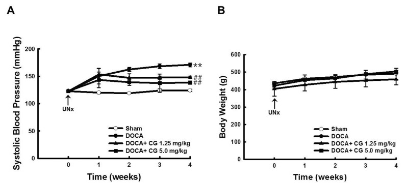
Using the tail-cuff method, systolic blood pressure (SBP) was measured and recorded for four weeks. Unilateral nephrectomy followed by drinking water containing 1% NaCl had little effect on SBP. DOCA injection resulted in significantly increased SBP (p<0.01 vs. sham group), which was reversed by administration of CG200745 (CG, Fig. 1A). Neither DOCA injection nor CG administration affected body weight gain (Fig. 1B).
CG200745 had little effect on vascular contraction and relaxation response
To determine whether CG affects vascular contraction and relaxation, we conducted an organ bath study. We investigated the contraction of aortic rings by treating with phenylephrine cumulatively. CG did not affect vascular contraction, whether endothelium was intact (Fig. 2A) or denuded (Fig. 2C). Furthermore, CG did not affect vascular relaxation induced by acetylcholine (Fig. 2B) or sodium nitroprusside (Fig. 2D), whether endothelium was intact or denuded, respectively.
CG200745 had no effect on blood chemistry
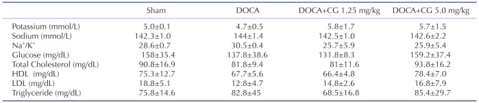
We investigated whether DOCA injection and CG administration had any effects on rat blood chemistry. The results of the blood components analysis showed no significant differences in blood component levels among all groups (Table 1).
CG200745 attenuated left heart weight in the DOCA plus CG administration groups
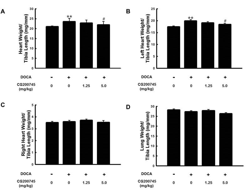
Ratios of heart weight (HW)/tibia length (TL) (Fig. 3A) were used to show phenotypic changes attributable to hypertrophy-induced heart mass increase because it is not affected by change of body weight. To determine whether the DOCA alone group and DOCA plus CG administration groups showed cardiac hypertrophy, we analyzed the HW/TL and left heart weight (LHW)/TL (Fig. 3B). The DOCA alone group showed increased HW/TL and LHW/TL ratios when compared with those of the sham group, which was attenuated by CG administration. The weights of the lung and right heart were similar among all groups. Neither DOCA injection nor CG administration affected right heart weight (RHW)/TL (Fig. 3C) or lung weight (LW)/TL (Fig. 3D) in the DOCA alone group and DOCA plus CG administration groups.
CG200745 attenuated cardiac hypertrophy
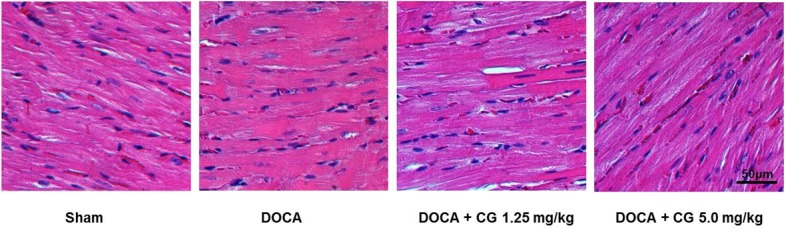
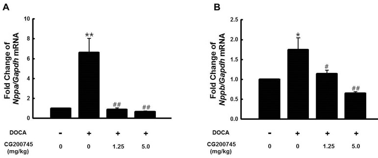
To confirm cardiac hypertrophy histologically in the DOCA groups, we performed a hematoxylin and eosin (H&E) stain (Fig. 4). DOCA groups had hypertrophy when compared with the sham group, which was restored by CG administration. Atrial natriuretic peptide A (Nppa, Fig. 5A) and atrial natriuretic peptide B (Nppb, Fig. 5B) mRNA expression were increased in the DOCA alone group, consistent with hypertrophy seen by histology. Nppa and Nppb mRNA expression were decreased with CG administration in the DOCA plus CG administration groups.
CG200745 attenuated cardiac fibrosis
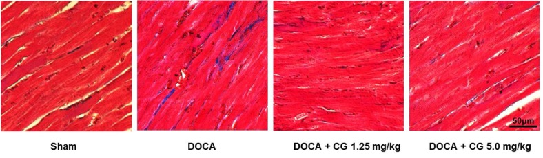
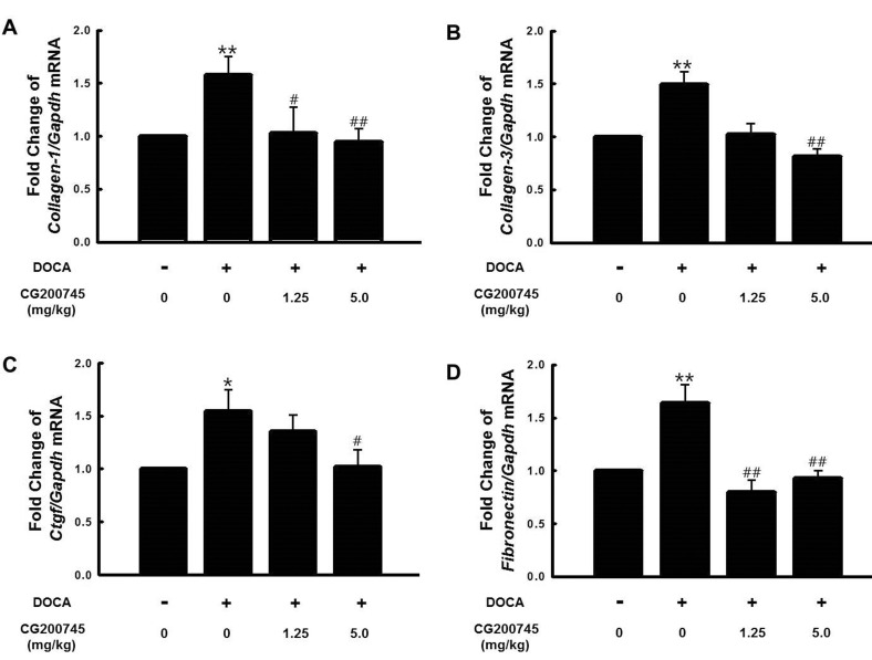
To confirm cardiac fibrosis histologically in the DOCA groups, we performed a trichrome stain (Fig. 6); collagen deposition was stained blue. The staining revealed that the DOCA increased cardiac fibrosis when compared with the sham group. Cardiac fibrosis was attenuated by CG administration. Collagen-1 (Fig. 7A), Collagen-3 (Fig. 7B), connective tissue growth factor (Ctgf, Fig. 7C), and Fibronectin (Fig. 7D) mRNA expression were increased in the DOCA alone group, consistent with the fibrosis seen by histology. Collagen-1, Collagen-3, Ctgf, and Fibronectin mRNA expression were decreased with CG administration.
DISCUSSION
In the present study, we demonstrate that CG200745 (CG) attenuates cardiac hypertrophy and fibrosis in DOCA-induced hypertensive rats. CG restores increased heart weight, histological hypertrophic changes, and the expression levels of hypertrophic genes as well as fibrosis in the hearts of DOCA-induced hypertensive rats.
HDAC is Known To Act As An Epigenetic Gene Regulator On Histone Or Non-histone Proteins And May Be A Potential Target For The Treatment Of Various Diseases Such As Cancer, Kidney Disease, Stroke, Hypertension, Cardiac Hypertrophy, And Heart Failure [13]. Hdac Inhibitors Have Profound Suppressive Effects On Pathological Cardiac Hypertrophy And Fibrosis [14]. Cardiovascular Diseases Are Inhibited By Pan-hdac Inhibitors Such As Suberanilohydroxamic Acid (Saha, Vorinostat) [15], Trichostatin A (Tsa) [16], And Sodium Valproate (Vpa) [8] In The Hypertrophied Hearts Of Hypertensive Rats. We Identified That Cg, A Pan-hdac Inhibitor, Attenuated Increased Heart Weight (Fig. 3A), Histological Hypertrophic Changes (Fig. 4), And Up-regulation Of Hypertrophic Biomarker Genes Such As Atrial Natriuretic Peptide A (Nppa, Fig. 5A) And Atrial Natriuretic Peptide (Nppb, Fig. 5B). Furthermore, We Confirmed That Cardiac Fibrosis Was Diminished By Cg Administration, Consistent With Down-regulation Of Collagen-1 (Fig. 7A), Collagen-3 (Fig. 7B), Connective Tissue Growth Factor (Ctgf, Fig. 7C), And Fibronectin (Fig. 7D) Gene Expression. Saha Not Only Attenuates Hypertension But Also Has An Effect On Vascular Endothelial Function In Doca-induced Hypertensive Rats [15]. Although Cg Attenuated Hypertension In Doca-induced Hypertensive Rats (Fig. 1A), Cg Had Little Effect On Vascular Contraction (Fig. 2A, C) And Relaxation (Fig. 2B, D) In The Same Rats.
HDAC is known to have two opposite functions in the heart: inhibition and induction of pathological cardiac hypertrophy. Class I HDACs (1, 2, 3, and 8) have been shown to induce pathological hypertrophy, whereas class IIa HDACs (4, 5, 7, and 9) block cardiac hypertrophy [17]. HDACs respond differently in response to distinct stresses in the hypertrophic heart. For instance, the intrinsic activity of HDAC2 is increased in the hearts of certain hypertrophic transgenic mice [18], whereas enzyme activity of HDAC6 and HDAC8 is increased in the hearts of DOCA-induced hypertensive rats [10]. Furthermore, global HDAC activity is shown to be increased in the hypertrophic hearts of SHRs [11]. The mechanisms by which specific HDACs are involved in pathological cardiac hypertrophy are still being elucidated. Class I HDACs help control cardiac fibrosis. Class I HDAC inhibition prevents the expansion of the pool of extracellular matrix (ECM)-producing fibroblasts in the myocardium of rats with congestive heart failure (CHF) [19], and reduces fibrosis in the hearts of angiotensin II-mediated heart dysfunction rats [20]. In contrast, HDAC6, a class IIb HDAC, takes part in regulating expression of fibrosis-related genes such as α-smooth muscle actin (SMA) in isoprenaline (ISO)-induced heart dysfunction rats [21]. Although HDAC inhibition appears to block many pathogenic mechanisms that control heart failure, the mechanistic details of these effects are still uncertain [14]. Therefore, it has been suggested that pan-HDAC inhibitors are effective for regulating cardiac hypertrophy and fibrosis.
CG has been studied for treatment of cancers such as non-small cell lung cancer [22] and prostate cancer [23], as well as induction of cell death such as lung and splenic apoptosis [24] and clonogenic cell death [25]. Our results show that CG has a beneficial effect on prevention of hypertension, cardiac hypertrophy, and fibrosis in DOCA-induced hypertensive rats. This is a novel finding that CG may be applied to prevent cardiovascular diseases.
CG attenuates heart weight (Fig. 3A, B), cardiac hypertrophy (Fig. 4, 5) and fibrosis (Fig. 6, 7), and hypertension (Fig. 1A), but not body weight (Fig. 1B), vascular contraction (Fig. 2A, C) and relaxation (Fig. 2B, D), and blood chemistry (Table 1). Although the exact mechanism by which CG prevents cardiovascular diseases remains elusive, CG could be utilized in new treatment strategies.
In summary, this study shows that CG attenuates cardiac hypertrophy, cardiac fibrosis, and blood pressure in DOCA-induced hypertensive rats. Therefore, CG could be used as a potential therapeutic drug for the treatment of cardiovascular diseases.

 XML Download