

 PDF
PDF ePub
ePub Citation
 Print
Print


INTRODUCTION
Diabetic nephropathy (DN), also known as nodular diabetic glomerulosclerosis, is characterised by mesangial cell (MC) proliferation and excessive accumulation of extracellular matrix (ECM), which may ultimately lead to chronic renal failure [1]. The main ECM proteins such as type IV collagen (ColIV) and fibronectin (FN) are often used as the markers of fibrogenesis in many kidney fibrosis diseases including diabetic glomerulosclerosis [234]. In the complicated pathogenesis, hyperglycemia is the main initiation factor in the etiology of DN, which can activate multiple intracellular signalling factors (such as TGF-β1, reactive oxygen species), resulting in abnormalities in blood flow and the accumulation of ECM [567]. In these factors, TGF-β1 and its downstream mediator connective tissue growth factor (CTGF) are recognized as fibrogenic cytokines which play a critical role in the glomerulosclerosis of DN [789]. Currently, MCs are recognized as the major cells to secrete ECM [9]. Therefore, inhibiting the proliferation of MCs and the accumulation of ECM can be applied as a practical approach for treating or relieving DN.
However, there is no treatment available to arrest the progression for the end-stage renal failure. Therefore new therapeutic strategies in the management of diabetic nephropathy are really in demand. Chinese medical herbs are considered to be one of the most promising sources due to their variety of species and applications. Rhubarb (Dahuang in Chinese), one of best-known Traditional Chinese Medicine, has been widely used for thousands of years in China for its treatment of many diseases. Rhubarb contains five free anthraquinones (FARs), including rhein, emodin, aloeemodin, chrysophanol, and physcion, which account for the main medicinal properties of rhubarb [10]. These FARs have been documented to have numerous therapeutic benefits, such as resisting high fat of blood [11], diminishing inflammation [12], accommodating immunity [13], exhibiting vasorelaxant effects [14] and antioxidant activities [15], suggesting its potential therapeutic value in the treatment of DN. Zheng et al. has found that rhein, one of anthraquinones, reversed the diabetic phenotype of mesangial cells over-expressing the glucose transporter (GLUT1) by inhibiting the hexosamine pathway [16]. Wang et al. reported that FARs extract had an effect on preventing Chronic Renal Failure [17]. However, seldom information about the FARs extract on the effects of glomerulosclerosis in DN could be found. Since cell proliferation and ECM accumulation play a vital role in the glomerulosclerosis of DN, it is essential to clarify the in vitro effects of the FARs extract on high glucose-cultured mesangial cell, which may be meaningful to the treatment of DN.
In this study, using gradient ethanol as the eluant, we purified the FARs extract by DM130 macroporous resin and tested its effects on the proliferation of MCs and the expression of main ECM (ColIV and FN) and ECM-related genes (TGF-β1 and CTGF) in high glucose-cultured mesangial cells. Our results will be beneficial to explore new drugs for the treatment of diabetic nephropathy.
Go to : 
METHODS
Materials
Rhubarb, purchased from Xuzhou traditional Chinese medicine factory (China) was identified as the dried roots of Rheum palmatum L. by Dr. Huankai Yao. Standard chemicals of five FARs (aloe-emodin, emodin, rhein, chrysophanol, and physcion) were obtained from Sichuan Weikeqi Biological Technology Co., Ltd (Sichuan, China) and resolved in methanol (MeOH) as the control solution. Rat MCs (No. HBZT-1) were obtained from Wuhan Boster Biological Technology Co. (Wuhan, China). DM130 macroporous resins were bought from Anhui Sanxing resin Biological Technology Co., Ltd (Anhui, China). Cell Counting Kit-8 (CCK-8) was purchased from DOJINDO Chemistry Institute (Japan). The TRIzol kit and cDNA reverse transcription kit were obtained from TAKARA Bio Inc (Japan). The rat TGF-β1, CTGF, FN and ColIV ELISA kits were purchased from R&D Systems Inc (USA). Methanol was chromatographic grade. All solvents used for preparation and separation of crude samples were of analytical grade.
Preparation and identification of FARs extract
Dried ground Rheum palmatum L. (100 g) was macerated in 1,000 ml 85% ethanol, followed by heating at 80℃ for 30 min. The supernatant solvent was filtered and the residue was repeatedly extracted twice. Then the supernatant solvents were combined and dried under the vacuum to give a dark brown powder. The power was dissolved in distilled water and purified by DM130 macroporous resin by using 30%, 60% and 90% ethanol as elution solvents. The eluents were evaporated and dissolved in MeOH to produce 0.5 mg/ml solution for analysis and identification by HPLC, using an Agilent 1100 HPLC-DAD system with a cosmosil-C18 column (250×4.6 mm, 5 µm) at room temperature. Linear gradient was employed using 0.3% H3PO4 (solution A) and methanol (solution B) as mobile phases processed as follows: 0~7.5 min, 25% A; 7.5~8 min, 25~15% A; 8~ 30 min,15% A; 30~32 min, 15~25% A; 32~35 min, 25% A at a flow rate of 0.8 ml/min. The detection wavelength was 220 nm at 25℃.
Mesangial cells culture and treatment
Rat MCs (No. HBZT-1) were provided by the China Center for Type Culture Collection (CCTCC) in Wuhan University. Cells were cultured in a Dulbecco's Modified Eagle's Medium (DMEM, Gibco, Grand Island, NY, USA) that was supplemented with 100 µg/ml streptomycin, 100 U/ml penicillin, and 10% (v/v) fetal bovine serum. The cells were incubated at 37℃ in a humidified atmosphere of 5% CO2. During the experiments, the cells were first exposed to a normal concentration of glucose (5.56 mmol/L) without serum for 12 h, and then treated with high glucose (HG, 25 mmol/L glucose), high glucose with 5 µg/ml FARs (FARsL), high glucose with 10 µg/ml FARs (FARsM), and high glucose with 20 µg/ml FARs (FARsH). The object drugs in the cell culture-FARs extractswere dissolved in 20 µmol/L Dimethyl sulfoxide (DMSO) (as a solvent). Mannitol (MA) was used as a control to rule out the effect of osmotic pressure. The cells were harvested for analysis after 24 h of treatment.
Cell proliferation assay
Cell proliferation was examined by Cell Counting Kit-8 colorimetric method [18]. MCs were seeded into 96-well plates at a density of 1.0×10⁶ per well. After 24 h, CCK-8 solution were added and cells continued to be cultured for another 2 h. Absorbance was read at 450 nm by visible spectrometry (Clinibio Co., Australia).
Reverse transcription-polumerase chain reaction
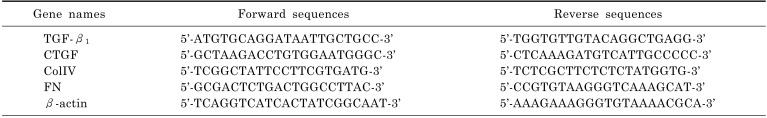
A reverse transcription polymerase chain reaction (RT-PCR) procedure was performed to determine the relative mRNA quantities of TGF-β1, CTGF, ColIV and FN in MCs. Total RNA was extracted from rat MCs with TRIzol Reagent according to the manufacturer's instructions. The total RNA (0.5 µg) obtained was converted into cDNA using the RevertAid™ First Strand cDNA Synthesis Kit with Random Hexamer primers. The upsteam and downstrean primers (Shanghai Sangon Company, China) of thoese genes were shown in Table 1. The reaction mixture was incubated at 48℃ for 45 min to reverse transcript, then went into cycles. The cycle conditions of these genes were all set to: initial denaturation for 5 min at 94℃, 30 cycles at 94℃ for 30 sec, 60℃ for 30 sec, 72℃ for 1 min, final elongation at 72℃ for 7 min. The RT-PCR products were separated by 2% agarose electrophoresis, and the band densities were analyzed using laser densitometry. The relative quantities of mRNA of these four genes in mesangial cells were represented by the ratio of band density of objective gene versus that of β-actin.
Enzyme-linked immunosorbent assay
The levels of TGF-β1, CTGF, ColIV and FN in the supernatant of MCs were determined by enzyme-linked immunosorbent assay (ELISA). The medium of MCs was collected and centrifuged at 13,000 g for 15 mins to pellet the debris. The levels of these four proteins were determined according to the manufacturer's instructions. The colorimetric reaction was measured at 450 nm.
Statistical analysis
Data were expressed as means±SD. Statistical analyses were performed using the paired t-test for two data comparison and one-way analysis of variance (ANOVA) with Dunnett's test for multiple data comparison. A value of p<0.05 was considered significant.
Go to :
RESULTS
The ingredient identification of FARs
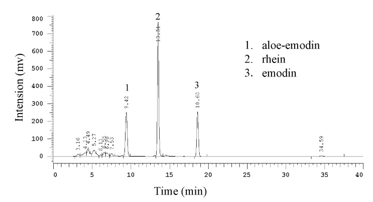
According to results of HPLC, the chromatographic peaks of the analytes were confirmed by comparing their retention time with those of the reference compounds. The main ingredients were identified as aloe-emodin, rhein, and emodin (Fig. 1).
Effects of FARs extract on the MC Proliferation
CCK-8 assay was adopted to assess the impact of the FARs extract on MC proliferation. The proliferation of MCs was enhanced after 24 h exposure to high glucose (p<0.01). The results showed that the cell proliferation rates in FARsL, FARsM and FARsH were significantly lower than those of HG group (p<0.01). Meanwhile, the cell proliferation in the MA group was almost identical to those in the NG group and the DMSO group was not significantly different from the HG group, indicating that no obvious impacts were generated by the osmotic pressure and the vehicle. The above results indicated that administration of FARs compound significantly suppressed high glucose induced-MC proliferation (Fig. 2).
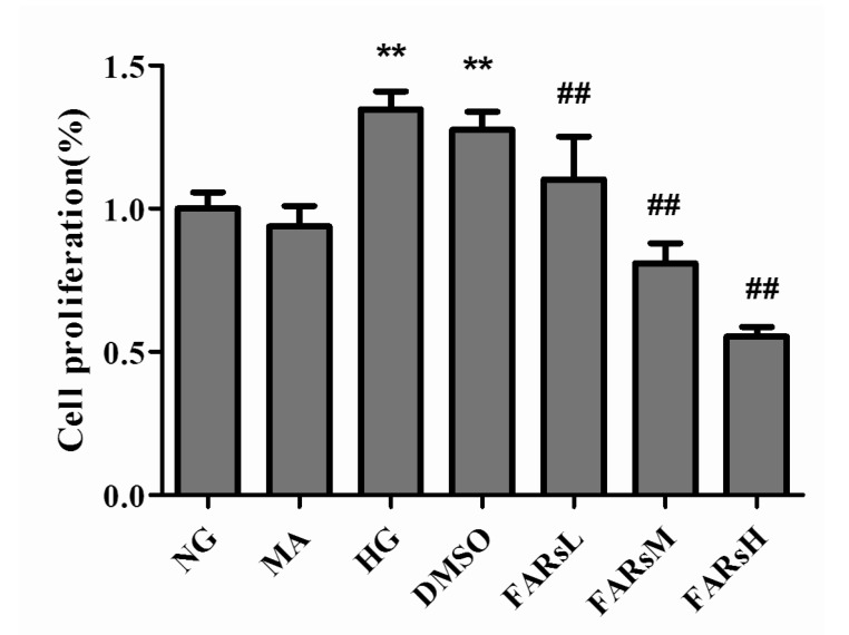 | Fig. 2The cell proliferation rates in FARs-treated MCs. The cells were exposed to DMEM culture media containing different substances, namely: 5.56 mmol/L glucose in the NG group, 5.56 mmol/L glucose+19.44 mmol/L mannitol in the MA group, 25 mmol/L glucose in the HG group, 25 mmol/L glucose+20 µmol/L DMSO in the DMSO group, 25 mmol/L glucose+5 µg/ml FARs in the FARsL group, 25 mmol/L glucose+10 µg/ml FARs in the FARsM group, 25 mmol/L glucose+20 µg/ml FARs in the FARsH group, respectively. Mean±SD, n=4. **p<0.01, compared with the NG group.
|
Effects of FARs compound on the mRNA levels of TGF-β 1, CTGF, ColIV and FN
The RT-PCR results showed that the mRNA levels of TGF-β1, CTGF, ColIV and FN in the HG groups were elevated compared to those of the NG groups. The CTGF, ColIV and FN mRNA levels in the groups treated with 5, 10, 20 µg/ml FARs were all lower than those of the HG group. The TGF-β1 mRNA levels in groups treated with 10, 20 µg/ml FARs were also significantly decreased compared with those in the HG-treated groups. Significant changes in the levels of TGF-β1, CTGF, ColIV and FN mRNA between MA group and NG group were not found. The four genes in the DMSO group were also not remarkably different from those in the HG group. All these indicated that administration of FARs extract significantly decreased high glucose-induced the increase of TGF-β1, CTGF, ColIV and FN mRNA quantities (Fig. 3).
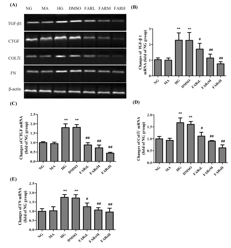 | Fig. 3The mRNA levels of TGF-β1, CTGF, ColIV and FN in the FARs-treated MCs. (A) Agarose electrophoresis of RT-PCR products amplified from the total RNA extracts of mesangial cells, β-actin was used as the internal standard in each sample. (B~E) RT-PCR data for the mRNA relative quantity of TGF-β1, CTGF, ColIV and FN performed by densitometric analysis. Mean±SD, n=4. **p<0.01, compared with the NG group; ##p<0.01, #p<0.05, compared with the HG group.
|
Effects of FARs compound on the proteins of TGF-β1, CTGF, ColIV and FN
The levels of these four proteins in the cell culture supernatant in the HG group were remarkably up-regulated compared to those in the NG group (p<0.01). Importantly, treatment with 5, 10, 20 µg/ml FARs significantly decreased the levels of TGF-β1, CTGF, ColIV and FN induced by high glucose. There is no significant difference between the NG group and MA group on the secretion of these four proteins. DMSO also had no significant effect on the proteins levels. All these indicated that administration of FARs significantly reduced high glucose-induced the secretion of TGF-β1, CTGF, ColIV and FN proteins (Fig. 4).
 | Fig. 4The relative quantity of TGF-β1, CTGF, ColIV and FN proteins in the supernatant of FARs-treated MCs. (A) The relative quantity of TGF-β1 and CTGF proteins. (B) The relative quantity of Col IV and FN proteins. Mean±SD, n=4. **p<0.01, compared with the NG group; ##p<0.01, #p<0.05, compared with the HG group.
|
Go to :
DISCUSSION
MC is one type of specialized vascular smooth muscle cells around the capillaries of the renal glomerulus, serving functions of structural support and regulating glomerular filtration rate. MC proliferation and the progressive accumulation of ECM in the mesangial areas are the major characteristics of diabetic glomerulosclerosis [1]. Therefore, reversing changes in the MCs proliferation and extracellular matrix might play a key role in delaying the progression of diabetic nephropathy. In the complicated mechanisms of DN, the pathway of hyperglcaemia-TGF-β1-ECM is important. TGF-β1, highly expressed in the kidneys of diabetes [78], contributes to the accumulation of ECM through the Smad pathway [48]. The administration of neutralizing antibodies against TGF-β1 to diabetic mice or mesangial cells has been shown to prevent the accumulation of ECM [19]. CTGF, acting as downstream of TGF-β1, has been shown to mediate expression of ECM proteins in response to various external perturbations [1920]. Moreover, CTGF facilitates TGF-β1 signaling and consequently promotes renal fibrosis [19]. It is becoming clear that the coordinated expression of TGF-β1 and CTGF is crucial for the induction of ECM proteins and thus, for the development of DN [7].
In the present study, we extracted free anthraquinones compounds, including rhein, emodin, aloe-emodin, from rhubarb and found that administration of FARs extract significantly suppressed the cell proliferation, the mRNA and protein levels of TGF-β1, CTGF, ColIV and FN induced by high glucose. The effects and mechanisms of FARs extract on DN remain to be fully understood. Rhein, one of FARs, was found to have an ability of resisting the renal fibrosis in DN rats [11]. Guo et al. found that rhein inhibits extracellular matrix accumulation induced by TGF-β1, which might partly account for the role of rhein in preventing and retarding the progression of diabetic nephropathy [21]. Emodin (another FARs) was found to ameliorate renal dysfunction in diabetic nephropathy rats by its inhibition of the activation of p38 MAPK pathway and downregulation of the expression of fibronectin [6]. And Li et al. found that emodin had the ability of suppressing cell proliferation and fibronectin expression in rat mesangial cells cultured under high glucose [22]. Meanwhile, aloe-emodin (the third FARs) was also reported to have antiproliferative activity in various cancer cell lines [2324], which might partly explain the inhibition of FARs on MC proliferation. The function of FARs compound on HG-cultured MCs may be attributed to the combination of the anthraquinone mixtures. However, seldom researches about the compound of FARs were found in DN, probably due to the pollution produced in the FARs' extraction and separation process. The traditional extraction of FARs tends to use the petroleum ether, benzene, chloroform, or ethyl ether as the solvents [252627], producing toxicity in some degree. In the present study, we adopted the ethanol as the solvent, improving the safety of extracts and reducing the pollution of environment, which was better able to satisfy the pharmaceutical requirement.
This study confirmed the effectiveness of our FARs extract on inhibiting ECM accumulation and cell proliferation in high glucose-cultured MCs, which may facilitate the finding of new drugs to prevent the progression of DN. Further researches are under way to comparing the effects of FARs extract with the single free anthraquinone extracted from the rhubarb and observing their effects on DN animals.
Go to :
 XML Download