Journal List > Korean J Physiol Pharmacol > v.18(6) > 1025978

Kang, Ryoo, Shin, Trojanowski, and Shaw: Role of Cerebrospinal Fluid Biomarkers in Clinical Trials for Alzheimer's Disease Modifying Therapies

Abstract

Until now, a disease-modifying therapy (DMT) that has an ability to slow or arrest Alzheimer's disease (AD) progression has not been developed, and all clinical trials involving AD patients enrolled by clinical assessment alone also have not been successful. Given the growing consensus that the DMT is likely to require treatment initiation well before full-blown dementia emerges, the early detection of AD will provide opportunities to successfully identify new drugs that slow the course of AD pathology. Recent advances in early detection of AD and prediction of progression of the disease using various biomarkers, including cerebrospinal fluid (CSF) Aβ1-42, total tau and p-tau181 levels, and imagining biomarkers, are now being actively integrated into the designs of AD clinical trials. In terms of therapeutic mechanisms, monitoring these markers may be helpful for go/no-go decision making as well as surrogate markers for disease severity or progression. Furthermore, CSF biomarkers can be used as a tool to enrich patients for clinical trials with prospect of increasing statistical power and reducing costs in drug development. However, the standardization of technical aspects of analysis of these biomarkers is an essential prerequisite to the clinical uses. To accomplish this, global efforts are underway to standardize CSF biomarker measurements and a quality control program supported by the Alzheimer's Association. The current review summarizes therapeutic targets of developing drugs in AD pathophysiology, and provides the most recent advances in the

ABBREVIATIONS

AD

Alzheimer's disease

amyloid beta

1-42

amyloid beta 1-42

NFT

neurofibrillary tangle

CSF

cerebrospinal fluid

MCI

mild cognitive impairment

NMDA

N-methyl-D-aspartate

FDA

Food & Drug Administration

APP

amyloid precursor protein

Abo

amyloid beta oligomer

PPAR-g

peroxisome proliferator-activated receptor gamma

BBB

blood-brain-barrier

RAGE

receptor for advanced glycation end products

LRP-1

low-density lipoprotein receptor-related protein 1

PET

positron emission tomography

IVIG

intravenous immunoglobulin

GSK-3

glycogen synthase kinase 3

ADNI

Alzheimer's Disease Neuroimaging Initiative

MRI

magnetic resonance imaging

FDG

2-[18F]-fluoro-2-deoxy-D-glucose

AICD

C-terminal fragment of amyloid precursor protein

INTRODUCTION

Alzheimer's disease (AD), a progressive neurodegenerative disorder, is the most common form of irreversible dementia, and it carries with it a considerable human, social and economic burden. Following the onset of pathological changes in the brain that include the progressive accumulation in the CNS of amyloid beta (Aβ) deposits and neurofibrillary tangles (NFTs) formed by pathological tau, which is thought to begin more than 15 (Aβ) to 10 (NFTs) years before cognitive impairments become clinically manifest, AD patients primarily develop progressive deterioration of episodic memory and a global decline in their cognitive functions. Among several mechanistic and pathological substrates that contribute to the gradual progression of AD over time, the major neuropathological substrates of AD are the aggregation and accumulation of misfolded Aβ and the intracellular deposition of fibrillized and hyperphosphorylated tau proteins (Fig. 1).
Currently, the greatest utility for biochemical cerebrospinal (CSF) biomarkers of AD may be for the early and more reliable diagnosis of AD, which places measures of the levels of CSF Aβ1-42 and tau proteins (total tau and tau phosphorylated at Thr181) in the revised version of diagnostic criteria for AD research [1,2,3,4]. Thus, abundant data from numerous studies carried out in centers across the globe over the past 20 years shows that the levels of Aβ1-42 in CSF of AD patients are significantly lower than in age-matched healthy elderly controls, whereas the levels of total tau (t-tau) and tau phosphorylated at threonine 181 (p-tau181) in AD CSF are significantly higher than those of controls. Moreover, it has been suggested that these CSF biomarkers are useful to differentiate those mild cognitive impairment (MCI) patients who progress to develop AD within the subsequent several years from the stable MCI patients [5,6,7,8,9,10].
The current standard pharmacotherapy for cognitive improvement in AD patients includes acetylcholinesterase inhibitors and the N-methyl-D-aspartate (NMDA) antagonist memantine. However, the approval of these drugs has not been based on their ability to slow disease progression but to improve the clinical symptomatology. Hence, only symptomatic drugs with transient benefits have been approved for clinical use in AD patients by the US Food and Drug Administration (FDA). Given the fact that genetic and pathological evidence strongly supports the "amyloid cascade hypothesis", several strategies of reducing Aβ accumulation in the brain have been applied to develop "disease-modifying therapy" to cure AD. In addition, a correlation between cognitive dysfunction and neurofibrillary tangle load that is more robust than that between Aβ load and cognitive impairments has led to a parallel strategy to develop tau focused therapies that inhibit and/or block tau aggregate formation, promote the clearance of tau pathology or correct for the loss of tau function when tau is sequestered in NFTs [11]. Other strategies for modifying disease progression include anti-inflammation, metabolic approaches, neurotrophin-based approaches and mitochondrial targets. It has been estimated that a disease-modifying therapy that could delay both dementia onset and progression by one year would reduce the prevalence by 9.2 million cases of the disease by the year 2050 [12,13]. To reduce the number of future AD cases, a variety of attempts to develop a drug that provides a disease-modifying effect against probable AD have been made; however, there are no approved disease-modifying therapies for AD at this time. Based on lessons from previous clinical trials in probable AD patients, there is a growing consensus that initiation of disease-modifying treatment before the onset or early phase of the disease (before the onset of clinical dementia) may be necessary. To maximize the power of clinical trials in patients without clinical dementia, valid biomarkers for the early detection of patients with AD pathology or the prediction of patients presenting a prodromal phase of AD (commonly referred to as MCI), who will likely develop AD in the future, would be helpful. Currently, Aβ1-42 and tau proteins (t-tau and p-tau181) in CSF are the most reliable biochemical biomarkers for the early detection of AD, the differentiation of AD from other forms of dementia, and the prediction of MCI progression to AD. In this review, the current development of AD therapeutics, particularly those drugs targeting amyloid and tau pathology, and the clinical performance of CSF biomarkers for AD diagnosis are summarized. The advantages and unresolved issues of CSF biomarkers in clinical trial design are also discussed.

TARGETS OF DEVELOPING DRUGS IN AD PATHOPHYSIOLOGY

Target related to amyloid production, aggregation and clearance

The molecular chain of events that ultimately results in synaptic and neuronal loss in the brain is complex and remains largely unresolved, even in the well-known pathologic hallmarks of AD, i.e. the deposits of Aβ peptides in amyloid plaques and other types of aggregates and NFTs formed by misfiled and fibrillized tau. A widely accepted hypothesis is that AD might be initiated by the abnormal (amyloidogenic) processing of amyloid precursor protein (APP) followed by the aggregation and accumulation of Aβ in the brain. The strategy to develop anti-Aβdrugs targeting the canonical amyloid cascade can be classified according to the mechanism of action: reduction of Aβ1-42 production, prevention of Aβ oligomer (Aβo) formation, and acceleration of Aβ clearance (Fig. 2). Based on the amyloid hypothesis, several drugs to reduce Aβ burden are being developed for patients with mild-to-moderate AD.
Considering that genetic mutations in the APP or PSEN1 genes cause familial AD and that β-secretase (β-site APP cleaving enzyme, BACE1) knock-out mice showed drastically reduced Aβ levels in the brain, the inhibition of β- and/or γ-secretases can be a strategy to block the initiation of the amyloid cascade. Several first-generation γ-secretase inhibitors (e.g., semagacestat) were tested in clinical studies; however, because γ-secretase is also involved in the processing of Notch, a Phase 3 trial for semagacestat in patients with mild-to-moderate AD not only failed to achieve its predetermined end points, but also worsened clinical measures and increased the incidence of skin cancer [14]. Other Notch-sparing, second-generation γ-secretase inhibitors (e.g., begacestat, avagacestat, PF-3804 014 and NIC5-15) are now in the early phase of clinical trials [15,16,17]. Because amyloidogenic Aβ species are generated by sequential activation of β-secretase and γ-secretase, the inhibition of β-secretase can be the second strategy to suppress the amyloidogenic pathway. However, β-secretase has many endogenous substrates that are not related to APP processing; therefore, no Phase 3 clinical trials of new β-secretase inhibitors are developing, but anti-β-secretase antibodies and the oral compound CTS- 21166 are under investigation. Interestingly, previous research found that the thiazolidinedione antidiabetic drugs (e.g., rosiglitazone and pioglitazone) are potentially beneficial in that they appear to suppress Aβ burden via peroxisome proliferator-activated receptor γ (PPAR-γ) activation [18,19]. PPAR-γ activators can suppress β-secretase expression and accelerate APP degradation by increasing its ubiquitination [20]. Although one clinical trial evaluating rosiglitazone demonstrated a beneficial effect on cognition, particularly in ApoE4 negative patients [21], the larger confirmatory clinical trials failed to demonstrate a beneficial effect on cognition in AD patients [22,23,24]. In addition, a recent warning by the US FDA about possible cardiac risks associated with rosiglitazone and the negative preliminary results led to a discontinuation of the further development of the rosiglitazone program for AD. There is a conflicting result regarding efficacy in AD for pioglitazone, another PPAR-γ activator [25,26]. Another strategy to reduce pathogenic Aβ production is the upregulation of α-secretase activity, leading to upregulation of neuroprotective sAPPα secretion. Several drugs have been tested in early phase clinical trials, but the results are not yet available.
Aβ, particularly Aβ1-42, is prone to aggregation and forms toxic Aβo. Given the evidence that the neurotoxic potency of Aβo is higher than the Aβ monomer or insoluble Aβ amyloid fibrils [27,28], compounds inhibiting Aβ aggregation or destabilizing Aβo seem to be promising drug candidates for AD. The initial anti-aggregant is tramiprosate (homotaurine), which binds preferentially to soluble Aβ; however, the outcome of a Phase 3 trial was not significant [29]. Another anti-aggregant that inhibits metal-induced Aβ oligomerization, PBT2, promotes Aβo clearance and improves cognition in an animal model [30]. In a Phase 2a clinical trial in patients with mild AD for 12 weeks, PBT2 was well-tolerated, reduced CSF Aβ1-42 concentrations and improved executive function [31]. However, recent news releases from Prana reported that PBT2 failed to meet its primary endopoint of reducing Aβ plaques in a 12-month phase 2 "IMAGINE" trial (http://pranabio.com/news/prana-biotechnology-announces-top-line-results-phase-2-imagine-trial-pbt2-alzheimers-disease/#.U4Ol1WdOVaQ). Based on the in vitro and in vivo results of stabilizing Aβ into non-toxic conformers and improving AD-related phenotypes in TgCRND8 transgenic mice [32,33], a cyclohexanehexol isomer, ELND005 (Scyllo-inositol), has been tested in a Phase 2 clinical trial [32]. Although the primary endpoints in the Phase 2 trial did not achieve statistical significance, ELND005 (250 mg, bid) demonstrated a biological effect on Aβ in CSF [34]. Currently, the sponsoring companies intend to advance this molecule into Phase 3 studies. To evaluate the biological effects of Aβo or Aβ aggregation inhibitors and to prove their mechanisms of action, a biofluid assay set-up would be a tool to find molecules and test their pharmacodynamic action. Previous research has reported that Aβo concentration measured by in-house ELISA method in CSF of AD patients is significantly higher than healthy controls [35]. Although there is criticism of the measurement of Aβo by single-antibody ELISA methodology, the effect of the anti-aggregant on Aβ oligomerization could be monitored by the measurement of Aβo in CSF. However, it is not clear that Aβo are detectable in CSF [36,37]. The recent development of tandem mass spectrometry (MS)-based quantitation of Aβ species in CSF may provide quantitative data for the effect not only of amyloidogenic secretases inhibitors but also Aβo inhibitors on APP metabolism in the brain [38].
Aβ clearance can be achieved by pumping Aβ out through the blood-brain-barrier (BBB) from the brain to the plasma and/or Aβ degradation by specific enzymes. There are two potential targets for enhancement of Aβ removal from the brain to the periphery: the receptor for advanced glycation end products (RAGE) and low-density lipoprotein receptor-related protein 1 (LRP-1) [39,40]. RAGE mediates the influx of Aβ into the brain, while LRP-1 mediates efflux of Aβ from the brain (Fig. 2). In addition to the possible role of RAGE-Aβ interaction for the activation of nuclear factor-κB signaling pathways, which may promote apoptosis and neuroinflammation [41], the RAGE-mediated influx of peripheral Aβ into the brain may increase amyloid load. A RAGE inhibitor or an LRP-1 activator may be a potential candidate for AD treatment based on amyloid clearance. In fact, an oral small molecule inhibiting RAGE activity (PF-04494700) has been tested in Phase 2 trials; however, the development was discontinued [42]. Another strategy to enhance Aβ clearance is the activation of Aβ degradation proteases, including neprilysin, insulin-degrading enzyme and plasmin [43,44]. However, specific activation of enzymes seems the more challenging approach than inhibition.

Immunotherapy against amyloid pathology

Two types of immunotherapy currently exist to enhance antibody-mediated Aβ clearance: active immunotherapy (anti-Aβ vaccine) and passive monoclonal anti-Aβ antibody treatment. The initial active immunotherapy with AN-1792 to induce anti-Aβ antibodies was not tolerable by the activation of cytotoxic T cells and autoimmune reaction followed by meningoencephalitis [45]. Subsequently, the more tolerable active immunotherapies using the improved design of an immunogen- containing the N-terminal, fragment of Aβ1-42 or N-terminus mimic peptide, including ACC-001, CAD106, V950, and Affitope AD02, are currently being tested in the early phases of clinical trials [46]. Passive immunotherapeutic approaches are also currently developing in parallel with active immunotherapy. To date, humanized monoclonal antibodies for passive immunotherapy are in clinical development. For example, an initial early phase clinical trial of bapineuzumab showed good tolerability with mild to moderate adverse events, including symptomatic and asymptomatic amyloid-related imaging abnormalities (ARIA) and improvement of exploratory efficacy measure (minimal mental status examination score; MMSE), when compared to the placebo group. However, the efficacy evaluated by prespecified primary endpoints (Alzheimer's Disease Assessment Scale-Cognitive; ADAS-Cog, or Disability Assessment for Dementia; DAD) in a Phase 2 trial did not show statistical significance, although a trend in favor of bapineuzumab was observed, particularly in apolipoprotein E4 noncarriers [47]. In addition, there were no observed treatment differences in either CSF Aβ or total tau, but the reduction of p-tau in the bapineuzumab group was significant when compared with the placebo group [48]. Although the clinical efficacy of bapineuzumab in the Phase 2 trial was not dramatic by week 52, amyloid removal from the brain was clearly observed in amyloid (PiB) PET scan [49]. In Phase 3 trial of bapineuzumab for 78 weeks, the clinical efficacy was disappointing although the changes of biomarkers were significant, particularly in ApoE carriers [50]. Another humanized monoclonal anti-Ab antibody, solanezumab (LY2062430) was applied to mild-to-moderate AD patients with an advantage of absence of significant ARIA [51]. Similar to bapineuzumab trial, however, solanezumab failed to have a success of clinical efficacy (phase 3 trial of EXPEDITION 1 and EXPEDITION 2 study) [52]. The disappointing results of intravenous bapineuzumab and solanezumab in phase III trial for mild to moderate AD patients would not necessarily exclude these from AD prevention trial, such as the Dominantly Inherited Alzheimer's Network (DIAN) study [53,54,55]. For passive immunotherapy using immunoglobulin, three small trials using intravenous immunoglobulin (IVIG) targeting multiple forms of Aβ suggest that IVIG can have favorable efficacy and will be tolerable [56,57,58]. However, two of these studies were not placebo-controlled studies, and another study was a retrospective case-control analysis. Recently, data of the clinical efficacy of IVIG in a placebo-controlled, multicenter Phase 2 clinical trial and the effect of IVIG on the concentration of plasma and CSF Aβ species have been published [59]. The results of this study showed an acceptable safety profile and a significant difference in plasma Aβ1-40 levels in the highest dose group when compared to the placebo group [60]. Recently, the disappointment of phase 3 clinical trial using IVIG in 390 patients with mild-to-moderate AD was announced, although a dose-dependent reduction in plasma Ab42, increase in plasma, CSF anti-oligomer and anti-fibril antibodies, and reduced brain fibrillar Ab42 was observed [61, and also see http://blog.alz.org/results-of-igiv-study-disappointing-but-not-discouraging].

Target related to tau hyperphosphorylation

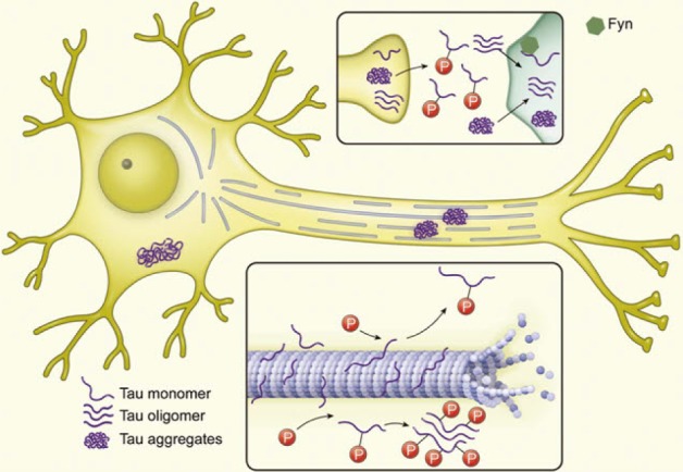
Intracellular fibrillary NFTs containing hyperphosphorylated and fibrillar species of tau are one of hallmarks of AD pathology, but they are not specific for AD. If tau is abnormally hyperphosphorylated and aggregated, it may be toxic due to gains of deleterious function or loss of the normal function of tau and microtubule instability followed by axonal transport failure (Fig. 3). There may be two approaches to inhibiting tau toxicity [11,62]. The first is the inhibition of abnormal hyperphosphorylation by targeting tau kinase and/or phosphatase. In addition, inhibitors of tau aggregation or disassemblers may be beneficial for protecting neurons from tau aggregate toxicity. There are several candidates of tau kinase and phosphatase related to tau hyperphosphorylation [63]. For example, glycogen synthase kinase 3 (GSK-3) is a well-known tau kinase, which is balanced with a phosphatase, protein phosphatase 2A. Valproic acid and lithium, well-known for the treatment of epilepsy and bipolar disorder, respectively, are GSK-3 inhibitors [64]. However, they didn't show consistent results for cognitive improvement or changes in CSF tau or p-tau181 concentration in clinical trials [65,66,67]. Several new GSK-3 inhibitors are being tested in clinical trials. NP031112 (tideglusib), which is a non-ATP competitive GSK3 inhibitor, reduces tau hyperphosphorylation and amyloid deposit, prevents neuronal death, and improves cognitive function in animal models [68]. This drug showed clinical benefits in a pilot, randomized, double-blind, placebo-controlled clinical trial, and it is currently being confirmed in a larger clinical trial [69]. Other multifunctional inhibitors of tau aggregation are also in development. Methylene blue, a famous histology dye, inhibits tau aggregation in addition to its antioxidant activity and the ability to enhance mitochondrial function. In a Phase 2, placebo-controlled study, a middle dose of methylene blue known as Rember improved cognitive function in mild-to-moderate AD patients and had evidence of slower progression of the disease [70]. Davunetide (an octapeptide) and nicotinamide are inhibitors of tau aggregation that showed prevention of cognitive deficit in AD animal models [71]. Rember is safe and tolerable and it continues in clinical studies but Davunetide failed in clinical trials for AD and other tauopathies (for a review, see http://www.alzforum.org/news/research-news/tau-targeting-drug-davunetide-washes-out-phase-3-trials).

CLINICAL PERFORMANCE OF CSF BIOMARKERS FOR EARLY DIAGNOSIS OF AD

By direct contact with extracellular space of the brain, CSF is the most useful biological fluid reflecting molecular events in the brain, which have driven intense research efforts to develop biochemical biomarkers for AD diagnosis in CSF. On the basis of prevailing scientific evidence and reliable clinical performance, CSF biomarkers have been involved in the recently published revision of AD diagnostic criteria for research purposes as supportive evidence for AD pathophysiology [3,4]. When we analyzed the overall clinical performance of CSF AD biomarkers (concentration of Aβ1-42, t-tau and p-tau181) for the diagnosis of AD in previously reported clinical studies, both mean sensitivity and specificity for Aβ1-42 and t-tau are over 80%, while those for p-tau181 are slightly lower than 80%. When Aβ1-42 and t-tau are combined (i.e., Aβ1-42/t-tau ratio or regression model using Aβ1-42 and t-tau concentration), both the mean sensitivity and specificity are higher than 85% [72]. Furthermore, longitudinal follow-up studies showed that CSF biomarker measurement has an ability to predict future development of AD in MCI patients [5,6,7,8,9,10]. The prediction of progression from MCI or preclinical AD to AD is important not only for the early diagnosis followed by therapeutic intervention but also for the design of clinical trials for developing disease-modifying therapies. In this context, large-scaled clinical studies are underway to test the clinical relevance of CSF biomarkers for early diagnosis of AD, even in the preclinical stage of AD development. For example, data from North American Alzheimer's Disease Neuroimaging Initiative (ADNI) showed the clinically reliable predictive performance of CSF AD biomarkers [8,73], using cut-off values determined by independent non-ADNI autopsy-confirmed samples [7]: the sensitivities of Aβ1-42 t-tau, p-tau181, and Aβ1-42/t-tau ratio are 96.4, 69.6, 67.9, and 85.7%, respectively, and the specificities are 76.9, 92.3, 73.1, and 84.6%, respectively,. North American ADNI is a multicenter, prospective, longitudinal, observation study for the evaluation of clinical characteristics, genetics, imaging biomarkers, and CSF AD biomarkers in healthy elderly subjects, MCI, and AD patients [74]. This study (ADNI-1) was completed in 2009, and ADNI-2 is ongoing. In ADNI-2, early amnestic MCI patients were added. The early amnestic MCI was defined as individuals meeting clinical criteria for amnestic MCI, who score between 0.5 and 1.5 standard deviation below the mean of normal healthy controls on delayed paragraph recall performance. Therefore, ADNI studies provided the long-term follow-up data for the changes in the initial diagnosis (e.g., early or late MCI to AD or normal to MCI). In addition to North American, European and Australian ADNI studies in western countries, Japanese, Chinese, and Korean ADNI studies are currently ongoing in Asia. Currently, numerous investigators are supporting the evidence that CSF biomarkers have a good diagnostic performance, particularly in combination with other biomarkers, including genetic biomarkers (e.g., ApoE genotype) and imaging biomarkers (e.g., hippocampal volume determined by MRI) [75,76,77,78]. However, there are several limitations for the application of CSF biomarkers for the diagnosis of AD in clinics distributed elsewhere. For example, the cut-off values for AD diagnosis are different across the studies due to interlaboratory variability in the measurement of CSF biomarker concentrations [73]. In other words, the causes of interlaboratory variability of CSF biomarker concentrations measured by immunoassay technologies were not yet completely elucidated (see below). To minimize the interlaboratory variability of CSF biomarker concentrations, global efforts, including the development of standardized protocols, reference materials and reference methods, are underway [79,80].
In addition, the amyloid hypothesis and chronological cascade relationships among amyloid and tau pathology, biomarkers of CSF level of Aβ1-42 and tau and imaging biomarkers, and clinical symptoms, still remain to be proven (Fig. 1), although no clear findings can negate this hypothesis. With the reliable clinical performance of CSF biomarkers, genetic and imaging biomarkers have a diagnostic value for early diagnosis of AD or prediction of disease progression. In particular, it is very important the elucidation of the neurodegenerative process and biomarkers reflecting the pathogenic process in preclinical stage of the disease, since it will accelerate the development of new therapeutics for AD treatment as well as the prevention of the disease [11,81]. To do this, the long-term longitudinal study observing the changes of clinical parameters, and CSF and imaging biomarkers in population with various degree of the disease will be required. Currently, ADNI-GO and ADNI-2 studies including early MCI subjects as well as normal healthy elderly subjects, MCI and early AD patients are underway.

CSF BIOMARKERS FOR AD TRIALS

The use of biomarkers in clinical trials depends on the mechanism of action of the developing therapy, goal of the trial, question to be solved, and stages of the trial. There are several purposes to include AD biomarkers in clinical trial design. First, they can be included in clinical trials as surrogate endpoints for the evaluation of the effects of a potential, new, disease-modifying therapy. Unlike cardiovascular or cancer clinical trials, in which the primary endpoint is typically the occurrence of a specific event, the use of mortality as an endpoint is not feasible for AD clinical trials due to the very large required sample size or follow- up period. If the measurement of biomarkers is valid, biomarker outcome measures can provide the evidence for the biochemical and/or molecular effects of a new therapy. In addition, biomarkers can be used as surrogate endpoints in which clinical measurements are not available, e.g., very early pre-symptomatic stage of AD. Although much work remains to be performed for the application of biomarkers as surrogate endpoints, the measurement of valid biomarkers has a potential to decrease the time that is required for evaluating clinical efficacy. To this end, it is necessary to determine whether the measurement of AD CSF or imaging biomarkers correlate with clinical endpoints and predict future clinical benefit or decline. In an AN1792 Aβ immunization Phase 1 trial, a small portion of antibody responders showed greater atrophy and progression of dementia despite the apparent reduction of amyloid load at autopsy [82,83]. However, it was evident in a larger Phase II trial that AN1792 immunization has a long-term functional benefit in antibody responders [84]. Among the current key unknowns are the time frames for affecting CSF biomarker measurement and what degree the biomarker change might relate to the clinical outcome. Therefore, we need to carefully consider the qualification and validation of AD biomarkers before using them as surrogate markers for clinical endpoints.
Second, biomarkers can be used diagnostically with clinical workup for inclusion and exclusion criteria. In particular, in early phase trials aiming to show mechanistic proof of concept, biomarker measurements can be used as a stratification tool for the pharmacodynamic evaluation of a drug. For example, the effects of a drug targeting brain amyloid reduction (e.g., anti-Aβ immunotherapy or secretase inhibitors) can be assessed in subjects with a high amyloid load who are differentiated by CSF or imaging biomarkers. In recent clinical trials, BMS-708163, a Notch-sparing γ-secretase, showed a good tolerance and a dose-dependent decrease of Aβ1-40 and Aβ1-42 in the CSF of healthy volunteers and AD patients [15,17,85]. As described above, the diagnostic performance of CSF biomarkers is highly reasonable for early diagnosis of AD and for the prediction of MCI progression, although further evidence should be accumulated for the diagnosis of preclinical AD. In fact, several recent disappointing results of clinical trials involving patients at the mild-to-moderate stage of AD emphasize the need for biomarkers that detect AD pathology in the early MCI stage or even at the pre-symptomatic stage. In the case of MCI, CSF AD biomarkers (e.g., diminished Aβ1-42 and elevated t-tau and p-tau181) can differentiate between those patients with underlying AD as the cause of the cognitive impairment from those MCI patients categorized in an early stage of non-AD dementia, patients with stable MCI for long periods of time, or patients in a group who will recover to normal cognition [86]. Disease-modifying drugs are likely to be most effective when they are given early in the pathogenic process of AD. Therefore, if patients in the early reversible stage of the disease can be differentiated from normal subjects by CSF and/or imaging biomarkers combined with clinical diagnosis, the possibility to have success in a clinical trial of disease-modifying therapy will increase.
Finally, biomarkers can be used for enrichment of study subjects, particularly in Phase 2 and 3 trials, and for increments of statistical power. In fact, AD patients with more extensive cortical atrophy, with low CSF Aβ1-42 level, or with genetic risk factors (e.g., ApoE4 allele) progress more rapidly than others [87]. A placebo-controlled trial including rapidly progressing subjects may enhance the opportunity to observe the drug-placebo difference within the follow-up time frame of the trial. In addition, the involvement of CSF and imaging biomarkers in clinical trial design may minimize the possible clinical variability in subjects who are recruited by clinical diagnosis alone. Furthermore, the statistical power of clinical trial design may increase by inclusion of biomarkers as baseline covariates to assess the treatment effects. As a consequence of low predictability of the progression to AD from MCI by clinical assessment alone, the inclusion of MCI patients in a treatment trial without the benefit of a biomarker-based assessment is likely to obscure a clinical outcome or require an increased sample size and longer observation. The enrichment and stratification strategy for recruitment of patients by inclusion of CSF AD biomarkers and/or other biomarkers will likely improve sample homogeneity and statistical power, and therefore, it would allow for a substantial reduction in sample size and cost-saving in clinical prevention trials or clinical trials of AD-modifying therapy in AD or MCI patients [69,88,89]. There are several prospective studies to evaluate the predictability of CSF AD biomarkers for AD progression from MCI with the realistic sample size and follow-up duration for what might be considered in Phase 2 or 3 clinical trials and with highly standardized protocols [5,7,90]. Despite the difference and lack of formal comparison, they agreed that CSF AD biomarkers showed a good diagnostic performance for MCI due to AD. Therefore, there is a trend to voluntarily include the CSF AD biomarkers in the design of current clinical trial for various purposes.

FUTURE PERSPECTIVES

Given the clinical performance of CSF AD biomarkers, many of the current AD clinical trials are focusing on the beneficial roles of biomarkers in the design of a cost-effective clinical trial in addition to the understanding of disease progression. Although recent results of a solanezumab trial argued the lack of correlation between CSF biomarker changes and treatment effect, biomarkers have the potential to be extremely useful for various purposes in clinical trials. We have learned from the massive amounts of data provided by ADNI and related projects around the world that there is still much to learn about how well CSF, imaging and genetic biomarkers reflect pathology and clinical manifestation. In addition, it might be important to determine the precision, reproducibility, and the causes of interlaboratory variability in the measurement of CSF AD biomarkers. For standardization, it will be important and consistent with the field to emphasize major standardization efforts like North American ADNI, started in 2004, and world-wide ADNI and other Alzheimer's Association-sponsored studies. The global collaborative efforts of investigators from academia, industry, and regulatory agencies to minimize the current analytical issues will likely position CSF biomarkers to become a contributing factor for successful clinical trials and development of new AD therapies.

ACKNOWLEDGEMENT

This work was financially supported by Mid-career Researcher Program (2013R1A2A2A01008223) through the National Research Foundation of Korea (NRF) funded by Ministry of Science, ICT and Future Planning for Ju-Hee Kang.

ABBREVIATIONS

AD

Alzheimer's disease

amyloid beta

1-42

amyloid beta 1-42

NFT

neurofibrillary tangle

CSF

cerebrospinal fluid

MCI

mild cognitive impairment

NMDA

N-methyl-D-aspartate

FDA

Food & Drug Administration

APP

amyloid precursor protein

Abo

amyloid beta oligomer

PPAR-g

peroxisome proliferator-activated receptor gamma

BBB

blood-brain-barrier

RAGE

receptor for advanced glycation end products

LRP-1

low-density lipoprotein receptor-related protein 1

PET

positron emission tomography

IVIG

intravenous immunoglobulin

GSK-3

glycogen synthase kinase 3

ADNI

Alzheimer's Disease Neuroimaging Initiative

MRI

magnetic resonance imaging

FDG

2-[18F]-fluoro-2-deoxy-D-glucose

AICD

C-terminal fragment of amyloid precursor protein

References

1. Dubois B, Feldman HH, Jacova C, Dekosky ST, Barberger-Gateau P, Cummings J, Delacourte A, Galasko D, Gauthier S, Jicha G, Meguro K, Obrien J, Pasquier F, Robert P, Rossor M, Salloway S, Stern Y, Visser PJ, Scheltens P. Research criteria for the diagnosis of Alzheimer's disease: revising the NINCDS-ADRDA criteria. Lancet Neurol. 2007; 6:734–746. PMID: 17616482.
crossref
2. Dubois B, Feldman HH, Jacova C, Cummings JL, Dekosky ST, Barberger-Gateau P, Delacourte A, Frisoni G, Fox NC, Galasko D, Gauthier S, Hampel H, Jicha GA, Meguro K, O'Brien J, Pasquier F, Robert P, Rossor M, Salloway S, Sarazin M, de Souza LC, Stern Y, Visser PJ, Scheltens P. Revising the definition of Alzheimer's disease: a new lexicon. Lancet Neurol. 2010; 9:1118–1127. PMID: 20934914.
crossref
3. Albert MS, DeKosky ST, Dickson D, Dubois B, Feldman HH, Fox NC, Gamst A, Holtzman DM, Jagust WJ, Petersen RC, Snyder PJ, Carrillo MC, Thies B, Phelps CH. The diagnosis of mild cognitive impairment due to Alzheimer's disease: recommendations from the National Institute on Aging-Alzheimer's Association workgroups on diagnostic guidelines for Alzheimer's disease. Alzheimers Dement. 2011; 7:270–279. PMID: 21514249.
crossref
4. McKhann GM, Knopman DS, Chertkow H, Hyman BT, Jack CR Jr, Kawas CH, Klunk WE, Koroshetz WJ, Manly JJ, Mayeux R, Mohs RC, Morris JC, Rossor MN, Scheltens P, Carrillo MC, Thies B, Weintraub S, Phelps CH. The diagnosis of dementia due to Alzheimer's disease: recommendations from the National Institute on Aging-Alzheimer's Association workgroups on diagnostic guidelines for Alzheimer's disease. Alzheimers Dement. 2011; 7:263–269. PMID: 21514250.
crossref
5. Hansson O, Zetterberg H, Buchhave P, Londos E, Blennow K, Minthon L. Association between CSF biomarkers and incipient Alzheimer's disease in patients with mild cognitive impairment: a follow-up study. Lancet Neurol. 2006; 5:228–234. PMID: 16488378.
crossref
6. Herukka SK, Helisalmi S, Hallikainen M, Tervo S, Soininen H, Pirttilä T. CSF Abeta42, Tau and phosphorylated Tau, APOE epsilon4 allele and MCI type in progressive MCI. Neurobiol Aging. 2007; 28:507–514. PMID: 16546302.
7. Shaw LM, Vanderstichele H, Knapik-Czajka M, Clark CM, Aisen PS, Petersen RC, Blennow K, Soares H, Simon A, Lewczuk P, Dean R, Siemers E, Potter W, Lee VM, Trojanowski JQ. Alzheimer's Disease Neuroimaging Initiative. Cerebrospinal fluid biomarker signature in Alzheimer's disease neuroimaging initiative subjects. Ann Neurol. 2009; 65:403–413. PMID: 19296504.
8. De Meyer G, Shapiro F, Vanderstichele H, Vanmechelen E, Engelborghs S, De Deyn PP, Coart E, Hansson O, Minthon L, Zetterberg H, Blennow K, Shaw L, Trojanowski JQ; Alzheimer's Disease Neuroimaging Initiative. Diagnosis-independent Alzheimer disease biomarker signature in cognitively normal elderly people. Arch Neurol. 2010; 67:949–956. PMID: 20697045.
crossref
9. Buchhave P, Minthon L, Zetterberg H, Wallin AK, Blennow K, Hansson O. Cerebrospinal fluid levels of β-amyloid 1-42, but not of tau, are fully changed already 5 to 10 years before the onset of Alzheimer dementia. Arch Gen Psychiatry. 2012; 69:98–106. PMID: 22213792.
10. Palmqvist S, Hertze J, Minthon L, Wattmo C, Zetterberg H, Blennow K, Londos E, Hansson O. Comparison of brief cognitive tests and CSF biomarkers in predicting Alzheimer's disease in mild cognitive impairment: six-year follow-up study. PLoS One. 2012; 7:e38639. PMID: 22761691.
crossref
11. Yoshiyama Y, Lee VM, Trojanowski JQ. Therapeutic strategies for tau mediated neurodegeneration. J Neurol Neurosurg Psychiatry. 2013; 84:784–795. PMID: 23085937.
crossref
12. Brookmeyer R, Johnson E, Ziegler-Graham K, Arrighi HM. Forecasting the global burden of Alzheimer's disease. Alzheimers Dement. 2007; 3:186–191. PMID: 19595937.
crossref
13. Scott TJ, O'Connor AC, Link AN, Beaulieu TJ. Economic analysis of opportunities to accelerate Alzheimer's disease research and development. Ann N Y Acad Sci. 2014; 1313:17–34. PMID: 24673372.
crossref
14. Doody RS, Raman R, Farlow M, Iwatsubo T, Vellas B, Joffe S, Kieburtz K, He F, Sun X, Thomas RG, Aisen PS; Alzheimer's Disease Cooperative Study Steering Committee, Siemers E, Sethuraman G, Mohs R; Semagacestat Study Group. A phase 3 trial of semagacestat for treatment of Alzheimer's disease. N Engl J Med. 2013; 369:341–350. PMID: 23883379.
crossref
15. Coric V, van Dyck CH, Salloway S, Andreasen N, Brody M, Richter RW, Soininen H, Thein S, Shiovitz T, Pilcher G, Colby S, Rollin L, Dockens R, Pachai C, Portelius E, Andreasson U, Blennow K, Soares H, Albright C, Feldman HH, Berman RM. Safety and tolerability of the γ-secretase inhibitor avagacestat in a phase 2 study of mild to moderate Alzheimer disease. Arch Neurol. 2012; 69:1430–1440. PMID: 22892585.
crossref
16. Dockens R, Wang JS, Castaneda L, Sverdlov O, Huang SP, Slemmon R, Gu H, Wong O, Li H, Berman RM, Smith C, Albright CF, Tong G. A placebo-controlled, multiple ascending dose study to evaluate the safety, pharmacokinetics and pharmacodynamics of avagacestat (BMS-708163) in healthy young and elderly subjects. Clin Pharmacokinet. 2012; 51:681–693. PMID: 23018531.
crossref
17. Tong G, Wang JS, Sverdlov O, Huang SP, Slemmon R, Croop R, Castaneda L, Gu H, Wong O, Li H, Berman RM, Smith C, Albright CF, Dockens RC. Multicenter, randomized, double-blind, placebo-controlled, single-ascending dose study of the oral γ-secretase inhibitor BMS-708163 (Avagacestat): tolerability profile, pharmacokinetic parameters, and pharmacodynamic markers. Clin Ther. 2012; 34:654–667. PMID: 22381714.
crossref
18. Yan Q, Zhang J, Liu H, Babu-Khan S, Vassar R, Biere AL, Citron M, Landreth G. Anti-inflammatory drug therapy alters beta-amyloid processing and deposition in an animal model of Alzheimer's disease. J Neurosci. 2003; 23:7504–7509. PMID: 12930788.
19. Escribano L, Simón AM, Gimeno E, Cuadrado-Tejedor M, López de Maturana R, García-Osta A, Ricobaraza A, Pérez-Mediavilla A, Del Río J, Frechilla D. Rosiglitazone rescues memory impairment in Alzheimer's transgenic mice: mechanisms involving a reduced amyloid and tau pathology. Neuropsychopharmacology. 2010; 35:1593–1604. PMID: 20336061.
crossref
20. Landreth G, Jiang Q, Mandrekar S, Heneka M. PPARgamma agonists as therapeutics for the treatment of Alzheimer's disease. Neurotherapeutics. 2008; 5:481–489. PMID: 18625459.
21. Risner ME, Saunders AM, Altman JF, Ormandy GC, Craft S, Foley IM, Zvartau-Hind ME, Hosford DA, Roses AD; Rosiglitazone in Alzheimer's Disease Study Group. Efficacy of rosiglitazone in a genetically defined population with mild-to-moderate Alzheimer's disease. Pharmacogenomics J. 2006; 6:246–254. PMID: 16446752.
crossref
22. Gold M, Alderton C, Zvartau-Hind M, Egginton S, Saunders AM, Irizarry M, Craft S, Landreth G, Linnamägi U, Sawchak S. Rosiglitazone monotherapy in mild-to-moderate Alzheimer's disease: results from a randomized, double-blind, placebo-controlled phase III study. Dement Geriatr Cogn Disord. 2010; 30:131–146. PMID: 20733306.
crossref
23. Tzimopoulou S, Cunningham VJ, Nichols TE, Searle G, Bird NP, Mistry P, Dixon IJ, Hallett WA, Whitcher B, Brown AP, Zvartau-Hind M, Lotay N, Lai RY, Castiglia M, Jeter B, Matthews JC, Chen K, Bandy D, Reiman EM, Gold M, Rabiner EA, Matthews PM. A multi-center randomized proof-of-concept clinical trial applying [18F]FDG-PET for evaluation of metabolic therapy with rosiglitazone XR in mild to moderate Alzheimer's disease. J Alzheimers Dis. 2010; 22:1241–1256. PMID: 20930300.
24. Harrington C, Sawchak S, Chiang C, Davies J, Donovan C, Saunders AM, Irizarry M, Jeter B, Zvartau-Hind M, van Dyck CH, Gold M. Rosiglitazone does not improve cognition or global function when used as adjunctive therapy to AChE inhibitors in mild-to-moderate Alzheimer's disease: two phase 3 studies. Curr Alzheimer Res. 2011; 8:592–606. PMID: 21592048.
crossref
25. Geldmacher DS1, Fritsch T, McClendon MJ, Landreth G. A randomized pilot clinical trial of the safety of pioglitazone in treatment of patients with Alzheimer disease. Arch Neurol. 2011; 68:45–50. PMID: 20837824.
crossref
26. Sato T, Hanyu H, Hirao K, Kanetaka H, Sakurai H, Iwamoto T. Efficacy of PPAR-γ agonist pioglitazone in mild Alzheimer disease. Neurobiol Aging. 2011; 32:1626–1633. PMID: 19923038.
crossref
27. Klein WL, Krafft GA, Finch CE. Targeting small Abeta oligomers: the solution to an Alzheimer's disease conundrum? Trends Neurosci. 2001; 24:219–224. PMID: 11250006.
28. Hardy J, Selkoe DJ. The amyloid hypothesis of Alzheimer's disease: progress and problems on the road to therapeutics. Science. 2002; 297:353–356. PMID: 12130773.
crossref
29. Aisen PS, Gauthier S, Ferris SH, Saumier D, Haine D, Garceau D, Duong A, Suhy J, Oh J, Lau WC, Sampalis J. Tramiprosate in mild-to-moderate Alzheimer's disease - a randomized, double-blind, placebo-controlled, multi-centre study (the Alphase Study). Arch Med Sci. 2011; 7:102–111. PMID: 22291741.
crossref
30. Adlard PA, Cherny RA, Finkelstein DI, Gautier E, Robb E, Cortes M, Volitakis I, Liu X, Smith JP, Perez K, Laughton K, Li QX, Charman SA, Nicolazzo JA, Wilkins S, Deleva K, Lynch T, Kok G, Ritchie CW, Tanzi RE, Cappai R, Masters CL, Barnham KJ, Bush AI. Rapid restoration of cognition in Alzheimer's transgenic mice with 8-hydroxy quinoline analogs is associated with decreased interstitial Abeta. Neuron. 2008; 59:43–55. PMID: 18614028.
31. Lannfelt L, Blennow K, Zetterberg H, Batsman S, Ames D, Harrison J, Masters CL, Targum S, Bush AI, Murdoch R, Wilson J, Ritchie CW; PBT2-201-EURO study group. Safety, efficacy, and biomarker findings of PBT2 in targeting Abeta as a modifying therapy for Alzheimer's disease: a phase IIa, double-blind, randomised, placebo-controlled trial. Lancet Neurol. 2008; 7:779–786. PMID: 18672400.
32. McLaurin J, Kierstead ME, Brown ME, Hawkes CA, Lambermon MH, Phinney AL, Darabie AA, Cousins JE, French JE, Lan MF, Chen F, Wong SS, Mount HT, Fraser PE, Westaway D, St George-Hyslop P. Cyclohexanehexol inhibitors of Abeta aggregation prevent and reverse Alzheimer phenotype in a mouse model. Nat Med. 2006; 12:801–808. PMID: 16767098.
33. Hawkes CA, Deng LH, Shaw JE, Nitz M, McLaurin J. Small molecule beta-amyloid inhibitors that stabilize protofibrillar structures in vitro improve cognition and pathology in a mouse model of Alzheimer's disease. Eur J Neurosci. 2010; 31:203–213. PMID: 20074226.
34. Salloway S, Sperling R, Keren R, Porsteinsson AP, van Dyck CH, Tariot PN, Gilman S, Arnold D, Abushakra S, Hernandez C, Crans G, Liang E, Quinn G, Bairu M, Pastrak A, Cedarbaum JM; ELND005-AD201 Investigators. A phase 2 randomized trial of ELND005, scyllo-inositol, in mild to moderate Alzheimer disease. Neurology. 2011; 77:1253–1262. PMID: 21917766.
crossref
35. Fukumoto H, Tokuda T, Kasai T, Ishigami N, Hidaka H, Kondo M, Allsop D, Nakagawa M. High-molecular-weight beta-amyloid oligomers are elevated in cerebrospinal fluid of Alzheimer patients. FASEB J. 2010; 24:2716–2726. PMID: 20339023.
36. Yang T, Hong S, O'Malley T, Sperling RA, Walsh DM, Selkoe DJ. New ELISAs with high specificity for soluble oligomers of amyloid β-protein detect natural Aβ oligomers in human brain but not CSF. Alzheimer's Dement. 2013; 9:99–112. PMID: 23375565.
crossref
37. Savage MJ, Kalinina J, Wolfe A, Tugusheva K, Korn R, Cash-Mason T, Maxwell JW, Hatcher NG, Haugabook SJ, Wu G, Howell BJ, Renger JJ, Shughrue PJ, McCampbell A. A sensitive aβ oligomer assay discriminates Alzheimer's and aged control cerebrospinal fluid. J Neurosci. 2014; 34:2884–2897. PMID: 24553930.
crossref
38. Lame ME, Chambers EE, Blatnik M. Quantitation of amyloid beta peptides Aβ(1-38), Aβ(1-40), and Aβ(1-42) in human cerebrospinal fluid by ultra-performance liquid chromatography-tandem mass spectrometry. Anal Biochem. 2011; 419:133–139. PMID: 21888888.
crossref
39. Shibata M, Yamada S, Kumar SR, Calero M, Bading J, Frangione B, Holtzman DM, Miller CA, Strickland DK, Ghiso J, Zlokovic BV. Clearance of Alzheimer's amyloid-ss(1-40) peptide from brain by LDL receptor-related protein-1 at the blood-brain barrier. J Clin Invest. 2000; 106:1489–1499. PMID: 11120756.
40. Deane R, Du Yan S, Submamaryan RK, LaRue B, Jovanovic S, Hogg E, Welch D, Manness L, Lin C, Yu J, Zhu H, Ghiso J, Frangione B, Stern A, Schmidt AM, Armstrong DL, Arnold B, Liliensiek B, Nawroth P, Hofman F, Kindy M, Stern D, Zlokovic B. RAGE mediates amyloid-beta peptide transport across the blood-brain barrier and accumulation in brain. Nat Med. 2003; 9:907–913. PMID: 12808450.
41. Srikanth V, Maczurek A, Phan T, Steele M, Westcott B, Juskiw D, Münch G. Advanced glycation endproducts and their receptor RAGE in Alzheimer's disease. Neurobiol Aging. 2011; 32:763–777. PMID: 19464758.
crossref
42. Galasko D, Bell J, Mancuso JY, Kupiec JW, Sabbagh MN, van Dyck C, Thomas RG, Aisen PS; Alzheimer's Disease Cooperative Study. Clinical trial of an inhibitor of RAGE-Aβ interactions in Alzheimer disease. Neurology. 2014; 82:1536–1542. PMID: 24696507.
crossref
43. Miners JS, Barua N, Kehoe PG, Gill S, Love S. Aβ-degrading enzymes: potential for treatment of Alzheimer disease. J Neuropathol Exp Neurol. 2011; 70:944–959. PMID: 22002425.
crossref
44. Nalivaeva NN, Beckett C, Belyaev ND, Turner AJ. Are amyloid-degrading enzymes viable therapeutic targets in Alzheimer's disease? J Neurochem. 2012; 120(Suppl 1):167–185. PMID: 22122230.
crossref
45. Orgogozo JM, Gilman S, Dartigues JF, Laurent B, Puel M, Kirby LC, Jouanny P, Dubois B, Eisner L, Flitman S, Michel BF, Boada M, Frank A, Hock C. Subacute meningoencephalitis in a subset of patients with AD after Abeta42 immunization. Neurology. 2003; 61:46–54. PMID: 12847155.
46. Winblad B, Andreasen N, Minthon L, Floesser A, Imbert G, Dumortier T, Maguire RP, Blennow K, Lundmark J, Staufenbiel M, Orgogozo JM, Graf A. Safety, tolerability, and antibody response of active Aβ immunotherapy with CAD106 in patients with Alzheimer's disease: randomised, double-blind, placebo-controlled, first-in-human study. Lancet Neurol. 2012; 11:597–604. PMID: 22677258.
crossref
47. Salloway S, Sperling R, Gilman S, Fox NC, Blennow K, Raskind M, Sabbagh M, Honig LS, Doody R, van Dyck CH, Mulnard R, Barakos J, Gregg KM, Liu E, Lieberburg I, Schenk D, Black R, Grundman M; Bapineuzumab 201 Clinical Trial Investigators. A phase 2 multiple ascending dose trial of bapineuzumab in mild to moderate Alzheimer disease. Neurology. 2009; 73:2061–2070. PMID: 19923550.
crossref
48. Blennow K, Zetterberg H, Rinne JO, Salloway S, Wei J, Black R, Grundman M, Liu E; AAB-001 201/202 Investigators. Effect of immunotherapy with bapineuzumab on cerebrospinal fluid biomarker levels in patients with mild to moderate Alzheimer disease. Arch Neurol. 2012; 69:1002–1010. PMID: 22473769.
crossref
49. Rinne JO, Brooks DJ, Rossor MN, Fox NC, Bullock R, Klunk WE, Mathis CA, Blennow K, Barakos J, Okello AA, Rodriguez Martinez de Liano S, Liu E, Koller M, Gregg KM, Schenk D, Black R, Grundman M. 11C-PiB PET assessment of change in fibrillar amyloid-beta load in patients with Alzheimer's disease treated with bapineuzumab: a phase 2, double-blind, placebo-controlled, ascending-dose study. Lancet Neurol. 2010; 9:363–372. PMID: 20189881.
50. Salloway S, Sperling R, Fox NC, Blennow K, Klunk W, Raskind M, Sabbagh M, Honig LS, Porsteinsson AP, Ferris S, Reichert M, Ketter N, Nejadnik B, Guenzler V, Miloslavsky M, Wang D, Lu Y, Lull J, Tudor IC, Liu E, Grundman M, Yuen E, Black R, Brashear HR; Bapineuzumab 301 and 302 Clinical Trial Investigators. Two phase 3 trials of bapineuzumab in mild-to-moderate Alzheimer's disease. N Engl J Med. 2014; 370:322–333. PMID: 24450891.
crossref
51. Farlow M, Arnold SE, van Dyck CH, Aisen PS, Snider BJ, Porsteinsson AP, Friedrich S, Dean RA, Gonzales C, Sethuraman G, DeMattos RB, Mohs R, Paul SM, Siemers ER. Safety and biomarker effects of solanezumab in patients with Alzheimer's disease. Alzheimers Dement. 2012; 8:261–271. PMID: 22672770.
52. Doody RS, Thomas RG, Farlow M, Iwatsubo T, Vellas B, Joffe S, Kieburtz K, Raman R, Sun X, Aisen PS, Siemers E, Liu-Seifert H, Mohs R; Alzheimer's Disease Cooperative Study Steering Committee; Solanezumab Study Group. Phase 3 trials of solanezumab for mild-to-moderate Alzheimer's disease. N Engl J Med. 2014; 370:311–321. PMID: 24450890.
crossref
53. Bateman RJ, Xiong C, Benzinger TL, Fagan AM, Goate A, Fox NC, Marcus DS, Cairns NJ, Xie X, Blazey TM, Holtzman DM, Santacruz A, Buckles V, Oliver A, Moulder K, Aisen PS, Ghetti B, Klunk WE, McDade E, Martins RN, Masters CL, Mayeux R, Ringman JM, Rossor MN, Schofield PR, Sperling RA, Salloway S, Morris JC; Dominantly Inherited Alzheimer Network. Clinical and biomarker changes in dominantly inherited Alzheimer's disease. N Engl J Med. 2012; 367:795–804. PMID: 22784036.
crossref
54. Worley S. After disappointments, Alzheimer's researchers seek out new paths: biomarkers and combination therapies may lead to disease-modifying treatments, experts say. P T. 2014; 39:365–374. PMID: 24883009.
55. National Institute on Aging. Preventing Alzheimer's disease; What do we know? http://www.nia.nih.gov/alzheimers/publication/preventing-alzheimers-disease/introduction.
56. Dodel RC, Du Y, Depboylu C, Hampel H, Frölich L, Haag A, Hemmeter U, Paulsen S, Teipel SJ, Brettschneider S, Spottke A, Nölker C, Möller HJ, Wei X, Farlow M, Sommer N, Oertel WH. Intravenous immunoglobulins containing antibodies against beta-amyloid for the treatment of Alzheimer's disease. J Neurol Neurosurg Psychiatry. 2004; 75:1472–1474. PMID: 15377700.
57. Fillit H, Hess G, Hill J, Bonnet P, Toso C. IV immunoglobulin is associated with a reduced risk of Alzheimer disease and related disorders. Neurology. 2009; 73:180–185. PMID: 19620605.
crossref
58. Relkin NR, Szabo P, Adamiak B, Burgut T, Monthe C, Lent RW, Younkin S, Younkin L, Schiff R, Weksler ME. 18-Month study of intravenous immunoglobulin for treatment of mild Alzheimer disease. Neurobiol Aging. 2009; 30:1728–1736. PMID: 18294736.
crossref
59. Dodel R, Rominger A, Bartenstein P, Barkhof F, Blennow K, Förster S, Winter Y, Bach JP, Popp J, Alferink J, Wiltfang J, Buerger K, Otto M, Antuono P, Jacoby M, Richter R, Stevens J, Melamed I, Goldstein J, Haag S, Wietek S, Farlow M, Jessen F. Intravenous immunoglobulin for treatment of mild-to-moderate Alzheimer's disease: a phase 2, randomised, double-blind, placebo-controlled, dose-finding trial. Lancet Neurol. 2013; 12:233–243. PMID: 23375965.
crossref
60. Weill Cornell Medical College. Updated results from phase 3 trial of IVIG for Alzheimer's disease. http://www.sciencedaily.com/releases/2013l07/130716092743.htm.
61. Knight EM, Gandy S. Immunomodulation and AD--down but not out. J Clin Immunol. 2014; 34(Suppl 1):S70–S73. PMID: 24781637.
62. Lee VM, Brunden KR, Hutton M, Trojanowski JQ. Developing therapeutic approaches to tau, selected kinases, and related neuronal protein targets. Cold Spring Harb Perspect Med. 2011; 1:a006437. PMID: 22229117.
crossref
63. Hanger DP, Anderton BH, Noble W. Tau phosphorylation: the therapeutic challenge for neurodegenerative disease. Trends Mol Med. 2009; 15:112–119. PMID: 19246243.
crossref
64. Jin N, Kovács AD, Sui Z, Dewhurst S, Maggirwar SB. Opposite effects of lithium and valproic acid on trophic factor deprivation-induced glycogen synthase kinase-3 activation, c-Jun expression and neuronal cell death. Neuropharmacology. 2005; 48:576–583. PMID: 15755485.
crossref
65. Profenno LA, Jakimovich L, Holt CJ, Porsteinsson A, Tariot PN. A randomized, double-blind, placebo-controlled pilot trial of safety and tolerability of two doses of divalproex sodium in outpatients with probable Alzheimer's disease. Curr Alzheimer Res. 2005; 2:553–558. PMID: 16375658.
crossref
66. Hampel H, Ewers M, Bürger K, Annas P, Mörtberg A, Bogstedt A, Frölich L, Schröder J, Schönknecht P, Riepe MW, Kraft I, Gasser T, Leyhe T, Möller HJ, Kurz A, Basun H. Lithium trial in Alzheimer's disease: a randomized, single-blind, placebocontrolled, multicenter 10-week study. J Clin Psychiatry. 2009; 70:922–931. PMID: 19573486.
67. Forlenza OV, Diniz BS, Radanovic M, Santos FS, Talib LL, Gattaz WF. Disease-modifying properties of long-term lithium treatment for amnestic mild cognitive impairment: randomised controlled trial. Br J Psychiatry. 2011; 198:351–356. PMID: 21525519.
68. Serenó L, Coma M, Rodríguez M, Sánchez-Ferrer P, Sánchez MB, Gich I, Agulló JM, Pérez M, Avila J, Guardia-Laguarta C, Clarimón J, Lleó A, Gómez-Isla T. A novel GSK-3beta inhibitor reduces Alzheimer's pathology and rescues neuronal loss in vivo. Neurobiol Dis. 2009; 35:359–367. PMID: 19523516.
69. del Ser T, Steinwachs KC, Gertz HJ, Andrés MV, Gómez-Carrillo B, Medina M, Vericat JA, Redondo P, Fleet D, León T. Treatment of Alzheimer's disease with the GSK-3 inhibitor tideglusib: a pilot study. J Alzheimers Dis. 2013; 33:205–215. PMID: 22936007.
crossref
70. Wischik CM, Bentham P, Wischik DJ, Seng KM. Tau aggregation inhibitor (TAI) therapy with remberTM arrests disease progression in mild and moderate Alzheimer's disease over 50 weeks. Alzheimers Dement. 2008; 4(Suppl 2):T167.
crossref
71. Schmechelemail DE, Gerard G, Vatakis NG, Harper L, Ross JS, Bari M, Walling D, Stedman M, Winston JL, Morimoto B, Keith JR. A phase 2, double-blind, placebo-controlled study to evaluate the safety, tolerability, and effect on cognitive function of AL-108 after 12 weeks of intranasal administration in subjects with mild cognitive impairment. Alzheimer's Dement. 2008; 4(4 Suppl 2):T483.
72. Kang JH, Korecka M, Toledo JB, Trojanowski JQ, Shaw LM. Clinical utility and analytical challenges in measurement of cerebrospinal fluid amyloid-β(1-42) and τ proteins as Alzheimer disease biomarkers. Clin Chem. 2013; 59:903–916. PMID: 23519967.
crossref
73. Shaw LM, Vanderstichele H, Knapik-Czajka M, Figurski M, Coart E, Blennow K, Soares H, Simon AJ, Lewczuk P, Dean RA, Siemers E, Potter W, Lee VM, Trojanowski JQ; Alzheimer's Disease Neuroimaging Initiative. Qualification of the analytical and clinical performance of CSF biomarker analyses in ADNI. Acta Neuropathol. 2011; 121:597–609. PMID: 21311900.
crossref
74. Weiner MW, Veitch DP, Aisen PS, Beckett LA, Cairns NJ, Green RC, Harvey D, Jack CR, Jagust W, Liu E, Morris JC, Petersen RC, Saykin AJ, Schmidt ME, Shaw L, Shen L, Siuciak JA, Soares H, Toga AW, Trojanowski JQ; Alzheimer's Disease Neuroimaging Initiative. The Alzheimer's Disease Neuroimaging Initiative: a review of papers published since its inception. Alzheimer's Dement. 2013; 9:e111–e194. PMID: 23932184.
crossref
75. Cui Y, Liu B, Luo S, Zhen X, Fan M, Liu T, Zhu W, Park M, Jiang T, Jin JS; Alzheimer's Disease Neuroimaging Initiative. Identification of conversion from mild cognitive impairment to Alzheimer's disease using multivariate predictors. PLoS One. 2011; 6:e21896. PMID: 21814561.
crossref
76. Hinrichs C, Singh V, Xu G, Johnson SC; Alzheimers Disease Neuroimaging Initiative. Predictive markers for AD in a multi-modality framework: an analysis of MCI progression in the ADNI population. Neuroimage. 2011; 55:574–589. PMID: 21146621.
crossref
77. Zhang D, Shen D; Alzheimer's Disease Neuroimaging Initiative. Predicting future clinical changes of MCI patients using longitudinal and multimodal biomarkers. PLoS One. 2012; 7:e33182. PMID: 22457741.
crossref
78. Lista S, Garaci FG, Ewers M, Teipel S, Zetterberg H, Blennow K, Hampel H. CSF Aβ1-42 combined with neuroimaging biomarkers in the early detection, diagnosis and prediction of Alzheimer's disease. Alzheimers Dement. 2014; 10:381–392. PMID: 23850330.
crossref
79. Mattsson N, Zetterberg H. What is a certified reference material? Biomark Med. 2012; 6:369–370. PMID: 22917138.
crossref
80. Mattsson N, Andreasson U, Persson S, Carrillo MC, Collins S, Chalbot S, Cutler N, Dufour-Rainfray D, Fagan AM, Heegaard NH, Robin Hsiung GY, Hyman B, Iqbal K, Lachno DR, Lleó A, Lewczuk P, Molinuevo JL, Parchi P, Regeniter A, Rissman R, Rosenmann H, Sancesario G, Schröder J, Shaw LM, Teunissen CE, Trojanowski JQ, Vanderstichele H, Vandijck M, Verbeek MM, Zetterberg H, Blennow K, Käser SA; Alzheimer's Association QC Program Work Group. CSF biomarker variability in the Alzheimer's Association quality control program. Alzheimers Dement. 2013; 9:251–261. PMID: 23622690.
crossref
81. Jack CR Jr, Knopman DS, Jagust WJ, Shaw LM, Aisen PS, Weiner MW, Petersen RC, Trojanowski JQ. Hypothetical model of dynamic biomarkers of the Alzheimer's pathological cascade. Lancet Neurol. 2010; 9:119–128. PMID: 20083042.
crossref
82. Fox NC, Black RS, Gilman S, Rossor MN, Griffith SG, Jenkins L, Koller M; AN1792(QS-21)-201 Study. QS-21)-201 Study. Effects of Abeta immunization (AN1792) on MRI measures of cerebral volume in Alzheimer disease. Neurology. 2005; 64:1563–1572. PMID: 15883317.
83. Holmes C, Boche D, Wilkinson D, Yadegarfar G, Hopkins V, Bayer A, Jones RW, Bullock R, Love S, Neal JW, Zotova E, Nicoll JA. Long-term effects of Abeta42 immunisation in Alzheimer's disease: follow-up of a randomised, placebocontrolled phase I trial. Lancet. 2008; 372:216–223. PMID: 18640458.
84. Vellas B, Black R, Thal LJ, Fox NC, Daniels M, McLennan G, Tompkins C, Leibman C, Pomfret M, Grundman M; AN1792 (QS-21)-251 Study Team. Long-term follow-up of patients immunized with AN1792: reduced functional decline in antibody responders. Curr Alzheimer Res. 2009; 6:144–151. PMID: 19355849.
crossref
85. Ereshefsky L, Jhee SS, Yen M, Moran SV. The role for CSF dynabridging studies in developing new therapies for Alzheimer's disease. Alzheimers Dement. 2009; 5(Suppl 4):P414–P415.
crossref
86. Jicha GA, Parisi JE, Dickson DW, Johnson K, Cha R, Ivnik RJ, Tangalos EG, Boeve BF, Knopman DS, Braak H, Petersen RC. Neuropathologic outcome of mild cognitive impairment following progression to clinical dementia. Arch Neurol. 2006; 63:674–681. PMID: 16682537.
crossref
87. Okonkwo OC, Mielke MM, Griffith HR, Moghekar AR, O'Brien RJ, Shaw LM, Trojanowski JQ, Albert MS; Alzheimer's Disease Neuroimaging Initiative. Cerebrospinal fluid profiles and prospective course and outcome in patients with amnestic mild cognitive impairment. Arch Neurol. 2011; 68:113–119. PMID: 21220682.
crossref
88. van Rossum IA, Vos S, Handels R, Visser PJ. Biomarkers as predictors for conversion from mild cognitive impairment to Alzheimer-type dementia: implications for trial design. J Alzheimer's Dis. 2010; 20:881–891. PMID: 20413876.
crossref
89. Holland D, McEvoy LK, Desikan RS, Dale AM; Alzheimer's Disease Neuroimaging Initiative. Enrichment and stratification for predementia Alzheimer disease clinical trials. PLoS One. 2012; 7:e47739. PMID: 23082203.
crossref
90. Mattsson N, Zetterberg H, Hansson O, Andreasen N, Parnetti L, Jonsson M, Herukka SK, van der Flier WM, Blankenstein MA, Ewers M, Rich K, Kaiser E, Verbeek M, Tsolaki M, Mulugeta E, Rosén E, Aarsland D, Visser PJ, Schröder J, Marcusson J, de Leon M, Hampel H, Scheltens P, Pirttilä T, Wallin A, Jönhagen ME, Minthon L, Winblad B, Blennow K. CSF biomarkers and incipient Alzheimer disease in patients with mild cognitive impairment. JAMA. 2009; 302:385–393. PMID: 19622817.
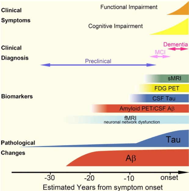
Fig. 1
Chronological relationship among pathology, clinical symptoms and biomarkers. Based on biomarker studies, Ab accumulation appears to start more than 20 years before the onset of dementia. Amyloid positron emission tomography (PET) or a decrease in CSF Aβ1-42 levels may indicate Ab accumulation in the brain, even in preclinical stage of AD. Neocortical tau pathology correlates with the timing of symptom onset and start approximately 10 years before the onset of dementia. However, these findings need to be reconciled with reports that tau pathology is observed in the prior to Ab pathology. FDG, 2-[18F]-fluoro-2-deoxy-D-glucose; MCI, mild cognitive impairment. Reproduced from [Therapeutic strategies for tau mediated neurodegneration, Yoshiyama Y, Lee VM and Trojanowski JQ, J Neruol Neurosurg Psychiatry 84:784-795, 2013] with permission from BMJ Publishing Group Ltd.
kjpp-18-447-g001
Fig. 2
Sequential process of amyloid precursor protein (APP) metabolism and production of amyloid beta species. Transmembrane APP is cleaved by α-secretase followed by γ-secretase to produce non-toxic amyloid species (non-amyloidogenic pathway, left blue thick arrow), while, through the amyloidogenic pathway (right red thick arrow), toxic Aβ species are generated by β-and γ-secretase. The toxic Aβ species including Aβ1-42 and Aβ1-40 are easily aggregated and produce highly toxic Aβ oligomers. Both processes produce soluble ectodomains [soluble APPα (sAPPα) and sAPPβ] and identical intracellular C-terminal fragment of APP (AICD). RAGE and LRP-1 located in bloodbrain barrier (BBB) are involved in the transport of the Ab between the brain and peripheral blood.
kjpp-18-447-g002
Fig. 3
Schematic presentation of tau mediated neurodegeneration. Phosphorylation and dephosphorylation of tau control the stability of microtubule. Hyperphosphorylation of tau induces disassembly of mitrotubules, causing axonal transport failure. Unbound tau produces oligomers or aggregates which congest axonal transport, and the tau pathology is synaptically transmitted. Reproduced from [Therapeutic strategies for tau mediated neurodegneration, Yoshiyama Y, Lee VM and Trojanowski JQ, J Neruol Neurosurg Psychiatry 84:784-795, 2013] with permission from BMJ Publishing Group Ltd.
kjpp-18-447-g003
TOOLS
Similar articles