Journal List > Korean J Physiol Pharmacol > v.18(5) > 1025973

Zainalabidin, Budin, Ramalingam, and Lim: Aortic Remodelling in Chronic Nicotine-Administered Rat

Abstract

Vascular remodelling is an adaptive mechanism, which counteracts pressure changes in blood circulation. Nicotine content in cigarette increases the risk of hypertension. The exact relationship between nicotine and vascular remodelling still remain unknown. Current study was aimed to determine the effect of clinically relevant dosage of nicotine (equivalent to light smoker) on aortic reactivity, oxidative stress markers and histomorphological changes. Twelve age-matched male Sprague-Dawley rats were randomly divided into two groups, i.e.: normal saline as control or 0.6 mg/kg nicotine for 28 days (i.p., n=6 per group). On day-29, the rats were sacrificed and the thoracic aorta was dissected immediately for further studies. Mean arterial pressure (MAP) and pulse pressure (PP) of nicotine-treated vs. control were significantly increased (p<0.05). Nicotine-treated group showed significant (p<0.05) increase tunica media thickness, and decrease in lumen diameter, suggesting vascular remodelling which lead to prior hypertension state. The phenylephrine (PE)-induced contractile response in nicotine group was significantly higher than control group (ED50=1.44×10⁵ M vs. 4.9×10⁶ M) (p<0.05~0.001). However, nicotine-treated rat showed significantly lower endothelium-dependent relaxation response to acetylcholine (ACh) than in control group (ED50=6.17×10⁷ M vs. 2.82×10⁷ M) (p<0.05), indicating loss of primary vascular function. Malondialdehyde (MDA), a lipid peroxidation marker was significantly higher in nicotine group. Superoxide dismutase (SOD) enzymatic activity and glutathione (GSH) were all reduced in nicotine group (p<0.05) vs. control, suggesting nicotine induces oxidative imbalance. In short, chronic nicotine administration impaired aortic reactivity, probably via redox imbalance and vascular remodelling mechanism.

ABBREVIATIONS

BW

body weight

BP

blood pressure

MSBP

mean systolic blood pressure

MAP

mean arterial pressure

PP

pulse pressure

PE

phenylephrine

ACh

acetylcholine

MDA

malondialdehyde

SOD

superoxide dismutase

GSH

glutathione

ROS

reactive oxygen species

IMT

intima-media thickness

IMA

intima-media area

CWT

circumferential wall tension

TS

tensile stress

INTRODUCTION

Exposure of nicotine, the addictive culprit component in cigarette via chronic smoking predispose individual towards vascular diseases. Nicotine induces oxidative stress, and therefore could be a reason for disease progression. It has been reported that nicotine disrupts the mitochondrial respiratory chain due to the increase in superoxide anions (O2-) and hydrogen peroxide (H2O2) production [1]. Subsequently, these reactive oxygen species (ROS) are able to induce oxidative stress in the organ through lipid peroxidation [2]. Aorta is one of the targets of nicotine-induced oxidative damage, shown by increase in malondialdehyde (MDA), the main degradation product of lipid peroxidation [3]. Nicotine-mediated oxidative damage also disturbs the capacities of endogenous antioxidant defence, in which the activity of antioxidant system was overwhelmed by ROS generation [4]. Previous in vivo studies have shown that chronic nicotine administration at 0.6 kg/mg which is equivalent to that of light smoker significantly elevated serum lipid peroxidation products in rat [5] and resulted in lowered endogenous glutathione (GSH) levels [6].
It is now increasingly clear that vessel may undergo continuous adaptive reconstruction (remodelling), known as angioadaptation in response to varying hemodynamic stimuli and functional demands [7,8]. It is believed that angioadaptation contribute to the pathophysiology of vascular diseases which is due to the failure of the arterial wall to maintain a suitable mesh size and allow normal blood flow. Pure nicotine products can increase heart rate acutely up to 10 to 15 beats/min, and increase blood pressure up to 5 to 10 mmHg, responses similar to the effects of cigarette smoking [9]. Nicotine can also indirectly act on the vessel by releasing catecholamine locally from adrenergic axon terminal which raise the blood pressure and hence induce hypertension [10,11]. This could lead to increased arterial wall thickness and alteration of arterial wall structural composition, i.e.: remodelling.
In hypertension condition, remodelling can be either eutrophic or hypertrophic remodelling. Eutrophic remodelling demonstrated decreased lumen diameter and increased media lumen ratio with the cross-sectional area of the tunica media remain unaltered [12]. In severe or longstanding hypertension, smooth muscle cell growth (in number or size) may predominate over apoptosis, and thus remodelling can be in the form of hypertrophic, which leads to the increase in media cross-sectional area and media-lumen ratio [13].
In spite of the abundant studies on nicotine's detrimental functional effect, there is still no clear evidence on its effect to the pathogenesis of the vascular function and structure, especially the aorta. Although in vivo studies had been intensely focused on the oxidant effects of nicotine on cardiovascular system, but the effect of chronic nicotine exposure on vessel remodelling and the relationship between oxidative stress and remodelling in vessel has not been investigated. Current work was aimed to demonstrate the effect of clinically-relevant dosage (equivalent to light smoker) of nicotine administration in mediating the process of aortic remodelling.

METHODS

Animals

A total of twelve age-matched (6 weeks) adult male Sprague Dawley rats were purchased from Synertec Scientific, Malaysia. All animals were housed under same laboratory conditions of ambient room temperature and lighting (12 hours light dark cycle) and were fed with standard laboratory pellet diet and water ad libitum. Animals were allowed to adapt to laboratory condition for 7 days prior to the experiment. All animal handling and experimental procedures were performed in accordance to the rules and regulations by the Universiti Kebangsaan Malaysia Animal Ethics Committee (UKMAEC). The animals were randomized into control and nicotine group. Nicotine group were treated intraperitoneally with nicotine at the dose of 0.6 mg/kg BW daily for 28 days. The dosage of nicotine used in current study mimics dosage exposure of a light smoker and was proven to induce oxidative stress in aorta of previous studies [3,14]. Control rats received injection of 0.9% normal saline. Body Weight (BW) and Blood Pressure (BP) Monitoring: BW and BP of each rat was determined on day-0 and day-28. Preparation of Aorta Tissue: On day-29, the rats were anesthetized with an intraperitoneal injection of heparin (1000 unit/kg BW, i.p) to prevent blood clot followed by sodium thiopental (50 mg/kg) [15]. After loss of all reflexes, the rats were sacrificed. Thoracic aorta was gently excised and placed in a petri dish filled with cold Krebs-Henseleit buffer. The aorta was dissected free of adhering fat and connective tissue and was carefully cleaned of debris and blood with care taken not to stretch the vessel excessively to ensure the integrity of endothelium layer. Aorta was immediately placed in organ bath for reactivity study. The rest of the tissue was stored in 10% formalin for histomorphological study and kept under deep freeze temperature (-80℃) for further biochemical investigation. In these tissue samples, superoxide dismutase (SOD) activity, MDA and GSH content were determined as marker of oxidative injury.

Blood pressure measurement in conscious rats

Blood pressure was measured in conscious restrained rats using non-invasive tail cuff method (CODA-2, Kent Scientific, Torrington, USA). The rats were trained for 7 days by measuring BP daily. Each session consisted of 5 acclimatization cycles followed by 15 BP measurements cycles. On the day of data collection, 2 sessions of 15 BP measurements were obtained. A set was accepted if the system identified >50% successful readings. The mean value from one session was used for mean arterial pressure (MAP) in each rat. Pulse pressure (PP) was derived as the difference between the systolic and diastolic pressure readings.

Aortic reactivity

The aorta was cut transversely into two to three rings of 4 mm in length based on method described before [16]. All tissue was mounted in 25 ml organ baths. Aortic rings were bathed in Krebs-Henseleit buffer (NaCl 118 mM, KCl 4.7 mM, CaCl2·2H2O 2.5 mM, MgSO4·7H2O 1.2 mM, KH2PO4 1.2 mM, NaHCO3 25.0 mM, and glucose 11.7 mM) of pH 7.4, continuously aerated with 95% O2 and 5% CO2, and maintained at 37℃ [17]. Aortic rings were mounted between stainless steel hooks. The hook anchoring the upper end of the ring was connected by a silk thread to the transducer. Aortic rings were allowed to equilibrate for 45 minutes with an initial resting tension of 1 g. Isometric contraction was recorded using FT03 force displacement transducer (Grass Instruments, Quincy, MA) coupled to Power-Lab/8sp multichannel data-acquisition system (ADInstrument, Australia) using ADI chart software (version 5.3) for digital processing and data analysis.
After equilibration, the ring was primed with 120 mM of KCl to check its functional integrity. Following washout of high K+ response, the contractile response of aortic rings to 10-9~10-4 M phenylephrine (PE) were determined cumulatively. After addition of each dose, a plateau response was observed before addition of subsequent dose. The contraction response was expressed as percentage of the maximal KCl-induced contraction. The tissues were then allowed for 30 minutes wash out period. The integrity of the vascular endothelium was accessed pharmacologically by ACh-induced relaxation of PE-precontracted ring. Cumulative dose response of acetylcholine (ACh) 10-9~10-4 M towards aortic ring relaxation response was evaluated and expressed as percentage of PE-induced pre-contraction.

Homogenate preparation and biochemical analysis

Aortic homogenate were prepared based on method described before [18]. The tissue was weighed and homogenized in 0.1 M pH 7.4 phosphate buffer solution (PBS). The homogenates were then centrifuged at 3000 rpm for 10 minutes at 4℃ and the supernatant were stored in at -80℃ until further biochemical analysis. Lipid peroxidation as evidenced by formation of MDA, one of the thiobarbituric acid reactive substances (TBARS), was measured by the method of [19] using 1,1,3,3-tetraethoxypropane (TEP) as the standard. Superoxide dismutase activity was estimated as described by [20]. In brief, SOD activities were measured using colorimetric assay by monitoring reduction of photochemical nitro blue tetrazolium (NBT). One unit of SOD was considered as amount of enzyme that causes 50% inhibition of NBT reduction. Glutathione measurements were performed based on [21]. In this colorimetric technique assay, assessment of GSH level was done through reaction of 5,5'-dithiobis-2-nitrobenzoic acid (DTNB) and GSH that produced a yellow compound was measured spectrophotometrically at 412 nm. The presence of protein in the homogenate was determined with [22].

Aortic histology

The formalin-fixed tissue specimens was sectioned into small pieces, dehydrated in ascending alcohol series and embedded in paraffin. Tissue was then sectioned to approximately 3~5 µm thickness. Aortic sections was fixed overnight on slides and subsequently stained with haematoxylin and eosin for observation of morphological changes.

Aortic morphometry measurement

Digital images of aortic sections were captured (JPEG format, 24-bit colour, 2560×1920 pixels) with a Micro-Publisher 5.0 RTV camera (Q Imaging, Surrey, BC, Canada) and a Nikon Eclipse 80i microscope (Nikon Corporation, Tokyo, Japan) and analysed with the software Image-Pro Plus version 7.0 (Media Cybernetics, Silver Spring, MD, USA). Morphometric measurements of aorta, which included intima-media thickness (IMT), intima-media area (IMA), lumen diameter, circumferential wall tension (CWT), and tensile stress (TS), were done according to previous protocol described by [23]. Four measurements of IMT per image were obtained at 0o, 90o, 180o and 270o by drawing a line across the tunica intima and media. The measurements acquired were averaged to get the value corresponding to the single image. Lumen area (α) was estimated by drawing a line over the circle at the boundary of the inner intima layer. Next, by using the values of α, the lumen diameter (d) was calculated as d=(2√α)/π, where α is expressed in mm2 and π is 3.14. The mean cross-sectional area of the tunica intima and tunica media (intima-media area, IMA) was calculated using the formula of IMA=[π(d/2+IMT)2]-[π(d/2)2]. CWT was calculated as CWT=MSBP×(d/2), where CWT was expressed in dyne/cm, MSBP (mean systolic blood pressure) as dynes/cm2, and d (lumen diameter) in cm. Tensile stress was calculated as TS=CWT/IMT. It was expressed in dyne/cm2 and IMT in cm.

Drugs and chemicals

Nicotine hydrogen bitartrate and heparin sodium salt was purchased from Merck Chemicals, USA. Sodium thiopental salt, phenylephrine hydrochloride and acetylcholine chloride was obtained from Sigma-Aldrich, St. Louis, USA.

Statistical analysis

Statistical analysis was carried out using Statistical Package for the Social Sciences (SPSS) version 21.0. All data were expressed as mean±standard error mean (SEM). Normality distribution of data were verified by Shapiro-Wilk normality test. Statistical analysis was performed using Independent t test. The significant levels of the data were determined at p<0.05. Pearson correlation test was performed to determine individual relationships between variables. A value of R>0 is considered as positive correlation.

RESULTS

Body weight and blood pressure

The changes in body weight (BW) and blood pressure (BP) of control and experimental rats are shown in Table 1. To determine the chronic effect of nicotine administration on systemic-vitals, BW and BP in conscious rat were recorded on day-0 and day-28. After 4 weeks of study, there was no significant difference in the percentage of body weight gain between groups (31.06±14.77 vs. 38.01±9.32 g; nicotine vs. control) (p>0.05). However, the BP indices showed significant difference where increase in MAP (49.13±1.38 vs. 19.62±3.25 mmHg) (p<0.001) and PP (40.5±4.02 vs. 5.23±2.57 mmHg) (p<0.001) of nicotine-treated rat was higher compared to control rats.

Aortic reactivity

In control rats, PE (10-9~10-4 M) showed concentration dependent contraction in the aortic rings precontracted with 120 mM KCl (ED50 of 4.9×10⁶ M). Surprisingly, rats treated with chronic low dose nicotine showed significantly higher contractile response compared to the control group (ED50=1.44×10⁵ M; p<0.05~0.001) (Fig. 1A).
Cumulative dose of ACh (10-9~10-4 M) with PE-precontracted aortic rings caused a dose-dependent relaxation response in control rats (ED50=2.82×10⁷ M). However, nicotine-treated rat showed significantly lower endothelium-dependent relaxation response of aortic rings to ACh than in control group with ED50 of 6.17×10⁷ M (p<0.05) (Fig. 1B), indicating loss of relaxation function in nicotine treated rats. The dosage causing 50% of the maximal response (ED50) of PE or ACh was derived from the concentration-response curves using a computer assisted probit transformation.

Oxidative stress marker and antioxidant levels

The degree of lipid peroxidation and aortic antioxidant status was presented in Fig. 2. The mean level of MDA, which is a lipid peroxidation marker, was found to be remarkably higher in aorta homogenate after chronic nicotine administration (6.12±0.19 nmol/mg protein) compared to control group (1.63±0.10 nmol/mg protein) (p<0.001). Meanwhile, SOD activity was significantly lower in nicotine group (1.61±0.13 U min-1/mg protein) compared to control (2.13±0.14 U min-1/mg protein) (p<0.05). Similarly, nicotine treated rats (0.025±0.001 mmol/mg protein) showed significantly reduced GSH level compared to control rats (0.030±0.001 mmol/mg protein) (p<0.05).

Histological analysis

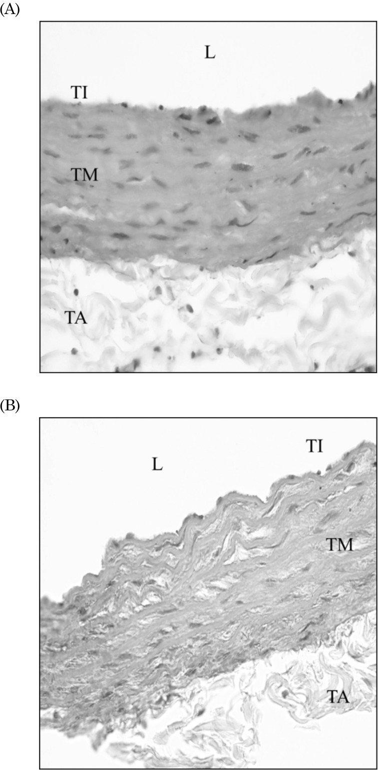
Fig. 3 shows the histology of aorta of control group (A) and nicotine group (B). Microscopic evaluation of aortic tissue in control group showed regular contour of all tunica layers with elastic lamella being well organized. Meanwhile, in the nicotine group, aortic architecture showed morphological degeneration in tunica media layer. Elastic lamella of nicotine group was observed to be disorganized, characterized by an increase in interlamellar space in the tunica media when compared to the control group.

Aortic morphometric study

The quantitative data of morphometric measurements is summarized in Table 2. Aortic sections from rats administered chronic nicotine (69.75±9.41 µm) show significant increased IMT compared to control (51.42±2.47 µm) (p<0.05). In addition, aortic IMA from nicotine groups (0.17±0.02 mm2) were also significantly greater than the control group (0.15±0.01 mm2) (p<0.05). Aortic lumen differ significantly by much narrower lumen in the nicotine group (0.73±0.05 mm) (p<0.05). Circumferential wall tension (CWT) was significantly increased nicotine-treated rats (0.69±0.06 104 dyne/cm) compared to control rats (0.61±0.05 104 dyne/cm) (p<0.05). No significant change was observed in TS between groups (p>0.05).

Relationship between oxidative stress, contraction & aortic remodelling

The co-relationship data is summarized in Table 3. The relationship of each variable was determined by Pearson's correlation test. In the nicotine treated rat, the oxidative stress marker, MDA was found significantly and positively related to the aortic contraction (R: 0.821; p<0.05) and wall thickness (R: 0.821; p<0.05). Besides that, nicotine treatment in rat showed highly significant correlation between contractile response and aortic remodelling (R: 0.976; p<0.001).

DISCUSSION

Alteration of aortic reactivity is clinically associated with several vascular disorders like hypertension and aneurysms. This transformation of aortic physiological response is believed to be due to vascular remodelling and redox imbalance. In our study, we evaluated the changes in aortic reactivity and the morphological changes in nicotine-treated rats and, subsequently whether these changes were caused by oxidative stress. We hypothesized that chronic administration of nicotine would generate harmful ROS and lead to changes in aortic hemodynamic, reactivity and morphological changes.
The rat's weight throughout the 28-days experiment was deteriorating in nicotine-treated group although it was not significant. Previous study has shown a much higher nicotine dose and longer period caused significant weight loss in rats given with nicotine at 2.5 mg/kg BW daily for 22 weeks [24]. The reduction in weight may be due to (i) augmented energy metabolism [25], (ii) activation of lipoprotein lipase [26] or (iii) alteration in expression of orexigenic and anorexigenic factor [27] by nicotine.
In our study, blood pressure in nicotine-treated rats showed a remarkable increase throughout the experiment. This result was consistent with earlier studies that have shown chronic exposure to cigarette smoking had elevated SBP, DBP and MAP compared to control groups of mice [28] and rat [29]. Pulse pressure indicates change in pressure from the diastolic level to the systolic level. Aortic compliance is responsible for the increase in pulse pressure. Flexible arteries (higher elasticity) that expand easily have high compliance while stiff arteries have low compliance. One of the main features of large artery remodelling is wall hypertrophy, due to changes in VSMC and extracellular matrix [30]. Extracellular matrix (ECM) consists of collagens, elastin, and proteoglycans. Collagen content reflects aortic stiffness while elastin contributes to the elasticity of a vessel. Previous in vitro work on cardiac fibroblast cell showed inhibitory effect of nicotine on collagen synthesis [31]. Nicotine also reported to be a powerful down-regulator of elastin production in skin fibroblast cell [32]. These findings suggest on future work of in vivo nicotine studies on extracellular matrix of main arteries.
Vascular reactivity has shown that the endothelium-dependent relaxation in nicotine-treated rats was reduced, whereas PE-induced contraction was enhanced in nicotine group, as compared to normal rats. The increment of aortic contraction could be due to the accumulation of oxidative stress markers. Chronic nicotine treatment in rats has caused a remarkable oxidative damage in the aorta as proven by increase in lipid peroxidation and concomitant decrease in SOD activity and also endogenous GSH level. Studies have demonstrated natural-occurring antioxidants such as taurine [14] and superoxide dismutase [33] played vasculo-protective role by reversing nicotine-induced arterial damages.
The present study demonstrated that aortas from nicotine-treated rats were more responsive to the contractile effect of α1-adrenoceptor agonist (PE) than the control. Previous studies have shown contradicting results of increased vasoconstriction [34] and also reduced vasoconstriction after chronic nicotine treatment [3,14]. Although the precise mechanism of altered aortic contractile response is not well understood, the thickening of aortic wall in our findings suggests aortic remodelling might be responsible in altering vascular response. Modification of agonist-mediated contractile response might be due to hypertrophy and hyperplasia of aortic smooth muscle cell (SMC), which is a form of angioadaptation. Previous in vitro studies on cultured SMC demonstrated potent mitogenic response of nicotine on SMC proliferation [35] and inhibitory effect on SMC apoptosis [36] which may in part contribute to the thickening of the aortic wall.
The vasorelaxant effect of ACh in vascular smooth muscle cell is dependent on the presence of intact functional endothelium. Both in vitro and in vivo studies on cheek pouch arterioles showed chronic nicotine selectively impaired endothelium-dependent arteriolar dilatation via mechanism of overproduction of oxygen-derived free radical generation [33,37]. Since it is known that aortic vasorelaxation is related to the production of NO from endothelium cell, endothelium dysfunction is typically the culprit for various vasculopathy events. An increased production and/or impaired scavenging action of ROS, i.e., oxidative stress, would perturb local production of NO [38]. Nitric oxide has been well-established as the most important endothelium-derived vasodilating substances which inhibit smooth muscle proliferation and migration [39]. The loss of endothelium integrity may promote hypertrophy of aortic media wall which composed mainly of smooth muscle cell. This decreased NO bioavailibility can subsequently contribute to vasoconstrictive remodelling. The impaired aortic endothelial function was concurrent with the elevated BP in nicotine-treated rats. The mechanisms involved in elevated BP in current study are, at least in part, due to inhibition of NO production with the loss of arterial elastic remodelling and compromised arterial compliance [40]. During systole, elasticity of the arterial wall facilitates healthy remodelling of cardiovascular system, reduces the left ventricular wall stress by alleviating the uprise of peak systolic pressure. Changes in aortic stiffness might account for the BP elevation as seen in our study.
The relationship between the amount of oxidative metabolism products and free radicals natural scavengers determines the outcome of tissue oxidative injury. We have observed a notably high level of lipid peroxidative indices, in agreement with previous study, in which the effect was due to nicotine-induced oxidative stress in the aortic tissue [14]. Lipid peroxidation lead to membrane permeability and fluidity changes, enhanced rates of degradation and eventually lead to cell lysis [41]. The increased concentration of lipid peroxidation product observed in nicotine-treated rats is also associated with disrupted SOD activity, a chain-breaking antioxidant. The depletion in SOD activity may be due to increased utilization to neutralize the high level of superoxide anions to a more stable hydrogen peroxide [42,43]. Endogenous antioxidant such as GSH is the frontier of intracellular antioxidant protective mechanism and function as reactive free radical scavenger. In line with past studies, our result also demonstrated that chronic nicotine administration significantly depleted GSH stores in the aorta [3,5], suggesting that GSH was used as an antioxidant for the detoxification of toxic oxygen metabolites, and therefore enhanced the susceptibility of the aortic tissues to oxidative injury. Nicotine results in redox imbalance through overproduction of lipid peroxidation product and depletion in antioxidant storage.
It is well established that artery remodelling is closely associated to the development and complication of hypertension. Morphometric study on aorta tissue showed increase of IMT and IMA, together with narrowed lumen diameter, suggesting the presence of hypertrophic inward remodelling. IMT thickening and vascular architectural changes are commonly associated with high blood pressure [12]. Cytotoxic ROS generation due to nicotine administration was found to be the cause for this vascular architectural change. Oxidative stress may contribute to endothelial dysfunction by decreasing the bioavailability of NO that normally reduces cellular proliferation. This may lead to an imbalance between the rate of growth and death, causing wall hypertrophy. On the other hand, nicotine-treated rat showed increase in CWT. Increased CWT could also be due to the prior elevated blood pressure. CWT is the force which acts in longitudinal and circumferential axis to oppose the distending effects of blood pressure. High blood pressure increases CWT, in which causing disturbance of vessel to perform its functional vascular tone [44]. Furthermore, TS, is the circumferential wall tension which acts perpendicularly to the wall divided by wall thickness and results from the extensional (dilating) effect of blood pressure on the vessel. Our result reported non-significant difference of TS among groups. This finding might be due to the thickening of IMT. The remodelling in the aorta was modelled by the Law of Laplace as T=pr/(2t), where T=wall tension, p=pressure, r=radius, t=wall thickness. Increased pressure requires increased thickness to maintain a stable wall stress. This explains the thickening of arteries to accommodate the high blood pressure. The changes in wall structure induced by haemodynamic alteration may initially be adaptive but can progress to maladaptive response, contributing to other cardiovascular complications.
In conclusion, our results suggest that chronic nicotine administration altered aortic haemodynamic and reactivity, possibly via redox imbalance and aortic remodelling. Current work is providing additional information on the use of in vivo nicotine model for studying vessel remodelling. In addition, the experimental model is clinically mimicking the dosage exposure of a light smoker. Intervention targeting nicotine toxicity on vessel should consider important of aortic remodelling as outcome.

ACKNOWLEDGEMENTS

This project was funded by a grant from the Universiti Kebangsaan Malaysia (GGPM-2011-095). Special thanks to the Department of Pharmacology and Department of Physiology, Faculty of Medicine, UKM, for their technical assistance.

ABBREVIATIONS

BW

body weight

BP

blood pressure

MSBP

mean systolic blood pressure

MAP

mean arterial pressure

PP

pulse pressure

PE

phenylephrine

ACh

acetylcholine

MDA

malondialdehyde

SOD

superoxide dismutase

GSH

glutathione

ROS

reactive oxygen species

IMT

intima-media thickness

IMA

intima-media area

CWT

circumferential wall tension

TS

tensile stress

References

1. Yildiz D, Ercal N, Armstrong DW. Nicotine enantiomers and oxidative stress. Toxicology. 1998; 130:155–165. PMID: 9865482.
crossref
2. Kovacic P, Cooksy A. Iminium metabolite mechanism for nicotine toxicity and addiction: Oxidative stress and electron transfer. Med Hypotheses. 2005; 64:104–111. PMID: 15533623.
crossref
3. Sener G, Kapucu C, Paskaloglu K, Ayanoglu-Dülger G, Arbak S, Ersoy Y, Alican I. Melatonin reverses urinary system and aorta damage in the rat due to chronic nicotine administration. J Pharm Pharmacol. 2004; 56:359–366. PMID: 15025861.
crossref
4. Halliwell B, Whiteman M. Measuring reactive species and oxidative damage in vivo and in cell culture: how should you do it and what do the results mean? Br J Pharmacol. 2004; 142:231–255. PMID: 15155533.
5. Sener G, Sehirli AO, Ipçi Y, Cetinel S, Cikler E, Gedik N. Chronic nicotine toxicity is prevented by aqueous garlic extract. Plant Foods Hum Nutr. 2005; 60:77–86. PMID: 16021835.
crossref
6. Toklu HZ, Şehirli Ö, Şahin H, Çetinel Ş, Yeğen BC, Şener G. Resveratrol supplementation protects against chronic nicotineinduced oxidative damage and organ dysfunction in the rat urogenital system. Marmara Pharm J. 2010; 14:29–40.
crossref
7. Price RJ, Less JR, Van Gieson EJ, Skalak TC. Hemodynamic stresses and structural remodeling of anastomosing arteriolar networks: design principles of collateral arterioles. Microcirculation. 2002; 9:111–124. PMID: 11932778.
crossref
8. Pries AR, Reglin B, Secomb TW. Remodeling of blood vessels: responses of diameter and wall thickness to hemodynamic and metabolic stimuli. Hypertension. 2005; 46:725–731. PMID: 16172421.
9. Benowitz NL, Gourlay SG. Cardiovascular toxicity of nicotine: implications for nicotine replacement therapy. J Am Coll Cardiol. 1997; 29:1422–1431. PMID: 9180099.
10. Benowitz NL. Safety of nicotine in smokers with hypertension. Am J Hypertens. 2001; 14:731–732. PMID: 11465662.
crossref
11. de Diego AM, Tapia L, Alvarez RM, Mosquera M, Cortés L, López I, Gutiérrez LM, Gandía L, García AG. A low nicotine concentration augments vesicle motion and exocytosis triggered by K+ depolarisation of chromaffin cells. Eur J Pharmacol. 2008; 598:81–86. PMID: 18831972.
12. Intengan HD, Schiffrin EL. Vascular remodeling in hypertension: roles of apoptosis, inflammation, and fibrosis. Hypertension. 2001; 38:581–587. PMID: 11566935.
13. Schiffrin EL. Remodeling of resistance arteries in essential hypertension and effects of antihypertensive treatment. Am J Hypertens. 2004; 17:1192–1200. PMID: 15607629.
crossref
14. Sener G, Ozer Sehirli A, Ipçi Y, Cetinel S, Cikler E, Gedik N, Alican I. Taurine treatment protects against chronic nicotine-induced oxidative changes. Fundam Clin Pharmacol. 2005; 19:155–164. PMID: 15810895.
crossref
15. Joukar S, Shahouzehi B, Najafipour H, Gholamhoseinian A, Joukar F. Ameliorative effect of black tea on nicotine induced cardiovascular pathogenesis in rat. EXCLI J. 2012; 11:309–317.
16. Baek I, Jeon SB, Song MJ, Yang E, Sohn UD, Kim IK. Flavone Attenuates vascular contractions by inhibiting RhoA/Rho kinase pathway. Korean J Physiol Pharmacol. 2009; 13:201–207. PMID: 19885038.
crossref
17. Muharis SP, Top AG, Murugan D, Mustafa MR. Palm oil tocotrienol fractions restore endothelium dependent relaxation in aortic rings of streptozotocin-induced diabetic and spontaneously hypertensive rats. Nutr Res. 2010; 30:209–216. PMID: 20417882.
crossref
18. Budin SB, Yusof K, Idris MH, Hamid ZA, Mohamed J. Tocotrienol-rich fraction of palm oil reduced pancreatic damage and oxidative stress in streptozotocin-induced diabetic rats. Aust J Basic Appl Sci. 2011; 5:2367–2374.
19. Stocks J, Dormandy TL. The autoxidation of human red cell lipids induced by hydrogen peroxide. Br J Haematol. 1971; 20:95–111. PMID: 5540044.
crossref
20. Beyer WF Jr, Fridovich I. Assaying for superoxide dismutase activity: some large consequences of minor changes in conditions. Anal Biochem. 1987; 161:559–566. PMID: 3034103.
crossref
21. Ellman GL. Tissue sulfhydryl groups. Arch Biochem Biophys. 1959; 82:70–77. PMID: 13650640.
crossref
22. Bradford MM. A rapid and sensitive method for the quantitation of microgram quantities of protein utilizing the principle of protein-dye binding. Anal Biochem. 1976; 72:248–254. PMID: 942051.
crossref
23. Fernandes-Santos C, de Souza Mendonça L, Mandarim-de-Lacerda CA. Favorable cardiac and aortic remodeling in olmesartan-treated spontaneously hypertensive rats. Heart Vessels. 2009; 24:219–227. PMID: 19466524.
crossref
24. Muthukumaran S, Sudheer AR, Menon VP, Nalini N. Protective effect of quercetin on nicotine-induced prooxidant and antioxidant imbalance and DNA damage in Wistar rats. Toxicology. 2008; 243:207–215. PMID: 18045763.
crossref
25. Frankish HM, Dryden S, Wang Q, Bing C, MacFarlane IA, Williams G. Nicotine administration reduces neuropeptide Y and neuropeptide Y mRNA concentrations in the rat hypothalamus: NPY may mediate nicotine's effects on energy balance. Brain Res. 1995; 694:139–146. PMID: 8974638.
crossref
26. Winders SE, Grunberg NE. Effects of nicotine on body weight, food consumption and body composition in male rats. Life Sci. 1990; 46:1523–1530. PMID: 2355798.
crossref
27. Jang MH, Shin MC, Kim KH, Cho SY, Bahn GH, Kim EH, Kim CJ. Nicotine administration decreases neuropeptide Y expression and increases leptin receptor expression in the hypothalamus of food-deprived rats. Brain Res. 2003; 964:311–315. PMID: 12576193.
crossref
28. Talukder MA, Johnson WM, Varadharaj S, Lian J, Kearns PN, El-Mahdy MA, Liu X, Zweier JL. Chronic cigarette smoking causes hypertension, increased oxidative stress impaired NO bioavailability endothelial dysfunction and cardiac remodeling in mice. Am J Physiol Heart Circ Physiol. 2011; 300:H388–H396. PMID: 21057039.
crossref
29. Guo X, Oldham MJ, Kleinman MT, Phalen RF, Kassab GS. Effect of cigarette smoking on nitric oxide, structural, and mechanical properties of mouse arteries. Am J Physiol Heart Circ Physiol. 2006; 291:H2354–H2361. PMID: 16815989.
crossref
30. Arribas SM, Hinek A, González MC. Elastic fibres and vascular structure in hypertension. Pharmacol Ther. 2006; 111:771–791. PMID: 16488477.
crossref
31. Lu D, Yokoyama U, Insel PA. Inhibitory effect of nicotine on collagen and alpha-smooth muscle actin expression in cardiac fibroblasts. FASEB J. 2008; 22:1130.20.
crossref
32. Kim JO, Ryoo YW, Lee KS. The effect of nicotine on elastin gene expression in cultured skin fibroblasts. Korean J Dermatol. 2001; 39:529–535.
33. Mayhan WG, Sharpe GM. Chronic exposure to nicotine alters endothelium-dependent arteriolar dilatation: effect of superoxide dismutase. J Appl Physiol (1985). 1999; 86:1126–1134. PMID: 10194193.
34. Xiao D, Huang X, Yang S, Zhang L. Direct effects of nicotine on contractility of the uterine artery in pregnancy. J Pharmacol Exp Ther. 2007; 322:180–185. PMID: 17403992.
crossref
35. Li JM, Cui TX, Shiuchi T, Liu HW, Min LJ, Okumura M, Jinno T, Wu L, Iwai M, Horiuchi M. Nicotine enhances angiotensin II-induced mitogenic response in vascular smooth muscle cells and fibroblasts. Arterioscler Thromb Vasc Biol. 2004; 24:80–84. PMID: 14592853.
crossref
36. Cucina A, Fuso A, Coluccia P, Cavallaro A. Nicotine inhibits apoptosis and stimulates proliferation in aortic smooth muscle cells through a functional nicotinic acetylcholine receptor. J Surg Res. 2008; 150:227–235. PMID: 18295799.
crossref
37. Mayhan WG, Sharpe GM. Superoxide dismutase restores endothelium-dependent arteriolar dilatation during acute infusion of nicotine. J Appl Physiol (1985). 1998; 85:1292–1298. PMID: 9760319.
38. Förstermann U. Nitric oxide and oxidative stress in vascular disease. Pflugers Arch. 2010; 459:923–939. PMID: 20306272.
crossref
39. Balakumar P, Koladiya RU, Ramasamy S, Rathinavel A, Singh M. Pharmacological interventions to prevent vascular endothelial dysfunction: future directions. J Health Sci. 2008; 54:1–16.
crossref
40. Kinlay S, Creager MA, Fukumoto M, Hikita H, Fang JC, Selwyn AP, Ganz P. Endothelium-derived nitric oxide regulates arterial elasticity in human arteries in vivo. Hypertension. 2001; 38:1049–1053. PMID: 11711496.
41. Reiter RJ, Tan DX, Manchester LC, Qi W. Biochemical reactivity of melatonin with reactive oxygen and nitrogen species: a review of the evidence. Cell Biochem Biophys. 2001; 34:237–256. PMID: 11898866.
crossref
42. Kalpana C, Menon VP. Modulatory effects of curcumin on lipid peroxidation and antioxidant status during nicotine-induced toxicity. Pol J Pharmacol. 2004; 56:581–586. PMID: 15591646.
43. Kalpana C, Menon VP. Inhibition of nicotine-induced toxicity by curcumin and curcumin analog: a comparative study. J Med Food. 2004; 7:467–471. PMID: 15671691.
crossref
44. Intengan HD, Thibault G, Li JS, Schiffrin EL. Resistance artery mechanics, structure, and extracellular components in spontaneously hypertensive rats: effects of angiotensin receptor antagonism and converting enzyme inhibition. Circulation. 1999; 100:2267–2275. PMID: 10578002.
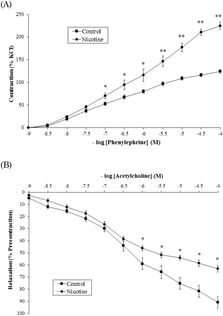
Fig. 1
Cumulative-response curves to phenylephrine and acetylcholine in thoracic aorta ring of control and nicotine groups. (A) PE-induced contractile response in nicotine administered rats showed to be significantly elevated compared to control (B) Nicotine significantly reduced ACh-induced relaxation. Values are means±SEM (n=6). *p<0.05 and **p<0.001 vs. control.
kjpp-18-411-g001
Fig. 2
Oxidative stress markers and antioxidant levels in the aortas of control vs. nicotine rats. (A) Nicotine group showed higher degree of lipid peroxidation. (B) SOD activity was lowered in nicotine-treated rats. (C) Nicotine significantly reduced the intracellular antioxidant, GSH level. Values are means±SEM (n=6). *p<0.05 and **p<0.001 vs. control.
kjpp-18-411-g002
Fig. 3
The histological features of H&E stained sections of aorta from control and nicotine groups. (A) Control group displays normal histology of aorta as indicated by regular arrangement of elastic lamella at tunica media layer. (B) Nicotine group showed disorganized tunica media layer with increase in interlamellar space. L, lumen; TI, tunica intima; TM, tunica media; TA, tunica adventitia. Same magnification applied to both pictures (Magnification: 400×).
kjpp-18-411-g003
Table 1
The body weight and blood pressure parameters
kjpp-18-411-i001

No significant changes in the level of body weight gain and significant increase in blood pressure indices. Values are means±SEM (n=6). **p<0.001 vs. control. MAP, mean arterial pressure; PP, pulse pressure.

Table 2
Aortic morphometric measurement
kjpp-18-411-i002

Nicotine treatment resulted in greater aortic wall thickness and tension but smaller lumen diameter. Values are means±SEM (n=6). *p<0.05 vs. control. IMT, intimal-media thickness; IMA, intimal-media area; CWT, circumferential wall tension; TS, tensile stress.

Table 3
Relationship between oxidative stress (MDA), aortic contraction (% KCl) and aortic remodelling (wall thickness)
kjpp-18-411-i003

Correlation coefficients whose magnitude between 0.8 and 1.0 indicate variables which can be considered positively and highly correlated. Oxidative stress was found significantly and positively related to the aortic contraction and wall thickness. Aortic contraction was positively and significantly correlated with aortic remodelling. Values are shown as Pearson Coefficient (R). *p<0.05 and **p<0.001 vs. different variables. MDA, malondialdehyde; MAP, mean arterial pressure.

TOOLS
Similar articles