Journal List > Korean J Physiol Pharmacol > v.18(3) > 1025941

Mangat, Jaggi, and Singh: Ameliorative Effect of a Selective Endothelin ETA Receptor Antagonist in Rat Model of L-Methionine-induced Vascular Dementia

Abstract

The present study was designed to investigate the efficacy of selective ETA receptor antagonist, ambrisentan on hyperhomocysteinemia-induced experimental vascular dementia. L-methionine was administered for 8 weeks to induce hyperhomocysteinemia and associated vascular dementia in male rats. Ambrisentan was administered to L-methionine-treated effect rats for 4 weeks (starting from 5th to 8th week of L-methionine treatment). On 52nd day onward, the animals were exposed to the Morris water maze (MWM) for testing their learning and memory abilities. Vascular endothelial function, serum nitrite/nitrate levels, brain thiobarbituric acid reactive species (TBARS), brain reduced glutathione (GSH) levels, and brain acetylcholinesterase (AChE) activity were also measured. L-methionine-treated animals showed significant learning and memory impairment, endothelial dysfunction, decrease in/serum nitrite/nitrate and brain GSH levels along with an increase in brain TBARS levels and AChE activity. Ambrisentan significantly improved hyperhomocysteinemia-induced impairment of learning, memory, endothelial dysfunction, and changes in various biochemical parameters. These effects were comparable to that of donepezil serving as positive control. It is concluded that ambrisentan, a selective ETA receptor antagonist may be considered as a potential pharmacological agent for the management of hyperhomocysteinemia-induced vascular dementia.

ABBREVIATIONS

AChE

acetylcholine esterase

AD

Alzheimer's disease

BSA

bovine serum albumin

DMSO

dimethyl sulfoxide

DTNB

5,5-dithiobis (2-nitrobenzoic acid)

ELT

escape latency time

GSH

reduced glutathione

p.o.

periorbital

MWM

Morris water maze

CMC

carboxymethylcellulose

NED

Naphthylethylenediamine

TBARS

thiobarbituric acid reactive substances

TSTQ

time spent in target quadrant

INTRODUCTION

Dementia is a syndrome due to disease of the brain, usually of a chronic or progressive nature, in which there is disturbance of multiple higher cortical and neuropsychological functions including memory, thinking, orientation, and comprehension, calculation, learning capacity, language and judgement [1]. Dementia of vascular origin (VaD) has gained much attention in the past years for being the second most common type of dementia after Alzheimer's disease (AD) [2,3]. It was estimated that 35.6 million people lived with dementia worldwide in 2010, with numbers expected to almost double every 20 years, to 65.7 million in 2030 and 115.4 million in 2050 [4]. VaD in turn has increased the risk of recurrent stroke, dependent living and death [5]. As the incidence rate of dementia increases rapidly with advancing age, a large increase in the number of patients is expected as a result of continuous aging of the population [6].
There is substantial evidence from observational studies that conventional risk factors such as hypertension [7], dyslipidemia [8], smoking [9], hyperhomocysteinemia [10] and diabetes [11] play a key role in the development of VaD and targeting these risk factors will minimize the burden. Our research group has recently reported that VaD can be induced in rats with the help of hyperhomocysteinemia, diabetes, experimental hypertension and hyperlipidemia [12,13].
Endothelin (ET) and nitric oxide (NO) are well known mediators produced by endothelial cells to maintain hemodynamic responses [14]. There are three main endothelial isoforms: ET-1, ET-2 and ET-3, of which ET-1 is the most potent vasoconstrictor agent. ET-1 binds to two receptors, endothelin A (ETA) and endothelin B (ETB) which are responsible for a variety of physiological functions, primarily blood flow control [15]. A key event in endothelial dysfunction is the reduction in bioavailability and biological activity of NO. Studies have demonstrated endothelial dysfunction in hyperhomocysteinemia leading to increased sensitivity to endothelin-1 and decreased relaxation in basilar artery [16]. Reduced levels of NO contribute to increased vascular tone, inflammation, platelet aggregation and oxidative stress which all are central features of atherosclerosis and hyperhomocysteinemia [17]. Endothelin receptor antagonists including ambrisentan are noted to exert their anti-inflammatory actions along with reduction in reactive oxygen species (ROS) generation which are subsequently responsible for endothelial dysfunction [18,19]. ET receptor antagonists have also been shown to provide a beneficial effect in various cerebrovascular disorders such as moyamoya disease [20], ischemic stroke [21] and subarachnoid hemorrhage [22]. Furthermore it has been recently reported that these antagonists have potential for the treatment of AD [23]. However, the potential of endothelin receptor antagonists in VaD is still unexplored.
The present study has been undertaken to investigate the efficacy of ambrisentan, a selective ETA endothelin receptor antagonist in a rat model of L-methionine-induced VaD.

METHODS

Animals

Adult male albino Wistar rats, weighing 200~250 g were employed in the present study and were housed in animal house with free access to water and standard chow (Kisan Feeds Ltd, Mumbai, India). The animals were exposed to 12 h light and 12 h dark cycle. The experiments were conducted between 9.00 and 18.00 h. The animals were acclimatized to laboratory conditions five days prior to behavioral study and were maintained in the laboratory until the completion of the study. The protocol of the study was duly approved by the Institutional Animal Ethics Committee (Approval No. 2012/14) and care of the animals was taken as per the guidelines of the Committee for the Purpose of Control and Supervision of Experiments on Animals (CPCSEA), Ministry of Environment and Forests, India (Reg No. 107/1999).

Drugs and reagents

All the drug solutions were freshly prepared before use. Donepezil was obtained as a gift sample from Wokhardt Ltd, Baddi, Himachal Pradesh, India. Ambrisentan was purchased from Atra Drugs Pvt Ltd, Hyderabad, India. Folin-Ciocalteu's Phenol and Acetylthiocholine were purchased from Merck Limited, Mumbai, India. L-methionine was purchased from SD fine chemicals limited, Mumbai, India. 5,5', dithiobis (2-nitrobenzoic acid) (DTNB), reduced glutathione (GSH), bovine serum albumin (BSA), sulfanilamide, N-naphthylethylenediamine (NED) and thiobarbituric acid were obtained from Loba Chem, Mumbai, India. Sodium nitroprusside was purchased from SD fine chemicals limited, Mumbai, India. Phenylephrine was obtained as a free sample from Aarti industries, Dombivli (East), Maharashtra, India. All the agents including ambrisentan, L-methionine and donepezil were administered orally. Ambrisentan and L-methionine were suspended in 0.5% w/v of CMC (Carboxymethylcellulose) whereas donepezil was dissolved in saline.

L-Methionine induced vascular dementia

L-methionine (1.7 g/kg/day, p.o.) was administered for 8 weeks to produce hyperhomocysteinemia induced vascular dementia in rats [12]. Body weight of rats was monitored weekly. Rats were subjected to Morris water maze test for the evaluation of their learning and memory status after a span of 8 weeks. L-methionine treatment was continued during acquisition trials on the Morris water maze. Analysis of serum homocysteine concentration was conducted on 1st day of the study before L-methionine treatment and on the 52nd day of the study before exposure on the Morris water maze. Behavioral and other assessments were done on the 52nd day of L-methionine treatment [3]. HPLC system was additionally used to measure levels of homocysteine.

Assessment of learning and memory by Morris water maze (MWM)

Memory was tested by practicing Morris water maze which is one of the most commonly used animal models to assess memory [24,25]. It is based on a principle where rats are placed in a large pool of water and their tendency to escape from the water is accomplished by finding an escape platform as animals dislike swimming. MWM consists of a large circular pool (150 cm in diameter, 45 cm in height) filled to a depth of 30 cm. The pool contained water maintained at a temperature of 22±2℃ and 1 kg of powdered skim milk to make the water opaque. Two threads, fixed at right angle to each other on the rim of the pool were used to divide the tank into four equal quadrants. A submerged white platform (15 cm in diameter) was fixed at 1.5 cm below the surface of the water in one of the four locations, approximately 50 cm from the side walls [26]. There was no alteration in the position of platform throughout the training session. Four consecutive trials were conducted daily on each rat with a gap of 5 min. The rat was placed centrally between the quadrants, facing the wall of the pool with the drop location changing for each trial, and was allowed a time period of 120 s to locate the submerged platform. Later, 20 s was accustomed to each rat while staying on the platform. If the rat failed to find the platform within 120 s, then it was guided gently onto the platform and allowed to remain there for 20 s. Escape latency time (ELT) was noted as an index of acquisition or learning to locate the hidden platform in the water maze was. The animal was subjected to acquisition trials for four consecutive days. Randomized daily starting positions were exercised and were not repeated in any of the quadrants. Quadrant (Q4) was maintained as a target quadrant in all of the acquisition trials.
kjpp-18-201-g005.jpg
The platform was removed and each rat was allowed to explore the pool for 120 s on the fifth day. Mean time spent in all four quadrants i.e. Q1, Q2, Q3 and Q4 were recorded. Index of retrieval was noted at the mean time spent with the animal in target quadrant i.e. Q4 in search of the hidden platform. Later, every rat was subjected to four such trials and each trial was started from a different quadrant. Same position was exercised by the experimenter during all trials while performing the MWM test. Prominent visual clues were not disturbed during the total duration of study as care was taken not to alter the relative location of the Morris water maze with respect to other objects in the laboratory. The proportion of time spent searching for the platform in the training quadrant, i.e. the previous location of the platform, was used as a measure of memory retention.

Biochemical assays

Blood samples for biochemical estimations were collected via retro-orbital bleeding just before sacrificing the animals. They were kept at room temperature for a period of 30 min followed by centrifugation at 4000 rpm for 15 min done to separate serum which was later used for estimation of homocysteine and nitrite/nitrate levels. The animals were sacrificed by cervical dislocation; thoracic aorta and brain tissue were carefully removed. Thoracic aorta was used to estimate endothelium dependent and independent relaxations, whereas brain was subjected to various biochemical estimations. Clear supernatant was first obtained after homogenizing the brain tissues in phosphate buffer (pH 7.4, 10% w/v) using teflon homogenizer followed by centrifugation at 3000 rpm for 15 min. This clear supernatant was then removed carefully from the centrifugation tube and it was later used for different biochemical estimations of TBARS, GSH, AChE, nitrite and proteins.
Determination of homocysteine was carried out using HPLC (Varian Inc., CA, USA) attached with fluorescent HPLC detector according to the method of Dimitrova et al. [27]. Serum nitrite concentration [28], whole brain AChE activity [29], TBARS levels [30], reduced glutathione (GSH) content [31] and total protein content [32] was assayed using standard methods by spectrophotometry (DU 640B spectrophotometer, Beckman Coulter Inc., CA, USA).

Assessment of vascular endothelial function using isolated rat aortic ring preparation

The rats were decapitated and descending thoracic aorta was removed. Thoracic aorta was cut into a ring of 3~5 mm width and was mounted in the tissue bath chamber containing Krebs-Henseleit solution (NaCl, 119 mM; KCl, 4.7 mM; NaHCO3, 25 mM; MgSO4, 1.0 mM; Glucose, 11.1 mM; KH2PO4, 1.2 mM and CaCl2, 2.5 mM) maintained at 37.8℃ and bubbled with carbogen (95% O2 and 5% CO2). The ring was held with the help of two opposite parallel L-shaped stainless steel loops in opposite directions. One loop was connected with force-displacement transducer (FT-2147) which was further coupled to physiograph (INCO, Ambala, India) whereas second loop was joined with a holder acting as an anchor submerged in bath chamber. The preparation was allowed to be stretched with 1.5 g tension followed by equilibration for 90 min with continuous washing with bath solution after every 10~15 min. The isometric contractile force was measured with the force-displacement transducer. The aortic ring preparation incubated with Krebs-Henseleit solution was primed with 80 mM KCl to check its functional integrity and to improve its contractility. The aortic ring preparation was stimulated with phenylephrine (3×10⁻⁶ M) until the contractile response reached a steady tension. Cumulative dose responses of acetylcholine (ACh; 10-8 to 10-4 M) and sodium nitroprusside (SNP; 10-8 to 10-4 M) were recorded with intact or denuded endothelium, respectively at 30 minute interval. The internal layer of aortic ring was rubbed gently with a moistened filter paper for 30 s to obtain an endothelial free preparation. The loss of ACh 10-6 M-induced relaxation confirmed the absence of endothelial layer.

Experimental protocol

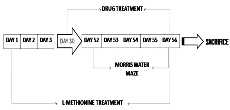
In total eight groups have been employed in the present study and each group consisted of six rats (Fig. 1).

1. Group I-Control group

Normal untreated rats were subjected to acquisition trial, conducted from day 1 to day 4 and retrieval trial, conducted on day 5 using MWM test.

2. Group II - Vehicle control group

Rats were administered 0.5% w/v CMC (10 ml/kg/day, p.o.) for 4 weeks and then subjected to MWM test. The treatment was continued during acquisition (from 24th to 27th day) and retrieval trials (on 28th day) on MWM.

3. Group III - L-Methionine treatment group

Rats were administered L-methionine (1.7 g/kg/day, p.o.) for 51 days and then subjected to MWM test. The treatment of L-methionine was continued during acquisition (from 52nd to 55th day) and retrieval trials (56th day) on the MWM.

4. Group IV - Ambrisentan

Rats were administered ambrisentan (10 mg/kg/day, p.o.) for 4 weeks followed by exposure to the MWM; the rest of the procedure was same as described in group II.

5. Group V - L-Methionine and Ambrisentan low dose

Ambrisentan (5 mg/kg/day, p.o.) was administered to L-methionine (1.7 g/kg/day, p.o.) treated rats, starting from the 30th day of L-methionine treatment followed by exposure to MWM on the 52nd day of L-methionine administration. The treatment was continued during acquisition (from 52nd to 55th day) and retrieval trials (on 56th day) on the MWM.

6. Group VI - L-Methionine and Ambrisentan high dose

Ambrisentan (10 mg/kg/day, p.o.) was administered to L-methionine (1.7 g/kg/day, p.o.) treated rats, starting from the 30th day of L-methionine treatment followed by exposure to MWM on the 52nd day of L-methionine administration. The treatment continued during acquisition (from 52nd to 55th day) and retrieval trials (on 56th day) on the MWM.

7. Group VII - Donepezil

Rats were administered donepezil (1 mg/kg/day, p.o.) for 4 weeks followed by exposure to MWM; the rest of the procedure was same as described in group II.

8. Group VIII - L-Methionine and Donepezil

Donepezil (1 mg/kg/day, p.o.) was administered to L-methionine (1.7 g/kg/day, p.o.) treated rats, starting from the 30th day of L-methionine treatment followed by exposure to MWM on the 52nd day of L-methionine administration. The treatment continued during acquisition (from 52nd to 55th day) and retrieval trials (on 56th day) on the MWM.

Statistical analysis

Statistical analyses were done using Sigma stat 3.5. All results were expressed as means±standard deviation. Data for isolated aortic ring preparation were statistically analyzed using repeated measures of analysis of variance (ANOVA) followed by Newman Keul's test. Data obtained from various groups was statistically analyzed using two-way ANOVA followed by Bonferroni Multiple Range test in case of ELT and TSTQ. The rest of the data obtained from various groups was statistically analyzed using oneway ANOVA followed by Tukey's test. p<0.05 was considered to be statistically significant.

RESULTS

Effect on escape latency time (ELT) and time spent in the target quadrant (TSTQ), using the Morris water maze (MWM)

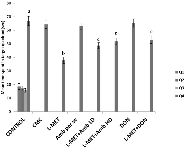
Control rats showed a downward trend in their ELT. There was a significant fall in day 4 ELT, when compared to day 1 ELT of these rats (Table 1). Further on day 5 a significant rise in TSTQ was observed, when compared to time spent in other quadrants (Fig. 2). Administration of 0.5% w/v CMC (10 ml/kg/day, p.o.) for 4 weeks did not show any significant effect on ELT and TSTQ. Administration of ambrisentan and donepezil did not show any significant effect on ELT and TSTQ (Table 1 and Fig. 2). Furthermore, L-methionine-treated rats showed a significant increase in day 4 ELT (55th day of L-methionine treatment), when compared to day 4 ELT of control animals (Table 1). Moreover, L-methionine administration produced a significant decrease in day 5 TSTQ (56th day of L-methionine treatment), when compared to day 5 TSTQ of control animals (Fig. 2), indicating impairment of memory as well.
Daily administration of low and high ambrisentan and donepezil prevented L-methionine induced rise in day 4 ELT significantly (p<0.0001, F (1, 40)=5210.38 for days, and p<0.0001, F (7, 40)=59.32 for treatment), indicating a reversal of L-methionine-induced impairment of acquisition (Table 1). Further treatment with these drugs also attenuated L-methionine-induced decrease in day 5 TSTQ (p<0.0001, F (1, 40)=545.63 for days, and p<0.0001, F (7, 40)=5.38 for treatment), indicating a reversal of L-methionine-induced impairment of memory (Fig. 2).

Effect on endothelium-dependent and independent relaxation

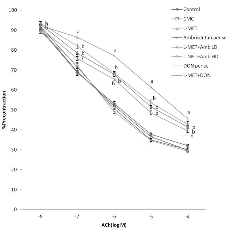
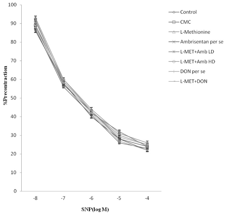
ACh and sodium nitroprusside (SNP) in a dose dependent manner produced endothelium-dependent and independent relaxation in phenylephrine precontracted aortic rings. L-methionine administration significantly attenuated ACh-induced endothelium-dependent relaxation (Fig 3), however it did not affect SNP-induced endothelium-independent relaxation (Fig. 4). Treatment of ambrisentan low and high dose and donepezil, significantly attenuated the effect of L-methionine on endothelial-dependent relaxation. However, ambrisentan and donepezil did not show any effect on endothelium-dependent relaxation.

Effect on homocysteine levels

Administration of L-methionine produced a significant increase in homocysteine levels when compared to control rats. Treatment with ambrisentan low and high dose and donepezil, significantly prevented (p<0.05, F (7, 40)=28.955) L-methionine-induced increase in homocysteine levels (Table 2). Further, ambrisentan or donepezil did not show any significant effect on homocysteine levels (Table 2).

Effect on serum nitrite, brain acetyl cholinesterase (AChE) activity and oxidative stress levels

Administration of L-methionine produced a significant decrease in serum nitrite & brain levels of the reduced form of glutathione (GSH) with a significant increase in AChE activity and brain TBARS, when compared to control rats. Treatment with ambrisentan low and high dose or donepezil, prevented L-methionine-induced decrease in serum nitrite (p<0.05, F (7, 40)=18.082), brain AChE activity (p<0.05, F (7, 40)=46.323), TBARS (p<0.05, F (7, 40)=32.648) and GSH (p<0.05, F (7, 40)=52.612) in a significant manner (Table 3). Further, ambrisentan low and high dose or donepezil, did not show any significant effect on any of the biochemical parameters (Table 3).

DISCUSSION

MWM employed in the present study is considered as one of the most effective testing paradigms to assess hippocampus-dependent spatial learning and memory of rodents. Control untreated animals in our study showed a marked reduction in day 4 ELT as compared to their day 1 ELT during acquisition trial, suggesting the normal acquisition or learning ability. Further, these animals have shown a significant increase in day 5 mean TSTQ when compared to time spent in other quadrants, indicating normal retrieval (memory) as well. These results are in compliance with the previous studies from our laboratory [12,33] as well as from other laboratories [34,35]. Vehicle (Carboxymethylcellulose) employed in the present study did not show any modification in the values of day 4 ELT and day 5 TSTQ as compared to control. Moreover, no effect was observed due to other drugs used in the study.
L-methionine treatment is a well documented and accepted model of hyperhomocysteinemia in rats [36]. Hyperhomocysteinemia, characterized by high plasma homocysteine levels, is recognized as an independent risk factor for endothelial dysfunction [37]. Hyperhomocysteinemia has been shown to induce endothelial dysfunction by decreasing the bioavailability of NO and by increasing vascular oxidative stress [17,38]. Moreover, endothelial dysfunction (vascular defects) has been reported to induce varying degree of memory impairment in animals and humans [39]. Furthermore, hyperhomocysteinemia is noted to produce a change in the structure and function of cerebral blood vessels as a consequence of cerebral vascular endothelial dysfunction [40]. The results of the present investigation also support the above contention, as a significant fall in endothelium-dependent relaxation and serum nitrite levels accompanied by an increase in oxidative stress (increased TBARS and decreased GSH) was noted in L-methionine treated rats.
Recent studies have indicated that experimental hyperhomocysteinemia resulting from chronic administration of L-methionine produces cognitive impairment [41,42]. This is further supported by earlier findings [12] and results of the present investigation whereby L-methionine treated rats have shown a significant decrease in MWM performance.
One of the likely mechanisms of the decreased NO bioavailability in hyperhomocysteinemia is increased concentration of Asymmetric dimethylarginine, an endogenous inhibitor of NO synthase by inhibiting the activity of dimethylarginine dimethylaminohydrolase, which in turn is followed by reducing the synthesis of NO [43]. High intracellular level of homocysteine in neurons has been found to enhance the vulnerability of hippocampal neurons and cortical neurons to excitotoxic insults [44]. It has been reported to produce neurotoxicity, DNA damage and apoptosis which are primarily responsible for various deformities that cause dementia [45]. Therefore, hyperhomocysteinemia induced as a consequence of chronic L-methionine treatment in our investigation has resulted in vascular dementia by virtue of its multiple effects.
In the present investigation treatment of ambrisentan attenuated the deleterious effect of L-methionine on learning and memory of rats. In addition it also improved L-methionine induced endothelial dysfunction and associated biochemical changes including reduction of serum nitrate levels, increased AChE activity and oxidative stress.
Endothelin (ET) being the most potent endothelium-derived contracting factor has been demonstrated to exist in three isoforms i.e. ET-1, ET-2 and ET-3. Out of the three isoforms, ET-1 is most widely studied and considered to be an important mediator of different physiological and pathophysiological functions [46]. The biological effects of ET-1 are transduced by two pharmacologically distinguishable receptor subtypes, ETA and ETB receptors [47]. In the vasculature, the ETA receptor is mainly located on vascular smooth muscle cells and mediates potent vasoconstriction, whereas ETB receptor is primarily located on endothelial cells where it results in the release of NO which causes vasodilatation [48]. Thus, the net effect produced by ET-1 is determined on the receptor localization and the balance between ETA and ETB receptors. Under physiological conditions, the net effect is vasoconstriction mediated by the ETA receptor, which is partly counteracted by the ETB receptor mediated release of NO.
During endothelial dysfunction, there is increased responsiveness to ET-1 along with increased expression of ETA receptors, which mediate a potent vasoconstrictor response in addition to decreased endothelial ETB receptor expression associated with functional decline of endothelium-dependent vasodilation [49,50]. Hyperhomocysteinemia has been demonstrated to induce endothelial dysfunction by activating ETA receptors and thereby shifting the balance towards enhanced contractility [51].
Vascular endothelial cells are accountable for generation of superoxide and hydrogen peroxide radicals by several cellular enzymes including eNOS [52]. ET-1 additionally is known to activate NADPH oxidases and thereby reiteratively increase superoxide production, resulting in oxidative stress via the ET receptor pathway and it also decreases NO bioavailability either by decreasing its production or by increasing its degradation (via formation of ROS) as noted in hyperhomocysteinemic rats [16]. Studies have also indicated a significant role of ET-1 and ETA receptors in particular in neuroinflammation and cytokine production [53]. On the other hand, endothelin receptor antagonists such as ambrisentan have been demonstrated to exert anti-inflammatory actions. Treatment with ambrisentan during induction of global cerebral ischemia additionally is reported to reduce the post ischemic inflammatory response by modulation of leukocyte-endothelium interactions besides imparting neuroprotection. Moreover, ambrisentan has also been documented to produce a reduction in the generation of ROS in the brain, which are subsequently responsible for the endothelial cell barrier dysfunction and increased cerebral vascular permeability. In consonance with these findings, ambrisentan in our study has shown a significant decrease in brain oxidative stress as indicated by an increase in brain TBARS and decrease in GSH levels. We further observed that ambrisentan elicited a decrease in L-methionine induced hyperhomocysteinemia and rise in brain AChE activity. The reason for this is not clear at the moment, perhaps this may be the first report of its kind and needs further investigation in this direction.
Therefore with support from literature and present data, it may be suggested that ambrisentan, a specific ETA endothelin receptor antagonist has shown an ameliorative effect in L-methionine-induced endothelial dysfunction and associated dementia by virtue of its multiple effects including ETA receptor blocking, anti-cholinesterase, antioxidative activity etc.
Acetylcholinesterase inhibitors are the main class of drugs frequently used for the management of memory deficits. Previous reports from our laboratory have shown beneficial effects of donepezil in dementia of AD [54] and that of vascular origin as well [55,56]. Donepezil is already in clinical use for memory deficits associated with AD and other neurodegenerative conditions. Therefore it has been used as a positive control in the present study.
From the results of the present study, it is concluded that hyperhomocysteinemia can induce endothelial dysfunction and subsequent VaD. Treatment with ambrisentan, a selective ETA receptor blocker has shown efficacy in a rat model of L-methionine-induced VaD. As this is the first report which suggests the beneficial effect of ambrisentan in hyperhomocysteinemia-induced VaD, further studies are required to explore the full potential of ET receptor antagonists in the management of VaD.

ACKNOWLEDGEMENTS

The authors are grateful to the Department of Pharmaceutical Sciences and Drug Research, Punjabi University, Patiala for providing funding and technical facilities for this work.

ABBREVIATIONS

AChE

acetylcholine esterase

AD

Alzheimer's disease

BSA

bovine serum albumin

DMSO

dimethyl sulfoxide

DTNB

5,5-dithiobis (2-nitrobenzoic acid)

ELT

escape latency time

GSH

reduced glutathione

p.o.

periorbital

MWM

Morris water maze

CMC

carboxymethylcellulose

NED

Naphthylethylenediamine

TBARS

thiobarbituric acid reactive substances

TSTQ

time spent in target quadrant

References

1. Pieper MJ, van Dalen-Kok AH, Francke AL, van der Steen JT, Scherder EJ, Husebø BS, Achterberg WP. Interventions targeting pain or behaviour in dementia: A systematic review. Ageing Res Rev. 2013; 12:1042–1055. PMID: 23727161.
crossref
2. Han HS, Jang JH, Jang JH, Choi JS, Kim YJ, Lee C, Lim SH, Lee HK, Lee J. Water extract of Triticum aestivum L. and its components demonstrate protective effect in a model of vascular dementia. J Med Food. 2010; 13:572–578. PMID: 20521983.
crossref
3. Sharma B, Singh N. Attenuation of vascular dementia by sodium butyrate in streptozotocin diabetic rats. Psychopharmacology (Berl). 2011; 215:677–687. PMID: 21225418.
crossref
4. Prince M, Bryce R, Albanese E, Wimo A, Ribeiro W, Ferri CP. The global prevalence of dementia: a systematic review and metaanalysis. Alzheimers Dement. 2013; 9:63–75. PMID: 23305823.
crossref
5. Catindig JA, Venketasubramanian N, Ikram MK, Chen C. Epidemiology of dementia in Asia: insights on prevalence, trends and novel risk factors. J Neurol Sci. 2012; 321:11–16. PMID: 22877510.
crossref
6. Bickel H, Ander KH, Brönner M, Etgen T, Gnahn H, Gotzler O, Poppert H, Pürner K, Sander D, Förstl H. Reduction of long-term care dependence after an 8-year primary care prevention program for stroke and dementia: the INVADE trial. J Am Heart Assoc. 2012; 1:e000786. PMID: 23130154.
crossref
7. Igoumenou A, Ebmeier KP. Diagnosing and managing vascular dementia. Practitioner. 2012; 256:13–16. 2PMID: 22720454.
8. Lüders S, Stöve S, Schrader J. Prevention of vascular dementia. Evidence and practice. Internist (Berl). 2012; 53:223–231. PMID: 22119909.
9. Peters R. Blood pressure, smoking and alcohol use, association with vascular dementia. Exp Gerontol. 2012; 47:865–872. PMID: 22664578.
crossref
10. Korczyn AD, Vakhapova V, Grinberg LT. Vascular dementia. J Neurol Sci. 2012; 322:2–10. PMID: 22575403.
crossref
11. Fei M, Yan Ping Z, Ru Juan M, Ning Ning L, Lin G. Risk factors for dementia with type 2 diabetes mellitus among elderly people in China. Age Ageing. 2013; 42:398–400. PMID: 23299282.
crossref
12. Koladiya RU, Jaggi AS, Singh N, Sharma BK. Ameliorative role of Atorvastatin and Pitavastatin in L-Methionine induced vascular dementia in rats. BMC Pharmacol. 2008; 8:14. PMID: 18691432.
crossref
13. Sharma B, Singh N. Salutary effect of NFκB inhibitor and folacin in hyperhomocysteinemia-hyperlipidemia induced vascular dementia. Prog Neuropsychopharmacol Biol Psychiatry. 2012; 38:207–215. PMID: 22510463.
crossref
14. Vaniotis G, Glazkova I, Merlen C, Smith C, Villeneuve LR, Chatenet D, Therien M, Fournier A, Tadevosyan A, Trieu P, Nattel S, Hébert TE, Allen BG. Regulation of cardiac nitric oxide signaling by nuclear β-adrenergic and endothelin receptors. J Mol Cell Cardiol. 2013; 62:58–68. PMID: 23684854.
crossref
15. Burnier M, Forni V. Endothelin receptor antagonists: a place in the management of essential hypertension? Nephrol Dial Transplant. 2012; 27:865–868. PMID: 22311858.
crossref
16. Zhen P, Zhao Q, Hou D, Liu T, Jiang D, Duan J, Lu L, Wang W. Genistein attenuates vascular endothelial impairment in ovariectomized hyperhomocysteinemic rats. J Biomed Biotechnol. 2012; 2012:730462. PMID: 23226943.
crossref
17. Norsidah KZ, Asmadi AY, Azizi A, Faizah O, Kamisah Y. Palm tocotrienol-rich fraction improves vascular proatherosclerotic changes in hyperhomocysteinemic rats. Evid Based Complement Alternat Med. 2013; 2013:976967. PMID: 23573162.
crossref
18. Knobloch J, Feldmann M, Wahl C, Jungck D, Behr J, Stoelben E, Koch A. Endothelin receptor antagonists attenuate the inflammatory response of human pulmonary vascular smooth muscle cells to bacterial endotoxin. J Pharmacol Exp Ther. 2013; 346:290–299. PMID: 23720457.
crossref
19. Lisk C, McCord J, Bose S, Sullivan T, Loomis Z, Nozik-Grayck E, Schroeder T, Hamilton K, Irwin DC. Nrf2 activation: a potential strategy for the prevention of acute mountain sickness. Free Radic Biol Med. 2013; 63:264–273. PMID: 23722164.
crossref
20. Day RW, Brockmeyer DL, Feola GP. Safe treatment of pulmonary hypertension with bosentan in a patient with moyamoya disease and cerebral ischemia. J Child Neurol. 2010; 25:504–507. PMID: 19808994.
crossref
21. Moldes O, Sobrino T, Blanco M, Agulla J, Barral D, Ramos-Cabrer P, Castillo J. Neuroprotection afforded by antagonists of endothelin-1 receptors in experimental stroke. Neuropharmacology. 2012; 63:1279–1285. PMID: 22975409.
crossref
22. Song JN, Yan WT, An JY, Hao GS, Guo XY, Zhang M, Li Y, Li DD, Sun P. Potential contribution of SOCC to cerebral vasospasm after experimental subarachnoid hemorrhage in rats. Brain Res. 2013; 1517:93–103. PMID: 23542055.
crossref
23. Palmer J, Love S. Endothelin receptor antagonists: potential in Alzheimer's disease. Pharmacol Res. 2011; 63:525–531. PMID: 21193044.
crossref
24. Lee B, Sur B, Shim I, Lee H, Hahm DH. Phellodendron amurense and its major alkaloid compound, berberine ameliorates scopolamine-induced neuronal impairment and memory dysfunction in rats. Korean J Physiol Pharmacol. 2012; 16:79–89. PMID: 22563252.
25. Kumar R, Jaggi AS, Singh N. Effects of erythropoietin on memory deficits and brain oxidative stress in the mouse models of dementia. Korean J Physiol Pharmacol. 2010; 14:345–352. PMID: 21165335.
crossref
26. Kim SJ, Lee JH, Chung HS, Song JH, Ha J, Bae H. Neuroprotective Effects of AMP-Activated protein kinase on scopolamine induced memory impairment. Korean J Physiol Pharmacol. 2013; 17:331–338. PMID: 23946693.
crossref
27. Dimitrova KR, DeGroot KW, Pacquing AM, Suyderhoud JP, Pirovic EA, Munro TJ, Wieneke JA, Myers AK, Kim YD. Estradiol prevents homocysteine-induced endothelial injury in male rats. Cardiovasc Res. 2002; 53:589–596. PMID: 11861029.
crossref
28. Sastry KV, Moudgal RP, Mohan J, Tyagi JS, Rao GS. Spectrophotometric determination of serum nitrite and nitrate by copper-cadmium alloy. Anal Biochem. 2002; 306:79–82. PMID: 12069417.
crossref
29. ELlman GL, Courtney KD, Andres V Jr, Feather-Stone RM. A new and rapid colorimetric determination of acetylcholinesterase activity. Biochem Pharmacol. 1961; 7:88–95. PMID: 13726518.
crossref
30. Ohkawa H, Ohishi N, Yagi K. Assay for lipid peroxides in animal tissues by thiobarbituric acid reaction. Anal Biochem. 1979; 95:351–358. PMID: 36810.
crossref
31. Beutler E, Duron O, Kelly BM. Improved method for the determination of blood glutathione. J Lab Clin Med. 1963; 61:882–888. PMID: 13967893.
32. Lowry OH, Rosebrough NJ, Farr AL, Randall RJ. Protein measurement with the Folin phenol reagent. J Biol Chem. 1951; 193:265–275. PMID: 14907713.
crossref
33. Gulati P, Muthuraman A, Jaggi AS, Singh N. Neuroprotective effect of gadolinium: a stretch-activated calcium channel blocker in mouse model of ischemia-reperfusion injury. Naunyn Schmiedebergs Arch Pharmacol. 2013; 386:255–264. PMID: 23229582.
crossref
34. Dong Z, Bai Y, Wu X, Li H, Gong B, Howland JG, Huang Y, He W, Li T, Wang YT. Hippocampal long-term depression mediates spatial reversal learning in the Morris water maze. Neuropharmacology. 2013; 64:65–73. PMID: 22732443.
crossref
35. Hashemi Nosrat Abadi T, Vaghef L, Babri S, Mahmood-Alilo M, Beirami M. Effects of different exercise protocols on ethanol-induced spatial memory impairment in adult male rats. Alcohol. 2013; 47:309–316. PMID: 23683528.
crossref
36. Sudduth TL, Powell DK, Smith CD, Greenstein A, Wilcock DM. Induction of hyperhomocysteinemia models vascular dementia by induction of cerebral microhemorrhages and neuroinflammation. J Cereb Blood Flow Metab. 2013; 33:708–715. PMID: 23361394.
crossref
37. Tlili A, Jacobs F, de Koning L, Mohamed S, Bui LC, Dairou J, Belin N, Ducros V, Dubois T, Paul JL, Delabar JM, De Geest B, Janel N. Hepatocyte-specific Dyrk1a gene transfer rescues plasma apolipoprotein A-I levels and aortic Akt/GSK3 pathways in hyperhomocysteinemic mice. Biochim Biophys Acta. 2013; 1832:718–728. PMID: 23429073.
crossref
38. Wang X, Cui L, Joseph J, Jiang B, Pimental D, Handy DE, Liao R, Loscalzo J. Homocysteine induces cardiomyocyte dysfunction and apoptosis through p38 MAPK-mediated increase in oxidant stress. J Mol Cell Cardiol. 2012; 52:753–760. PMID: 22227328.
crossref
39. Asif M, Soiza RL, McEvoy M, Mangoni AA. Asymmetric dimethylarginine: a possible link between vascular disease and dementia. Curr Alzheimer Res. 2013; 10:347–356. PMID: 23036019.
crossref
40. Rodionov RN, Dayoub H, Lynch CM, Wilson KM, Stevens JW, Murry DJ, Kimoto M, Arning E, Bottiglieri T, Cooke JP, Baumbach GL, Faraci FM, Lentz SR. Overexpression of dimethylarginine dimethylaminohydrolase protects against cerebral vascular effects of hyperhomocysteinemia. Circ Res. 2010; 106:551–558. PMID: 20019334.
crossref
41. Rhodehouse BC, Erickson MA, Banks WA, Bearden SE. Hyperhomocysteinemic mice show cognitive impairment without features of Alzheimer's disease phenotype. J Alzheimers Dis. 2013; 35:59–66. PMID: 23334704.
crossref
42. Scherer EB, Loureiro SO, Vuaden FC, Schmitz F, Kolling J, Siebert C, Savio LE, Schweinberger BM, Bogo MR, Bonan CD, Wyse AT. Mild hyperhomocysteinemia reduces the activity and immunocontent, but does not alter the gene expression, of catalytic α subunits of cerebral Na+,K+-ATPase. Mol Cell Biochem. 2013; 378:91–97. PMID: 23467881.
43. Emeksiz HC, Serdaroglu A, Biberoglu G, Gulbahar O, Arhan E, Cansu A, Arga M, Hasanoglu A. Assessment of atherosclerosis risk due to the homocysteine-asymmetric dimethylarginine-nitric oxide cascade in children taking antiepileptic drugs. Seizure. 2013; 22:124–127. PMID: 23266348.
crossref
44. Schaub C, Uebachs M, Beck H, Linnebank M. Chronic homocysteine exposure causes changes in the intrinsic electrophysiological properties of cultured hippocampal neurons. Exp Brain Res. 2013; 225:527–534. PMID: 23307157.
crossref
45. Ho YS, Yu MS, Yang XF, So KF, Yuen WH, Chang RC. Neuroprotective effects of polysaccharides from wolfberry, the fruits of Lycium barbarum, against homocysteine-induced toxicity in rat cortical neurons. J Alzheimers Dis. 2010; 19:813–827. PMID: 20157238.
crossref
46. Potts LB, Ren Y, Lu G, Kuo E, Ngo E, Kuo L, Hein TW. Constriction of retinal arterioles to endothelin-1: requisite role of rho kinase independent of protein kinase C and L-type calcium channels. Invest Ophthalmol Vis Sci. 2012; 53:2904–2912. PMID: 22427601.
crossref
47. Bae EH, Kim SW. Changes in endothelin receptor type B and neuronal nitric oxide synthase in puromycin aminonucleoside-induced nephrotic syndrome. Korean J Physiol Pharmacol. 2010; 14:223–228. PMID: 20827336.
crossref
48. Ohkita M, Tawa M, Kitada K, Matsumura Y. Pathophysiological roles of endothelin receptors in cardiovascular diseases. J Pharmacol Sci. 2012; 119:302–313. PMID: 22863667.
crossref
49. Guo QH, Tian YL, Wang Z, Li AY, Ma ZH, Guo YJ, Weiss JW, Ji ES, Chu L. Endothelin receptors in augmented vasoconstrictor responses to endothelin-1 in chronic intermittent hypoxia. Clin Exp Pharmacol Physiol. 2013; 40:449–457. PMID: 23662699.
crossref
50. Wang Z, Li AY, Guo QH, Zhang JP, An Q, Guo YJ, Chu L, Weiss JW, Ji ES. Effects of cyclic intermittent hypoxia on ET-1 responsiveness and endothelial dysfunction of pulmonary arteries in rats. PLoS One. 2013; 8:e58078. PMID: 23555567.
crossref
51. de Andrade CR, Leite PF, Montezano AC, Casolari DA, Yogi A, Tostes RC, Haddad R, Eberlin MN, Laurindo FR, de Souza HP, Correa FM, de Oliveira AM. Increased endothelin-1 reactivity and endothelial dysfunction in carotid arteries from rats with hyperhomocysteinemia. Br J Pharmacol. 2009; 157:568–580. PMID: 19371338.
crossref
52. Choi S, Na HY, Kim JA, Cho SE, Suh SH. Contradictory effects of superoxide and hydrogen peroxide on KCa3.1 in human endothelial cells. Korean J Physiol Pharmacol. 2013; 17:181–187. PMID: 23776393.
crossref
53. Lin CC, Hsieh HL, Shih RH, Chi PL, Cheng SE, Yang CM. Up-regulation of COX-2/PGE2 by endothelin-1 via MAPK-d ependent NF-κB pathway in mouse brain microvascular endothelial cells. Cell Commun Signal. 2013; 11:8. PMID: 23343326.
crossref
54. Singh B, Sharma B, Jaggi AS, Singh N. Attenuating effect of lisinopril and telmisartan in intracerebroventricular streptozotocin induced experimental dementia of Alzheimer's disease type: possible involvement of PPAR-γ agonistic property. J Renin Angiotensin Aldosterone Syst. 2013; 14:124–136. PMID: 23060470.
crossref
55. Sharma B, Singh N. Defensive effect of natrium diethyldithiocarbamate trihydrate (NDDCT) and lisinopril in DOCA-salt hypertension-induced vascular dementia in rats. Psychopharmacology (Berl). 2012; 223:307–317. PMID: 22526544.
crossref
56. Sharma B, Singh N. Pharmacological inhibition of inducible nitric oxide synthase (iNOS) and nicotinamide adenine dinucleotide phosphate (NADPH) oxidase, convalesce behavior and biochemistry of hypertension induced vascular dementia in rats. Pharmacol Biochem Behav. 2013; 103:821–830. PMID: 23201648.
crossref
Fig. 1
Schematic presentation of experimental protocol.
kjpp-18-201-g001
Fig. 2
Effect of pharmacological interventions on mean time spent in the target quadrant (TSTQ) using Morris water-maze test. L-MET, L-Methionine; Amb LD, Ambrisentan low dose; Amb HD, Ambrisentan high dose; DON, Donepezil; CMC, Carboxymethylcellulose. Each group (n=6) represents mean±standard deviation. Two way ANOVA followed by Bonferroni post hoc test. ap<0.05 versus mean time spent in other quadrants in control; bp<0.05 versus mean time spent in the target quadrant in control group; cp<0.05 versus mean time spent in the target quadrant in L-Methionine treated group.
kjpp-18-201-g002
Fig. 3
Effect of pharmacological interventions on Ach-induced endothelium-dependent relaxation using an aortic ring preparation. L-MET, L-Methionine; Amb LD, Ambrisentan low dose; Amb HD, Ambrisentan high dose; DON, Donepezil; CMC, Carboxymethylcellulose. Each group (n=6) represents mean±standard deviation. Responses are expressed as percentage of precontraction induced by 3×10⁻⁶ M phenylephrine. Repeated measure ANOVA followed by Newman Keul's test. ap<0.05 versus control; bp<0.05 versus L-Methionine treated group.
kjpp-18-201-g003
Fig. 4
Effect of pharmacological interventions on endothelium independent relaxation using an aortic ring preparation. L-MET, L-Methionine; Amb LD, Ambrisentan low dose; Amb HD, Ambrisentan high dose; DON, Donepezil; CMC, Carboxymethylcellulose. Each group (n=6) represents mean±standard deviation. Responses are expressed as percentage of precontraction induced by 3×10⁻⁶ M phenylephrine. Repeated measure ANOVA followed by Newman Keul's test.
kjpp-18-201-g004
Table 1
Effect of pharmacological interventions on escape latency time (ELT) using Morris water maze
kjpp-18-201-i001

Each group (n=6) represents mean±standard deviation. Two way ANOVA followed by Bonferroni post hoc test. F (1, 40)=5210.38 for days, p<0.0001 and F (7, 40)=59.32 for treatment, p<0.0001. ap<0.01 versus Day 1 ELT in the respective group. bp<0.05 versus Day 4 ELT in the control group. cp<0.05 versus Day 4 ELT in L-Methionine treated group.

Table 2
Effect of various pharmacological interventions on serum homocysteine level of animals
kjpp-18-201-i002

Each group (n=6) represented by mean±SD.

One way ANOVA followed by Tukey's multiple range test. F (7, 40)=28.955. ap<0.05 versus basal values in L-Methionine group. bp<0.05 versus final values in L-Methionine group.

Table 3
Effect of various agents on serum nitrite/nitrate levels; brain acetylcholinesterase cholinesterase (AChE) activity and oxidative stress (TBARS, GSH levels) of animals
kjpp-18-201-i003

Each group (n=6) represents mean±standard deviation.

TBARS - Thiobarbituric acid reactive species; GSH - Reduced form of glutathione; AChE - Acetylcholinesterase; CMC - Carboxymethylcellulose.

One way ANOVA followed by Tukey's test. Serum nitrite/nitrate - F (7, 40)=18.082; ap<0.05 versus control group; bp<0.05 versus L-Methionine treated group. Brain TBARS - F (7, 40)=32.648; ap<0.05 versus control group; bp<0.05 versus L-Methionine treated group. Brain AChE activity - F (7, 40)=46.323; ap<0.05 versus control group; bp<0.05 versus L-Methionine treated group. Brain GSH activity - F (7, 40)=52.612; ap<0.05 versus control group; bp<0.05 versus L-Methionine treated group.

TOOLS
Similar articles