Journal List > Korean J Physiol Pharmacol > v.17(5) > 1025892

Lee: Cyclophilin A as a New Therapeutic Target for Hepatitis C Virus-induced Hepatocellular Carcinoma

Abstract

Hepatocellular carcinoma (HCC) related to hepatitis B virus (HBV) and hepatitis C virus (HCV) infections is thought to account for more than 80% of primary liver cancers. Both HBV and HCV can establish chronic liver inflammatory infections, altering hepatocyte and liver physiology with potential liver disease progression and HCC development. Cyclophilin A (CypA) has been identified as an essential host factor for the HCV replication by physically interacting with the HCV non structural protein NS5A that in turn interacts with RNA-dependent RNA polymerase NS5B. CypA, a cytosolic binding protein of the immunosuppressive drug cyclosporine A, is overexpressed in many cancer types and often associated with malignant transformation. Therefore, CypA can be a good target for molecular cancer therapy. Because of antiviral activity, the CypA inhibitors have been tested for the treatment of chronic hepatitis C. Nonimmunosuppressive Cyp inhibitors such as NIM811, SCY-635, and Alisporivir have attracted more interests for appropriating CypA for antiviral chemotherapeutic target on HCV infection. This review describes CypA inhibitors as a potential HCC treatment tool that is contrived by their obstructing chronic HCV infection and summarizes roles of CypA in cancer development.

ABBREVIATIONS

HCC

hepatocellular carcinoma

HBV

hepatitis B virus

HCV

hepatitis C virus

CypA

cyclophilin A

Cyps

cyclophilins

CsA

cyclosporine A

PPIase

peptidyl prolyl isomerase

ER

endoplasmic reticulum

MPTP

mitochondrial permeability transition pore

HIV-1

human immunodeficiency virus-1

IFN

interferon

IL

interleukin

INTRODUCTION

Cyclophilins (Cyps) constitute a subgroup of large family proteins called immunophilins. Cyps are well conserved and are found throughout evolution from E. coli to humans. Cyclosporine A (CsA) is a reversible inhibitor for all Cyps and works at nanomolar concentrations [1,2]. Cyps possess a peptidyl prolyl isomerase (PPIase) that catalyzes the conversion between cis-trans isomers of proline on the peptide. PPIases are regarded as a chaperone [3] and work with other chaperone proteins such as the heat shock protein and protein disulfide isomerases [4,5]. PPIases not only function in folding of de novo synthesized proteins but also accelerate slow steps in the refolding of denatured proteins [6]. There are 18 human Cyp family proteins which include Cyp isoforms such as CypA, CypB, CypC, CypD, CypE, Cyp40, Cyp20 and CypNK. Although complete isoform characterization is required to define precise functions for the individual Cyp proteins, multiple Cyps display separate functions by localizing through unique domains for particular cellular compartments such as CypA in the cytosol and nucleus, CypB in the endoplasmic reticulum (ER), CypD in mitochondria. The substrates or specific roles of individual Cyps in their subcellular locales or during critical cellular processes are still largely unknown. Chaperone activity of Cyps has been demonstrated first time when inhibition of the PPIase significantly delays maturation of collagen [7,8]. Cyps are also involved in repairing stress-induced damaged proteins like other molecular chaperon proteins. Environmental stresses can induce some Cyp expressions to carry out the repair function [9,10]. Recently, human Cyps emerge to be recognized as an important protein by participating in important physiological tasks like muscle differentiation [11] and immune response [12].
CypA forms a ternary complex with CsA, which inhibits calcineurin phosphatase activity and exerts an immunosuppressive action. CypB interacts with Stat3 and influences its transcriptional activity [13]. The prevention of mitochondrial permeability transition pore (MPTP) opening and anti-apoptotic effect by CsA is from its CypD inhibition. CypD is found in mitochondria and plays a role in apoptosis, being a member of the MPTP complex with ANT, VDAC, and hexokinase [14,15]. Cyp20 is important for folding of mitochondrial proteins as well as for transporting newly synthesized proteins between intracellular compartments [16,17]. In addition to the intracellular functions, extracellular Cyps such as CypA and CypB may contribute to intercellular communication inducing signaling responses in target cells [18]. CypB as well as CypA can be secreted from cells and those secretory Cyps can be a chemoattractant to immune cells [19,20]. It is reported that the secreted forms of CypA and CypB may function as a ligand and activate signal transduction pathways in various cell types [21]. CD147, an Ig-like membrane receptor, is involved in the signaling and has been proposed as a receptor for both CypA and CypB [22,23]. This binding of CypA to CD147 can become a pathological factor for rheumatoid arthritis [24]. Furthermore, CypA can be secreted in response to oxidative stress, which triggers the pathogenesis of vascular diseases [25].
CypA, found in almost all tissues, is the most abundant among the Cyps. The cytosolic CypA concentration may account for as much as 0.1~0.4% of the total protein in a cell [26]. CypA as a multifunctional chaperone has been demonstrated to function in intracellular trafficking [27], signal transduction and transcription regulation [28]. CypA's function in pathophysiological conditions has currently attracted attentions of researchers in the field of oncogenic development, cardiovascular diseases as well as viral infections [29,30]. Overexpression of CypA often correlates with severity of cancers including pancreatic and hepatic cancers, suggesting its role in the tumor malignancy. CypA can help infections of viruses including the clinically important ones such as human immunodeficiency virus-1 (HIV-1) [31], HBV and HCV [32,33].
The role of CypA in helping viral infection has primarily been studied in the case of HIV infection. CypA helps HIV-1 assembly by binding to HIV-1 capsid proteins [34]. Cyps, especially CypA, has been studied extensively and found to play essential roles during HCV infection. Recent studies have demonstrated that CypA is the one to interact with HCV nonstructural protein NS5A and essential for the viral genome replication although interaction of CypB and RNA polymerase, NS5B, was proposed first [35]. Likewise, the three nonimmunosuppressive Cyp inhibitors (NIM811, SCY-635, and Alisporivir) that are now under clinical trials on HCV infections are believed to be effective through CypA inhibition [36]. It is found that infections with hepatotropic viruses are linked to the majority of cases of HCC and in fact, chronic infections with HBV and HCV account for up to 80% of HCCs [37,38]. HCC development follows the sequential steps of chronic inflammation, liver regeneration, fibrosis, and cirrhosis [39,40]. While the molecular mechanisms of hepatocyte transformation are not fully understood, it is believed to involve alterations of multiple cellular signal transduction pathways and changes in micro (mi)RNAs and cytokines. Inflammatory cytokines appear to be an important component of the immune system and play an important role in promoting HCC development. In this review, the role of CypA in HCV infection and subsequent HCC development will be discussed.

ROLE OF HCV IN THE DEVELOPMENT OF HCC

HCC is one of the most common cancers and the third leading cause of cancer-associated death [41]. It takes 30~40 years for HCV-infected patients to develop HCC. Recent investigations of HCV reveal several cellular signal transduction pathways that are altered by HCV infections [39]. HCV possesses four structural proteins (core, E1, E2, and p7) and six nonstructural (NS2, NS3, NS4A, NS4B, NS5A, and NS5B) viral proteins [42]. The HCV non-structural proteins play important roles in virus-host interactions and thus may help avoid the immune response. HCV translation occurs on the rough ER and host signal peptidases play important roles in processing the structural proteins. Core composes the viral nucleocapsid by interactions with the HCV RNA. The E1 and E2 are envelope glycoproteins which form heterodimers in the ER. E1 and E2 together may mediate entry of the viral particles into cells. Viroporin is the name for p7, a small transmembrane protein with ion channel activity. However, the exact role of p7 in the HCV life cycle is poorly understood. NS2 is a cysteine protease while NS3 is a complex serine protease with helicase and adenosine triphosphatase activities. Importantly, the nonstructural proteins are also required for RNA replication and these NS2 and NS3 viral proteases cleave HCV polyprotein, generating NS4A, NS4B, NS5A, and NS5B. NS3 with NS4A, an NS3 cofactor, in necessity form an NS3/NS4A complex and embedded in the ER and the mitochondrial outer membrane. The NS3A/4A complex is crucial in viral resistance to the innate immune response by inactivation of the interferon (IFN) regulatory factor 3 and cleavage of the Toll-like receptor 3 adaptor protein, TRIF. NS4B is also required for HCV replication and NS5A is involved in HCV replication and virion assembly. NS5B is an RNA-dependent RNA polymerase. HCV core, NS3 and NS5A proteins are known to interrupt p53 actions and some HCV structural proteins may have a direct oncogenic role [43].
In addition to the viral proteins, modulation in the host factors such as miRNA and cytokines also play important roles in viral pathogenesis. Connection of miRNA functions to HCV-related hepatocarcinogenesis draws an attention because modulation of miRNA profiles can be associated with hepatocarcinogenesis [44]. miRNAs result in gene silencing via translational repression or mRNA degradation [45,46]. Many miRNAs that are differentially expressed in HCC appear to play roles in both viral replication and HCC development [47]. miRNA-122 is known to enhance HCV RNA translation [48-51]. miRNA-122 binds to the 5' UTR of HCV and is required for HCV replication in hepatocytes [52]. Besides, downregulation of miRNA-145 and miRNA-199b as well as upregulation of miRNA-224 are detected during HCC development [53]. Downregulation of miRNA-26 is also detected in patients with HCC [54]. Since expression profiles of miRNAs are modulated by HCV and are found to be altered in human HCC, miRNAs may become a diagnostic and prognostic tool and potential therapeutic targets for the treatment of HCV-associated HCC.
HCV completes its life cycle in the cytoplasm and does not integrates its RNA genome in the host DNA. Therefore, HCV-related HCC chromosomal aberrations cannot be found and genomic instability and oncogene activation are not driven by insertional mutagenesis [55]. Instead, HCV infection appears to promote carcinogenesis through the induction of chronic inflammatory states [56]. Cirrhosis is developed among the 25% of chronically HCV-infected patients and ultimately may lead the disease state into HCC [57]. Therefore, development of HCV-associated HCC is closely associated with liver inflammation, fibrosis and chirrohsis after chronic infection. During chronic HCV infection, HCC accompanies altered intracellular signaling pathways. HCV infection influences the host immune system, modulating cytokines in the microenvironment, further promoting HCV infection, and possibly leading to HCC development [58]. Chronic infection can signify persistent cytokine production and influence the cytokine balance. Cytokines play important roles against viral infections and viral polypeptides and interact with cells involved in innate and adaptive immunity [59]. While many cytokines have pleiotropic effects, they can be categorized as proinflammatory or anti-inflammatory. CD4 Th cells secrete mainly interleukin (IL) cytokines. IL cytokines can be divided into Th1 cytokines like IL-1α, IL-1β, IL-2, IL-12p35, IL-12p40, IL-15, TNF-α and IFN-γ that generally induces proinflammatory responses and the other Th2 cytokines like IL-4, IL-8, IL-10, and IL-5 that induce anti-inflammatory responses. Aberrant immune responses after chronic infection accompanying imbalance of proinflammatory Th1 and anti-inflammatory Th2 cytokines can initiate carcinogenic cascade in liver. The proinflammatory Th1 and anti-inflammatory Th2 cytokine balances are impaired in the microenvironment of HCC patients. The pro-and anti-inflammatory cytokines that are deviated from normal immune responses contribute to chronic infection. TNF-α is the most important mediator of liver inflammation, which has multiple functions in the development of HCC [60]. TNF-α together with tumor growth factor-β, IL 1, and IL-6 is an important factor, inducing the production of fibrogenic factors and therefore contributing to fibrosis in chronic liver disease [61]. The proinflammatory cytokine IFN-α is currently used for treatment of chronic viral hepatitis and to prevent development of HCC while proinflammatory TNF-α is associated with an increase in cell-cycle progression [62]. Cytotoxic effect of TNF-α is inhibitory to the HCC development but TNF-α can help hepatocytes to be carcinogenic. The repetitive cell death and regeneration of hepatocytes cause accumulation of mutations [63-65]. IL-8, anti-inflammatory cytokine, shows an angiogenic function and can accelerate the proliferation of various HCC cell lines [66]. Increased IL-10 expression is associated with the development of HCC [67-69]. HCV contributes to destroy cellular immunity by inducing IL-10. Interestingly, obesity-induced HCC undergoes similar processes which include lipid accumulation and chronic inflammation with modulations in cytokines and adipokines [70]. Among the cytokines, IL-6 is a marker for obesity-associated inflammation. The activation of genes involved in the IL-6 signaling pathway has been reported during the development of HCC [71]. Activation of TNF-α has an important factor for the inflammatory process and TNF-α levels correlate with the degree of inflammation [72,73]. HCV core proteins can directly modulate several cytokines. NS5A upregulates the expression of TGF-β as well as TGF-β2 and VEGF proteins [74,75], and modulates TGF-β signaling through interaction with TGF-β receptor I [76,77]. Virus proteins such as HCV p21core, E2 envelope protein and nonstructural NS5A may directly alter cytokine productions in the liver. HCV p21core decreases the production of IFN-γ and IL-2 by suppressing HCV-specific CTL responses [78]. Furthermore, p21core directly interacts with TNF-type receptors and p21core increases Fas-mediated apoptosis and liver infiltration of peripheral T cells [79,80]. The E2 protein inhibits NK cells and NS5A inhibit IFN-α-induced protein kinase [81,82]. IL-10 is highly expressed in HCC tumors, suggesting that high levels of IL-10 or other Th2 cytokines may be associated with HCC progression [83-87]. Proinflammatory Th1 cytokines such as IL-1β, IL-15, IL-18, TNF-α, IL-12 and IL-2 expression are also known to be elevated in HCC patients [88-90]. Increased TNF-α/IL-10 ratio is associated with cirrhosis and HCC and thus high TNF-α/IL-10 ratio may show an increased risk to develop severe hepatic disease [91]. All these suggest the importance of the microenvironmental factors such as host-viral interactions in triggering HCC after chronic inflammation. In addition, HBV- and HCV-encoded proteins can directly act to alter the hepatocyte physiology and hepatocellular signal transduction pathways [92,93]. Therefore, hepatocarcinogenesis may include both indirect, namely inflammation-mediated, and direct HBV or HCV protein-induced alterations in hepatocyte physiology.

CYPA FUNCTION IN CANCER

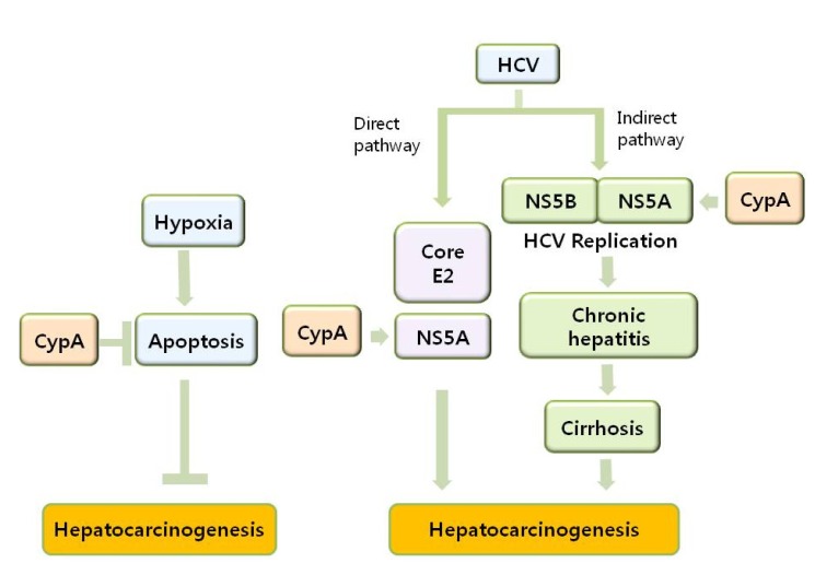
The inflammatory responses after viral infections such as destruction and regeneration of hepatocytes and expression of HCV-encoded proteins will contribute to hepatocyte transformation. The mechanisms of CypA as a host factor during HCV infection and HCC development include anti-apoptotic effect as well as direct and indirect pathways (Fig. 1). CypA, predominantly expressed in human cells, was initially found to be upregulated in hepatocellular carcinoma [94,95]. Overexpression of various Cyps has been repeatedly observed in many cancers and their expression levels and patterns in different cancer types have been considerably well documented. However, the process of how each Cyps participate in the biochemical mechanisms of tumourigenesis is far from being understood. CypA has been reported to be overexpressed in diverse cancer types of small cell lung cancer [96,97], pancreatic cancer [98-101], breast cancer [102,103], colorectal cancer [104-106], squamous cell carcinoma [107,108], melanoma [109], and glioblastoma multiform [110]. Upregulation of CypA in small cell lung cancer stimulates cancer cell growth [111,112]. Subsequently, Qi et al. suggested that CypA may positively function during malignant transformation of esophageal squamous cells [108]. The fold increase of CypA can be found up to 5 times and CypA is also involved in metastasis [109]. Since the proteins with chaperone activity are often stress proteins that protect cells from stress-induced protein damage or denaturation, CypA up-regulation in response to stresses may be regarded naturally as other chaperones. Studying if the CypA up-regulation in cancer is controlled by the same regulatory mechanisms of stress induction can help to understand CypA-involved cancer development. Upregulation of CypA in cancers by hypoxic inducible transcription factor-1α (HIF-1α) has been reported. HIF-1α induces CypA expression by HIF-1α binding to hypoxia response elements in the CypA promoter region under hypoxic conditions [113]. CypA can also be overexpressed during p53-induced apoptosis [114,115]. It will be intriguing to know if interaction of CypA with p53 may lead to stabilization of p53.
Clinical implications of the overexpressed CypA in cancers are currently investigated with an expectation of CypA being a therapeutic target. During development of solid tumors, reactive oxygen species are continuously generated in the central hypoxic region. CypA may exert antioxidant effects on the hypoxic regions in solid tumors through its PPIase activity [11]. Overexpression of CypA in cancer cells renders resistance to hypoxia- and cisplatin-induced cell death in a p53 independent manner [113]. CypA are also regulated by chemotherapeutic agents; for example, cisplatin causes CypA overexpression and induces resistance to several chemotherapeutic agents [98]. Treatment with chemotherapeutic agents such as celecoxib and 5-fluorouracil, on the contrary, is reported to lower CypA expression [105,106].
Human Cyps appear to show pleiotropic effects via conformational interconversions of prolyl cis-trans isomerization on the backbones of key proteins in signaling pathways. Recent studies indicate that PPIases utilize isomerization catalysis on the substrate proteins under both physiological and pathophysiological conditions. Then, the development of isoenzyme-, tissue- or organelle-specific PPIase inhibitors will open a new road for cancer intervention strategies.

CYPA AS A THERAPEUTIC TARGET FOR HCC

Targeting CypA for cancer therapeutics has recently begun to emerge because CypA overexpression is often found in many different cancer types and CypA appears to be involved in malignant transformation in some cancer types. In parallel, CypA inhibitors show antiviral activity and anti HCC effect as replication of HCV RNA genome depends on the expression of CypA. Pegylated IFN-α and ribavirin that directly target the virally-encoded proteins are a current treatment regimen for chronic HCV infection [116]. Viral protein inhibitors, however, show a limited response with severe side effects and also develop high viral resistance [117-119]. The CypA inhibitors are advantageous because host targeting can provide a higher barrier to resistance than viral inhibitors. Both CypA and CypB are important host factors for the HCV replication process since interactions of both CypA with NS5A and CypB with the HCV RNA-dependent RNA polymerase NS5B contribute to form a multi-protein complex [120]. Treatment of CypA inhibitors cause dissociation from the protein complex that is formed between CypA and NS5A [121-123]. The dissociation by CypA inhibitors decreases the affinity of NS5A for viral RNA and inhibits viral replication [124-128]. CsA was first tested to be effective against HCV and afterwards, Cyp inhibitors without immunosuppressive activity were developed for treatment of HCV infection [129-133]. CsA derivatives exert anti-HCV activity by inhibiting host CypA and CypA has not been implicated in the mechanism of action of either pegylated IFN-α or ribavirin [134]. Therefore, synergistic effects against HCV of are expected when CypA inhibitors and conventional drugs are used in combination. Three CsA derivatives, NIM811, SCY-635, and Alisporivir have shown promising anti-HCV responses in pre-clinical and clinical trials and the resistance level was substantially lower than viral protein inhibitors [135].
The anti-HCV mechanisms of action of these CypA inhibitors are not clearly defined yet. However, recent studies unravel some fundamental findings. The inhibitors inhibit PPIase activity of CypA and the isomerase active site of CypA is critical for hepatitis C virus replication [136-138]. CypA binds to prolines located in the domain II of the HCV NS5A protein and Cyp inhibitors interfere the Cyp A-NS5A interactions in a dose-dependent manner [139-141]. While PPIase activity of CypA suggests the function of CypA in structural interconversion of the interacting protein, how the interaction between CypA and NS5A influences HCV replication remains to be elucidated. With the finding that CypA specifically promotes NS5A binding to viral RNA, it has been suggested that cis-trans isomerization of NS5A proline bonds by CypA enhances NS5A binding to viral RNA and somehow facilitates HCV replication [142-144]. Since NS5A binds the HCV polymerase NS5B directly, interaction of CypA with NS5A may affect NS5B polymerase activity. In addition, interaction of CypB with NS5B is important for HCV replication and the anti-HCV effects of Cyp inhibitors may be the outcome of blocking modulations of both NS5A and NS5B by CypA and CypB [145,146].
NIM811 was discovered as 2-5-fold higher binding affinity to Cyps than CsA and inhibited HCV replication in vitro with an EC50 of 0.12 µM in the absence of human serum and 0.63 µM in the presence of 40% human serum. NIM811 was well tolerated and there appeared to be no drug-related adverse event during evaluation. NIM811 was limited by its significant side effect in decreasing platelet [147]. Alisporivir is another CsA analogue without immunosuppressive action [147]. Alisporivir is more potent than NIM811 in inhibiting HCV with an EC50 of 0.045 µM and a serum (100%) adjusted EC50 of 0.33 µM [148]. While it was initially developed for HIV-1, Alisporivir displayed high antiviral activity against HCV RNA [149]. In behalf of its higher barrier to resistance, an IFN-free regimen was carried out to treat IFN non-responders in a Phase II trial and HCV became undetectable in 75.4% of the patients [150]. Alisporivir is the first time trial for a host-targeting antiviral alone and effective both in vitro and in patients, being considered as an ideal candidate inhibitor as an IFN-free, all-oral regimen [151]. The main side effect was a jaundice, however, without any increase in ALT, AST or γ-GT, or with hemolysis [152]. Unfortunately, six cases of severe pancreatitis during phase III trial were reported [153]. SCY-635 was another CsA analog with an EC50 of 0.03 µM [154]. It showed a different side-effect of elevated serum creatinine phosphokinase and ALT/AST, but there was no signs of bilirubin increase or platelet decrease. Recent clinical studies suggest antiviral combination therapies toward IFN-free and ribavirin-free regimens and Alisporivir possessing prominent features as described is under clinical trials for combination with specific direct-acting antiviral agents.

SUMMARY

HCV infections frequently advance into the development of HCC and considerable evidence suggests that 1) inflammatory responses, 2) associated destruction and regeneration of hepatocytes, and 3) activities of HCV-encoded proteins contribute to hepatocyte transformation. Viral proteins and inflammatory responses are clearly associated with the alterations in hepatocyte physiology and the liver environment. However understanding the mechanisms involved is still incomplete and further studies warrant effective strategies for treating HCV infections and preventing HCC. Current knowledge of the proteomics of HCV-induced liver disease is still developing and protein profiles from recent discoveries will be important for diagnosis and treatment of HCV-induced HCC. In the meantime, targeting CypA for cancer therapeutics has been earnestly sought because CypA overexpression is common in malignant cancer cells. Simultaneously, CypA inhibitors can show antiviral activity and thus be benefited as HCC therapeutic agents. CsA suppresses HCV genome replication in HCV replicon cells and human hepatocytes infected with HCV. However, the inhibition of the phosphatase calcineurin by CsA and CypA complex display immunosuppresive action which hinders its use as an antiviral agent. Thereupon, specific CsA derivatives devoid of immune suppressive effects such as NIM811, SCY-635, and Alisporivir have been developed for HCV treatment. In fact, SCY-635 and Alisporivir have emerged as powerful antiviral agents since they inhibit the in vitro replication of HCV subgenomic replicons. Furthermore, clinical use of Alisporivir as an anti-HCV agent has been confirmed in patient studies. In conclusion, the CypA inhibitors may represent a candidate as therapeutic agents for the HCV-induced HCC by preventing both HCV infection and hepatocarcinogenesis.

ACKNOWLEDGEMENTS

This work was supported by the National Research Foundation of Korea (NRF) grant (No. 2010-0024592) from the Korea Ministry of Education, Science and Technology (MEST) and 2011 supporting grants for research science development from Dongseo University.

ABBREVIATIONS

HCC

hepatocellular carcinoma

HBV

hepatitis B virus

HCV

hepatitis C virus

CypA

cyclophilin A

Cyps

cyclophilins

CsA

cyclosporine A

PPIase

peptidyl prolyl isomerase

ER

endoplasmic reticulum

MPTP

mitochondrial permeability transition pore

HIV-1

human immunodeficiency virus-1

IFN

interferon

IL

interleukin

References

1. Friedman J, Weissman I. Two cytoplasmic candidates for immunophilin action are revealed by affinity for a new cyclophilin: one in the presence and one in the absence of CsA. Cell. 1991; 66:799–806. PMID: 1652374.
crossref
2. Rush DN. Cyclosporine toxicity to organs other than the kidney. Clin Biochem. 1991; 24:101–105. PMID: 2060127.
crossref
3. Luban J. Absconding with the chaperone: essential cyclophilin-Gag interaction in HIV-1 virions. Cell. 1996; 87:1157–1159. PMID: 8980222.
crossref
4. Klappa P, Freedman RB, Zimmermann R. Protein disulphide isomerase and a lumenal cyclophilin-type peptidyl prolyl cistrans isomerase are in transient contact with secretory proteins during late stages of translocation. Eur J Biochem. 1995; 232:755–764. PMID: 7588713.
crossref
5. Naylor DJ, Hoogenraad NJ, Høj PB. Characterisation of several Hsp70 interacting proteins from mammalian organelles. Biochim Biophys Acta. 1999; 1431:443–450. PMID: 10350619.
crossref
6. Castro AP, Carvalho TM, Moussatché N, Damaso CR. Redistribution of cyclophilin A to viral factories during vaccinia virus infection and its incorporation into mature particles. J Virol. 2003; 77:9052–9068. PMID: 12885921.
crossref
7. Steinmann B, Bruckner P, Superti-Furga A. Cyclosporin A slows collagen triple-helix formation in vivo: indirect evidence for a physiologic role of peptidyl-prolyl cis-trans-isomerase. J Biol Chem. 1991; 266:1299–1303. PMID: 1985948.
crossref
8. Kruse M, Brunke M, Escher A, Szalay AA, Tropschug M, Zimmermann R. Enzyme assembly after de novo synthesis in rabbit reticulocyte lysate involves molecular chaperones and immunophilins. J Biol Chem. 1995; 270:2588–2594. PMID: 7852322.
9. Craig EA, Gambill BD, Nelson RJ. Heat shock proteins: molecular chaperones of protein biogenesis. Microbiol Rev. 1993; 57:402–414. PMID: 8336673.
crossref
10. Parsell DA, Lindquist S. The function of heat-shock proteins in stress tolerance: degradation and reactivation of damaged proteins. Annu Rev Genet. 1993; 27:437–496. PMID: 8122909.
crossref
11. Hong F, Lee J, Song JW, Lee SJ, Ahn H, Cho JJ, Ha J, Kim SS. Cyclosporin A blocks muscle differentiation by inducing oxidative stress and inhibiting the peptidyl-prolyl-cis-trans isomerase activity of cyclophilin A: cyclophilin A protects myoblasts from cyclosporin A-induced cytotoxicity. FASEB J. 2002; 16:1633–1635. PMID: 12207006.
12. Wiederrecht G, Lam E, Hung S, Martin M, Sigal N. The mechanism of action of FK-506 and cyclosporin A. Ann N Y Acad Sci. 1993; 696:9–19. PMID: 7509138.
crossref
13. Bauer K, Kretzschmar AK, Cvijic H, Blumert C, Löffler D, Brocke-Heidrich K, Schiene-Fischer C, Fischer G, Sinz A, Clevenger CV, Horn F. Cyclophilins contribute to Stat3 signaling and survival of multiple myeloma cells. Oncogene. 2009; 28:2784–2795. PMID: 19503092.
crossref
14. Haworth RA, Hunter DR. Allosteric inhibition of the Ca2+-activated hydrophilic channel of the mitochondrial inner membrane by nucleotides. J Membr Biol. 1980; 54:231–236. PMID: 6248646.
15. Halestrap AP, Davidson AM. Inhibition of Ca2+-induced large-amplitude swelling of liver and heart mitochondria by cyclosporin is probably caused by the inhibitor binding to mitochondrial-matrix peptidyl-prolyl cis-trans isomerase and preventing it interacting with the adenine nucleotide translocase. Biochem J. 1990; 268:153–160. PMID: 2160810.
16. Bhattacharyya T, Karnezis AN, Murphy SP, Hoang T, Freeman BC, Phillips B, Morimoto RI. Cloning and subcellular localization of human mitochondrial hsp70. J Biol Chem. 1995; 270:1705–1710. PMID: 7829505.
crossref
17. Thomas PJ, Qu BH, Pedersen PL. Defective protein folding as a basis of human disease. Trends Biochem Sci. 1995; 20:456–459. PMID: 8578588.
crossref
18. Price ER, Zydowsky LD, Jin MJ, Baker CH, McKeon FD, Walsh CT. Human cyclophilin B: a second cyclophilin gene encodes a peptidyl-prolyl isomerase with a signal sequence. Proc Natl Acad Sci USA. 1991; 88:1903–1907. PMID: 2000394.
crossref
19. Sherry B, Yarlett N, Strupp A, Cerami A. Identification of cyclophilin as a proinflammatory secretory product of lipopolysaccharide-activated macrophages. Proc Natl Acad Sci USA. 1992; 89:3511–3515. PMID: 1565646.
crossref
20. Yurchenko V, Zybarth G, O'Connor M, Dai WW, Franchin G, Hao T, Guo H, Hung HC, Toole B, Gallay P, Sherry B, Bukrinsky M. Active site residues of cyclophilin A are crucial for its signaling activity via CD147. J Biol Chem. 2002; 277:22959–22965. PMID: 11943775.
crossref
21. Sherry B, Zybarth G, Alfano M, Dubrovsky L, Mitchell R, Rich D, Ulrich P, Bucala R, Cerami A, Bukrinsky M. Role of cyclophilin A in the uptake of HIV-1 by macrophages and T lymphocytes. Proc Natl Acad Sci U S A. 1998; 95:1758–1763. PMID: 9465090.
crossref
22. Jin ZG, Melaragno MG, Liao DF, Yan C, Haendeler J, Suh YA, Lambeth JD, Berk BC. Cyclophilin A is a secreted growth factor induced by oxidative stress. Circ Res. 2000; 87:789–796. PMID: 11055983.
crossref
23. Yurchenko V, O'Connor M, Dai WW, Guo H, Toole B, Sherry B, Bukrinsky M. CD147 is a signaling receptor for cyclophilin B. Biochem Biophys Res Commun. 2001; 288:786–788. PMID: 11688976.
crossref
24. Konttinen YT, Li TF, Mandelin J, Liljeström M, Sorsa T, Santavirta S, Virtanen I. Increased expression of extracellular matrix metalloproteinase inducer in rheumatoid synovium. Arthritis Rheum. 2000; 43:275–280. PMID: 10693866.
crossref
25. Jin ZG, Melaragno MG, Liao DF, Yan C, Haendeler J, Suh YA, Lambeth JD, Berk BC. Cyclophilin A is a secreted growth factor induced by oxidative stress. Circ Res. 2000; 87:789–796. PMID: 11055983.
crossref
26. Sarris AH, Harding MW, Jiang TR, Aftab D, Handschumacher RE. Immunofluorescent localization and immunochemical determination of cyclophilin-A with specific rabbit antisera. Transplantation. 1992; 54:904–910. PMID: 1440857.
crossref
27. Zhu C, Wang X, Deinum J, Huang Z, Gao J, Modjtahedi N, Neagu MR, Nilsson M, Eriksson PS, Hagberg H, Luban J, Kroemer G, Blomgren K. Cyclophilin A participates in the nuclear translocation of apoptosis-inducing factor in neurons after cerebral hypoxia-ischemia. J Exp Med. 2007; 204:1741–1748. PMID: 17635954.
crossref
28. Krummrei U, Bang R, Schmidtchen R, Brune K, Bang H. Cyclophilin-A is a zinc-dependent DNA binding protein in macrophages. FEBS Lett. 1995; 371:47–51. PMID: 7664883.
crossref
29. Satoh K, Shimokawa H, Berk BC. Cyclophilin A: promising new target in cardiovascular therapy. Circ J. 2010; 74:2249–2256. PMID: 20962430.
30. Obchoei S, Wongkhan S, Wongkham C, Li M, Yao Q, Chen C. Cyclophilin A: potential functions and therapeutic target for human cancer. Med Sci Monit. 2009; 15:RA221–RA232. PMID: 19865066.
31. Towers GJ, Hatziioannou T, Cowan S, Goff SP, Luban J, Bieniasz PD. Cyclophilin A modulates the sensitivity of HIV-1 to host restriction factors. Nat Med. 2003; 9:1138–1143. PMID: 12897779.
crossref
32. Wohlfarth C, Efferth T. Natural products as promising drug candidates for the treatment of hepatitis B and C. Acta Pharmacol Sin. 2009; 30:25–30. PMID: 19060918.
crossref
33. Satoh K, Nigro P, Berk BC. Oxidative stress and vascular smooth muscle cell growth: a mechanistic linkage by cyclophilin A. Antioxid Redox Signal. 2010; 12:675–682. PMID: 19747062.
crossref
34. Sherry B, Zybarth G, Alfano M, Dubrovsky L, Mitchell R, Rich D, Ulrich P, Bucala R, Cerami A, Bukrinsky M. Role of cyclophilin A in the uptake of HIV-1 by macrophages and T lymphocytes. Proc Natl Acad Sci USA. 1998; 95:1758–1763. PMID: 9465090.
crossref
35. Heitman J, Cullen BR. Cyclophilin B escorts the hepatitis C virus RNA polymerase: a viral achilles heel? Mol Cell. 2005; 19:145–146. PMID: 16039584.
crossref
36. Membreno FE, Espinales JC, Lawitz EJ. Cyclophilin inhibitors for hepatitis C therapy. Clin Liver Dis. 2013; 17:129–139. PMID: 23177289.
crossref
37. El-Serag HB. Epidemiology of viral hepatitis and hepatocellular carcinoma. Gastroenterology. 2012; 142:1264–1273. PMID: 22537432.
crossref
38. Bouchard MJ, Navas-Martin S. Hepatitis B and C virus hepatocarcinogenesis: lessons learned and future challenges. Cancer Lett. 2011; 305:123–143. PMID: 21168955.
crossref
39. Sherman M. Hepatocellular carcinoma: epidemiology, risk factors, and screening. Semin Liver Dis. 2005; 25:143–154. PMID: 15918143.
crossref
40. Roberts LR, Gores GJ. Hepatocellular carcinoma: molecular pathways and new therapeutic targets. Semin Liver Dis. 2005; 25:212–225. PMID: 15918149.
crossref
41. El-Serag HB, Rudolph KL. Hepatocellular carcinoma: epidemiology and molecular carcinogenesis. Gastroenterology. 2007; 132:2557–2576. PMID: 17570226.
crossref
42. Suzuki T, Aizaki H, Murakami K, Shoji I, Wakita T. Molecular biology of hepatitis C virus. J Gastroenterol. 2007; 42:411–423. PMID: 17671755.
crossref
43. Dustin LB, Rice CM. Flying under the radar: the immunobiology of hepatitis C. Annu Rev Immunol. 2007; 25:71–99. PMID: 17067278.
crossref
44. Ura S, Honda M, Yamashita T, Ueda T, Takatori H, Nishino R, Sunakozaka H, Sakai Y, Horimoto K, Kaneko S. Differential microRNA expression between hepatitis B and hepatitis C leading disease progression to hepatocellular carcinoma. Hepatology. 2009; 49:1098–1112. PMID: 19173277.
crossref
45. Nieto N. Oxidative-stress and IL-6 mediate the fibrogenic effects of Kupffer cells on stellate cells. Hepatology. 2006; 44:1487–1501. PMID: 17133487.
crossref
46. Bartel DP. MicroRNAs: target recognition and regulatory functions. Cell. 2009; 136:215–233. PMID: 19167326.
crossref
47. Varnholt H, Drebber U, Schulze F, Wedemeyer I, Schirmacher P, Dienes HP, Odenthal M. MicroRNA gene expression profile of hepatitis C virus-associated hepatocellular carcinoma. Hepatology. 2008; 47:1223–1232. PMID: 18307259.
crossref
48. Jopling CL, Yi M, Lancaster AM, Lemon SM, Sarnow P. Modulation of hepatitis C virus RNA abundance by a liver-specific MicroRNA. Science. 2005; 309:1577–1581. PMID: 16141076.
crossref
49. Kutay H, Bai S, Datta J, Motiwala T, Pogribny I, Frankel W, Jacob ST, Ghoshal K. Downregulation of miR-122 in the rodent and human hepatocellular carcinomas. J Cell Biochem. 2006; 99:671–678. PMID: 16924677.
crossref
50. Jopling CL, Norman KL, Sarnow P. Positive and negative modulation of viral and cellular mRNAs by liver-specific microRNA miR-122. Cold Spring Harb Symp Quant Biol. 2006; 71:369–376. PMID: 17381319.
crossref
51. Randall G, Panis M, Cooper JD, Tellinghuisen TL, Sukhodolets KE, Pfeffer S, Landthaler M, Landgraf P, Kan S, Lindenbach BD, Chien M, Weir DB, Russo JJ, Ju J, Brownstein MJ, Sheridan R, Sander C, Zavolan M, Tuschl T, Rice CM. Cellular cofactors affecting hepatitis C virus infection and replication. Proc Natl Acad Sci U S A. 2007; 104:12884–12889. PMID: 17616579.
crossref
52. Nunnari G, Schnell MJ. MicroRNA-122: a therapeutic target for hepatitis C virus (HCV) infection. Front Biosci (Schol Ed). 2011; 3:1032–1037. PMID: 21622252.
crossref
53. Gao P, Wong CC, Tung EK, Lee JM, Wong CM, Ng IO. Deregulation of microRNA expression occurs early and accumulates in early stages of HBV-associated multistep hepatocarcinogenesis. J Hepatol. 2011; 54:1177–1184. PMID: 21145831.
crossref
54. Ji J, Shi J, Budhu A, Yu Z, Forgues M, Roessler S, Ambs S, Chen Y, Meltzer PS, Croce CM, Qin LX, Man K, Lo CM, Lee J, Ng IO, Fan J, Tang ZY, Sun HC, Wang XW. MicroRNA expression, survival, and response to interferon in liver cancer. N Engl J Med. 2009; 361:1437–1447. PMID: 19812400.
crossref
55. McGivern DR, Lemon SM. Tumor suppressors, chromosomal instability, and hepatitis C virus-associated liver cancer. Annu Rev Pathol. 2009; 4:399–415. PMID: 18928409.
crossref
56. Finch CE, Crimmins EM. Inflammatory exposure and historical changes in human life-spans. Science. 2004; 305:1736–1739. PMID: 15375259.
crossref
57. Bréchot C, Jaffredo F, Lagorce D, Gerken G, Meyer zum Büschenfelde K, Papakonstontinou A, Hadziyannis S, Romeo R, Colombo M, Rodes J, Bruix J, Williams R, Naoumov N. Impact of HBV, HCV and GBV-C/HGV on hepatocellular carcinomas in Europe: results of a European concerted action. J Hepatol. 1998; 29:173–183. PMID: 9722197.
crossref
58. Wang YY, Lo GH, Lai KH, Cheng JS, Lin CK, Hsu PI. Increased serum concentrations of tumor necrosis factor-alpha are associated with disease progression and malnutrition in hepatocellular carcinoma. J Chin Med Assoc. 2003; 66:593–598. PMID: 14703276.
59. Mondelli MU, Barnaba V. Viral and host immune regulatory mechanisms in hepatitis C virus infection. Eur J Gastroenterol Hepatol. 2006; 18:327–331. PMID: 16538102.
crossref
60. Tsai JF, Jeng JE, Ho MS, Chang WY, Lin ZY, Tsai JH. Hepatitis B and C virus infection as risk factors for hepatocellular carcinoma in Chinese: a case-control study. Int J Cancer. 1994; 56:619–621. PMID: 8314335.
crossref
61. Stroffolini T, Chiaramonte M, Tiribelli C, Villa E, Simonetti RG, Rapicetta M, Stazi MA, Bertin T, Crocè SL, Trande P, et al. Hepatitis C virus infection, HBsAg carrier state and hepatocellular carcinoma: relative risk and population attributable risk from a case-control study in Italy. J Hepatol. 1992; 16:360–363. PMID: 1336785.
62. Wheelhouse NM, Chan YS, Gillies SE, Caldwell H, Ross JA, Harrison DJ, Prost S. TNF-alpha induced DNA damage in primary murine hepatocytes. Int J Mol Med. 2003; 12:889–894. PMID: 14612962.
63. Rehermann B. Intrahepatic T cells in hepatitis B: viral control versus liver cell injury. J Exp Med. 2000; 191:1263–1268. PMID: 10770794.
64. Freeman AJ, Marinos G, Ffrench RA, Lloyd AR. Immunopathogenesis of hepatitis C virus infection. Immunol Cell Biol. 2001; 79:515–536. PMID: 11903612.
crossref
65. Rapicetta M, Ferrari C, Levrero M. Viral determinants and host immune responses in the pathogenesis of HBV infection. J Med Virol. 2002; 67:454–457. PMID: 12116045.
crossref
66. Akiba J, Yano H, Ogasawara S, Higaki K, Kojiro M. Expression and function of interleukin-8 in human hepatocellular carcinoma. Int J Oncol. 2001; 18:257–264. PMID: 11172590.
crossref
67. Ishido S, Fujita T, Hotta H. Complex formation of NS5B with NS3 and NS4A proteins of hepatitis C virus. Biochem Biophys Res Commun. 1998; 244:35–40. PMID: 9514871.
crossref
68. Zekri AR, Ashour MS, Hassan A, Alam El-Din HM, El-Shehaby AM, Abu-Shady MA. Cytokine profile in Egyptian hepatitis C virus genotype-4 in relation to liver disease progression. World J Gastroenterol. 2005; 11:6624–6630. PMID: 16425355.
crossref
69. Brady MT, MacDonald AJ, Rowan AG, Mills KH. Hepatitis C virus non-structural protein 4 suppresses Th1 responses by stimulating IL-10 production from monocytes. Eur J Immunol. 2003; 33:3448–3457. PMID: 14635055.
crossref
70. Sun B, Karin M. Obesity, inflammation, and liver cancer. J Hepatol. 2012; 56:704–713. PMID: 22120206.
crossref
71. Zekri AR, Bahnassy AA, Abdel-Wahab SA, Khafagy MM, Loutfy SA, Radwan H, Shaarawy SM. Expression of pro- and anti-inflammatory cytokines in relation to apoptotic genes in Egyptian liver disease patients associated with HCV-genotype-4. J Gastroenterol Hepatol. 2009; 24:416–428. PMID: 19054267.
crossref
72. Ait-Goughoulte M, Banerjee A, Meyer K, Mazumdar B, Saito K, Ray RB, Ray R. Hepatitis C virus core protein interacts with fibrinogen-beta and attenuates cytokine stimulated acute-phase response. Hepatology. 2010; 51:1505–1513. PMID: 20162731.
73. Tilg H, Wilmer A, Vogel W, Herold M, Nölchen B, Judmaier G, Huber C. Serum levels of cytokines in chronic liver diseases. Gastroenterology. 1992; 103:264–274. PMID: 1612333.
crossref
74. Torre D, Zeroli C, Giola M, Ferrario G, Fiori GP, Bonetta G, Tambini R. Serum levels of interleukin-1 alpha, interleukin-1 beta, interleukin-6, and tumor necrosis factor in patients with acute viral hepatitis. Clin Infect Dis. 1994; 18:194–198. PMID: 8161626.
75. Taniguchi H, Kato N, Otsuka M, Goto T, Yoshida H, Shiratori Y, Omata M. Hepatitis C virus core protein upregulates transforming growth factor-beta 1 transcription. J Med Virol. 2004; 72:52–59. PMID: 14635011.
76. Shin JY, Hur W, Wang JS, Jang JW, Kim CW, Bae SH, Jang SK, Yang SH, Sung YC, Kwon OJ, Yoon SK. HCV core protein promotes liver fibrogenesis via up-regulation of CTGF with TGF-beta1. Exp Mol Med. 2005; 37:138–145. PMID: 15886528.
77. Battaglia S, Benzoubir N, Nobilet S, Charneau P, Samuel D, Zignego AL, Atfi A, Brechot C, Bourgeade MF. Liver cancer-derived hepatitis C virus core proteins shift TGF-beta responses from tumor suppression to epithelial-mesenchymal transition. PLoS One. 2009; 4:e4355. PMID: 19190755.
crossref
78. Large MK, Kittlesen DJ, Hahn YS. Suppression of host immune response by the core protein of hepatitis C virus: possible implications for hepatitis C virus persistence. J Immunol. 1999; 162:931–938. PMID: 9916717.
79. You LR, Chen CM, Lee YH. Hepatitis C virus core protein enhances NF-kappaB signal pathway triggering by lymphotoxin-beta receptor ligand and tumor necrosis factor alpha. J Virol. 1999; 73:1672–1681. PMID: 9882379.
80. Soguero C, Joo M, Chianese-Bullock KA, Nguyen DT, Tung K, Hahn YS. Hepatitis C virus core protein leads to immune suppression and liver damage in a transgenic murine model. J Virol. 2002; 76:9345–9354. PMID: 12186917.
crossref
81. Chan-Fook C, Jiang WR, Clarke BE, Zitzmann N, Maidens C, McKeating JA, Jones IM. Hepatitis C virus glycoprotein E2 binding to CD81: the role of E1E2 cleavage and protein glycosylation in bioactivity. Virology. 2000; 273:60–66. PMID: 10891408.
82. Reyes GR. The nonstructural NS5A protein of hepatitis C virus: an expanding, multifunctional role in enhancing hepatitis C virus pathogenesis. J Biomed Sci. 2002; 9:187–197. PMID: 12065893.
crossref
83. Kitaoka S, Shiota G, Kawasaki H. Serum levels of interleukin-10, interleukin-12 and soluble interleukin-2 receptor in chronic liver disease type C. Hepatogastroenterology. 2003; 50:1569–1574. PMID: 14571788.
84. Chia CS, Ban K, Ithnin H, Singh H, Krishnan R, Mokhtar S, Malihan N, Seow HF. Expression of interleukin-18, interferon-gamma and interleukin-10 in hepatocellular carcinoma. Immunol Lett. 2002; 84:163–172. PMID: 12413732.
85. Kakumu S, Okumura A, Ishikawa T, Yano M, Enomoto A, Nishimura H, Yoshioka K, Yoshika Y. Serum levels of IL-10, IL-15 and soluble tumour necrosis factor-alpha (TNF-alpha) receptors in type C chronic liver disease. Clin Exp Immunol. 1997; 109:458–463. PMID: 9328122.
86. Zekri AR, Ashour MS, Hassan A, Alam El-Din HM, El-Shehaby AM, Abu-Shady MA. Cytokine profile in Egyptian hepatitis C virus genotype-4 in relation to liver disease progression. World J Gastroenterol. 2005; 11:6624–6630. PMID: 16425355.
crossref
87. Beckebaum S, Zhang X, Chen X, Yu Z, Frilling A, Dworacki G, Grosse-Wilde H, Broelsch CE, Gerken G, Cicinnati VR. Increased levels of interleukin-10 in serum from patients with hepatocellular carcinoma correlate with profound numerical deficiencies and immature phenotype of circulating dendritic cell subsets. Clin Cancer Res. 2004; 10:7260–7269. PMID: 15534100.
crossref
88. Huang YS, Hwang SJ, Chan CY, Wu JC, Chao Y, Chang FY, Lee SD. Serum levels of cytokines in hepatitis C-related liver disease: a longitudinal study. Zhonghua Yi Xue Za Zhi (Taipei). 1999; 62:327–333. PMID: 10389289.
89. Nakazaki H. Preoperative and postoperative cytokines in patients with cancer. Cancer. 1992; 70:709–713. PMID: 1320454.
crossref
90. Ikeguchi M, Hirooka Y. Interleukin-2 gene expression is a new biological prognostic marker in hepatocellular carcinomas. Onkologie. 2005; 28:255–259. PMID: 15867481.
crossref
91. Aroucha DC, do Carmo RF, Moura P, Silva JL, Vasconcelos LR, Cavalcanti MS, Muniz MT, Aroucha ML, Siqueira ER, Cahú GG, Pereira LM, Coêlho MR. High tumor necrosis factor-α/interleukin-10 ratio is associated with hepatocellular carcinoma in patients with chronic hepatitis C. Cytokine. 2013; 62:421–425. PMID: 23602201.
crossref
92. Bartosch B, Thimme R, Blum HE, Zoulim F. Hepatitis C virus-induced hepatocarcinogenesis. J Hepatol. 2009; 51:810–820. PMID: 19545926.
crossref
93. Bouchard MJ, Schneider RJ. The enigmatic X gene of hepatitis B virus. J Virol. 2004; 78:12725–12734. PMID: 15542625.
crossref
94. Corton JC, Moreno ES, Merritt A, Bocos C, Cattley RC. Cloning genes responsive to a hepatocarcinogenic peroxisome proliferator chemical reveals novel targets of regulation. Cancer Lett. 1998; 134:61–71. PMID: 10381131.
crossref
95. Lim SO, Park SJ, Kim W, Park SG, Kim HJ, Kim YI, Sohn TS, Noh JH, Jung G. Proteome analysis of hepatocellular carcinoma. Biochem Biophys Res Commun. 2002; 291:1031–1037. PMID: 11866469.
crossref
96. Yang H, Chen J, Yang J, Qiao S, Zhao S, Yu L. Cyclophilin A is upregulated in small cell lung cancer and activates ERK1/2 signal. Biochem Biophys Res Commun. 2007; 361:763–767. PMID: 17678621.
crossref
97. Campa MJ, Wang MZ, Howard B, Fitzgerald MC, Patz EF Jr. Protein expression profiling identifies macrophage migration inhibitory factor and cyclophilin a as potential molecular targets in non-small cell lung cancer. Cancer Res. 2003; 63:1652–1656. PMID: 12670919.
98. Cecconi D, Astner H, Donadelli M, Palmieri M, Missiaglia E, Hamdan M, Scarpa A, Righetti PG. Proteomic analysis of pancreatic ductal carcinoma cells treated with 5-aza-2'-deoxycytidine. Electrophoresis. 2003; 24:4291–4303. PMID: 14679576.
crossref
99. Shen J, Person MD, Zhu J, Abbruzzese JL, Li D. Protein expression profiles in pancreatic adenocarcinoma compared with normal pancreatic tissue and tissue affected by pancreatitis as detected by two-dimensional gel electrophoresis and mass spectrometry. Cancer Res. 2004; 64:9018–9026. PMID: 15604267.
crossref
100. Li M, Wang H, Li F, Fisher WE, Chen C, Yao Q. Effect of cyclophilin A on gene expression in human pancreatic cancer cells. Am J Surg. 2005; 190:739–745. PMID: 16226951.
crossref
101. Mikuriya K, Kuramitsu Y, Ryozawa S, Fujimoto M, Mori S, Oka M, Hamano K, Okita K, Sakaida I, Nakamura K. Expression of glycolytic enzymes is increased in pancreatic cancerous tissues as evidenced by proteomic profiling by two-dimensional electrophoresis and liquid chromatography-mass spectrometry/mass spectrometry. Int J Oncol. 2007; 30:849–855. PMID: 17332923.
crossref
102. Zheng J, Koblinski JE, Dutson LV, Feeney YB, Clevenger CV. Prolyl isomerase cyclophilin A regulation of Janus-activated kinase 2 and the progression of human breast cancer. Cancer Res. 2008; 68:7769–7778. PMID: 18829531.
crossref
103. Hathout Y, Riordan K, Gehrmann M, Fenselau C. Differential protein expression in the cytosol fraction of an MCF-7 breast cancer cell line selected for resistance toward melphalan. J Proteome Res. 2002; 1:435–442. PMID: 12645915.
crossref
104. Melle C, Osterloh D, Ernst G, Schimmel B, Bleul A, von Eggeling F. Identification of proteins from colorectal cancer tissue by two-dimensional gel electrophoresis and SELDI mass spectrometry. Int J Mol Med. 2005; 16:11–17. PMID: 15942672.
crossref
105. Lou J, Fatima N, Xiao Z, Stauffer S, Smythers G, Greenwald P, Ali IU. Proteomic profiling identifies cyclooxygenase-2-independent global proteomic changes by celecoxib in colorectal cancer cells. Cancer Epidemiol Biomarkers Prev. 2006; 15:1598–1606. PMID: 16985019.
crossref
106. Wong CS, Wong VW, Chan CM, Ma BB, Hui EP, Wong MC, Lam MY, Au TC, Chan WH, Cheuk W, Chan AT. Identification of 5-fluorouracil response proteins in colorectal carcinoma cell line SW480 by two-dimensional electrophoresis and MALDI-TOF mass spectrometry. Oncol Rep. 2008; 20:89–98. PMID: 18575723.
crossref
107. Chen J, He QY, Yuen AP, Chiu JF. Proteomics of buccal squamous cell carcinoma: the involvement of multiple pathways in tumorigenesis. Proteomics. 2004; 4:2465–2475. PMID: 15274141.
crossref
108. Qi YJ, He QY, Ma YF, Du YW, Liu GC, Li YJ, Tsao GS, Ngai SM, Chiu JF. Proteomic identification of malignant transformation-related proteins in esophageal squamous cell carcinoma. J Cell Biochem. 2008; 104:1625–1635. PMID: 18320592.
crossref
109. Al-Ghoul M, Brück TB, Lauer-Fields JL, Asirvatham VS, Zapata C, Kerr RG, Fields GB. Comparative proteomic analysis of matched primary and metastatic melanoma cell lines. J Proteome Res. 2008; 7:4107–4118. PMID: 18698805.
crossref
110. Han X, Yoon SH, Ding Y, Choi TG, Choi WJ, Kim YH, Kim YJ, Huh YB, Ha J, Kim SS. Cyclosporin A and sanglifehrin A enhance chemotherapeutic effect of cisplatin in C6 glioma cells. Oncol Rep. 2010; 23:1053–1062. PMID: 20204291.
crossref
111. Howard BA, Zheng Z, Campa MJ, Wang MZ, Sharma A, Haura E, Herndon JE 2nd, Fitzgerald MC, Bepler G, Patz EF Jr. Translating biomarkers into clinical practice: prognostic implications of cyclophilin A and macrophage migratory inhibitory factor identified from protein expression profiles in non-small cell lung cancer. Lung Cancer. 2004; 46:313–323. PMID: 15541816.
crossref
112. Howard BA, Furumai R, Campa MJ, Rabbani ZN, Vujaskovic Z, Wang XF, Patz EF Jr. Stable RNA interference-mediated suppression of cyclophilin A diminishes non-small-cell lung tumor growth in vivo. Cancer Res. 2005; 65:8853–8860. PMID: 16204056.
crossref
113. Choi KJ, Piao YJ, Lim MJ, Kim JH, Ha J, Choe W, Kim SS. Overexpressed cyclophilin A in cancer cells renders resistance to hypoxia- and cisplatin-induced cell death. Cancer Res. 2007; 67:3654–3662. PMID: 17440077.
crossref
114. Gu S, Liu Z, Pan S, Jiang Z, Lu H, Amit O, Bradbury EM, Hu CA, Chen X. Global investigation of p53-induced apoptosis through quantitative proteomic profiling using comparative amino acid-coded tagging. Mol Cell Proteomics. 2004; 3:998–1008. PMID: 15284338.
crossref
115. Yu X, Harris SL, Levine AJ. The regulation of exosome secretion: a novel function of the p53 protein. Cancer Res. 2006; 66:4795–4801. PMID: 16651434.
crossref
116. Tong MJ, Reddy KR, Lee WM, Pockros PJ, Hoefs JC, Keeffe EB, Hollinger FB, Hathcote EJ, White H, Foust RT, Jensen DM, Krawitt EL, Fromm H, Black M, Blatt LM, Klein M, Lubina J. Consensus Interferon Study Group. Treatment of chronic hepatitis C with consensus interferon: a multicenter, randomized, controlled trial. Hepatology. 1997; 26:747–754. PMID: 9303508.
crossref
117. Cross TJ, Antoniades CG, Harrison PM. Current and future management of chronic hepatitis C infection. Postgrad Med J. 2008; 84:172–176. PMID: 18424572.
crossref
118. Simmonds P, Bukh J, Combet C, Deléage G, Enomoto N, Feinstone S, Halfon P, Inchauspé G, Kuiken C, Maertens G, Mizokami M, Murphy DG, Okamoto H, Pawlotsky JM, Penin F, Sablon E, Shin-I T, Stuyver LJ, Thiel HJ, Viazov S, Weiner AJ, Widell A. Consensus proposals for a unified system of nomenclature of hepatitis C virus genotypes. Hepatology. 2005; 42:962–973. PMID: 16149085.
crossref
119. Manns MP, Wedemeyer H, Cornberg M. Treating viral hepatitis C: efficacy, side effects, and complications. Gut. 2006; 55:1350–1359. PMID: 16905701.
crossref
120. Fernandes F, Poole DS, Hoover S, Middleton R, Andrei AC, Gerstner J, Striker R. Sensitivity of hepatitis C virus to cyclosporine A depends on nonstructural proteins NS5A and NS5B. Hepatology. 2007; 46:1026–1033. PMID: 17600342.
crossref
121. Chatterji U, Lim P, Bobardt MD, Wieland S, Cordek DG, Vuagniaux G, Chisari F, Cameron CE, Targett-Adams P, Parkinson T, Gallay PA. HCV resistance to cyclosporin A does not correlate with a resistance of the NS5A-cyclophilin A interaction to cyclophilin inhibitors. J Hepatol. 2010; 53:50–56. PMID: 20451281.
crossref
122. Coelmont L, Hanoulle X, Chatterji U, Berger C, Snoeck J, Bobardt M, Lim P, Vliegen I, Paeshuyse J, Vuagniaux G, Vandamme AM, Bartenschlager R, Gallay P, Lippens G, Neyts J. DEB025 (Alisporivir) inhibits hepatitis C virus replication by preventing a cyclophilin A induced cis-trans isomerisation in domain II of NS5A. PLoS One. 2010; 5:e13687. PMID: 21060866.
crossref
123. Fernandes F, Ansari IU, Striker R. Cyclosporine inhibits a direct interaction between cyclophilins and hepatitis C NS5A. PLoS One. 2010; 5:e9815. PMID: 20352119.
crossref
124. Yang F, Robotham JM, Grise H, Frausto S, Madan V, Zayas M, Bartenschlager R, Robinson M, Greenstein AE, Nag A, Logan TM, Bienkiewicz E, Tang H. A major determinant of cyclophilin dependence and cyclosporine susceptibility of hepatitis C virus identified by a genetic approach. PLoS Pathog. 2010; 6:e1001118. PMID: 20886100.
crossref
125. Hanoulle X, Badillo A, Wieruszeski JM, Verdegem D, Landrieu I, Bartenschlager R, Penin F, Lippens G. Hepatitis C virus NS5A protein is a substrate for the peptidyl-prolyl cis/trans isomerase activity of cyclophilins A and B. J Biol Chem. 2009; 284:13589–13601. PMID: 19297321.
crossref
126. Verdegem D, Badillo A, Wieruszeski JM, Landrieu I, Leroy A, Bartenschlager R, Penin F, Lippens G, Hanoulle X. Domain 3 of NS5A protein from the hepatitis C virus has intrinsic alpha-helical propensity and is a substrate of cyclophilin A. J Biol Chem. 2011; 286:20441–20454. PMID: 21489988.
127. Gregory MA, Bobardt M, Obeid S, Chatterji U, Coates NJ, Foster T, Gallay P, Leyssen P, Moss SJ, Neyts J, Nur-e-Alam M, Paeshuyse J, Piraee M, Suthar D, Warneck T, Zhang MQ, Wilkinson B. Preclinical characterization of naturally occurring polyketide cyclophilin inhibitors from the sanglifehrin family. Antimicrob Agents Chemother. 2011; 55:1975–1981. PMID: 21383094.
crossref
128. Foster TL, Gallay P, Stonehouse NJ, Harris M. Cyclophilin A interacts with domain II of hepatitis C virus NS5A and stimulates RNA binding in an isomerase-dependent manner. J Virol. 2011; 85:7460–7464. PMID: 21593166.
crossref
129. Inoue K, Sekiyama K, Yamada M, Watanabe T, Yasuda H, Yoshiba M. Combined interferon alpha2b and cyclosporin A in the treatment of chronic hepatitis C: controlled trial. J Gastroenterol. 2003; 38:567–572. PMID: 12825133.
130. Inoue K, Yoshiba M. Interferon combined with cyclosporine treatment as an effective countermeasure against hepatitis C virus recurrence in liver transplant patients with end-stage hepatitis C virus related disease. Transplant Proc. 2005; 37:1233–1234. PMID: 15848679.
crossref
131. Jang YY, Han ES, Lee CS, Kim YK, Song JH, Shin YK. Enhancement of cyclosporine-induced oxidative damage of kidney mitochondria by iron. Korean J Physiol Pharmacol. 1999; 3:631–640.
132. Paeshuyse J, Kaul A, De Clercq E, Rosenwirth B, Dumont JM, Scalfaro P, Bartenschlager R, Neyts J. The nonimmunosuppressive cyclosporin DEBIO-025 is a potent inhibitor of hepatitis C virus replication in vitro. Hepatology. 2006; 43:761–770. PMID: 16557546.
133. Hopkins S, Scorneaux B, Huang Z, Murray MG, Wring S, Smitley C, Harris R, Erdmann F, Fischer G, Ribeill Y. SCY-635, a novel nonimmunosuppressive analog of cyclosporine that exhibits potent inhibition of hepatitis C virus RNA replication in vitro. Antimicrob Agents Chemother. 2010; 54:660–672. PMID: 19933795.
134. Feld JJ, Hoofnagle JH. Mechanism of action of interferon and ribavirin in treatment of hepatitis C. Nature. 2005; 436:967–972. PMID: 16107837.
crossref
135. Puyang X, Poulin DL, Mathy JE, Anderson LJ, Ma S, Fang Z, Zhu S, Lin K, Fujimoto R, Compton T, Wiedmann B. Mechanism of resistance of hepatitis C virus replicons to structurally distinct cyclophilin inhibitors. Antimicrob Agents Chemother. 2010; 54:1981–1987. PMID: 20176894.
crossref
136. Chatterji U, Bobardt M, Selvarajah S, Yang F, Tang H, Sakamoto N, Vuagniaux G, Parkinson T, Gallay P. The isomerase active site of cyclophilin A is critical for hepatitis C virus replication. J Biol Chem. 2009; 284:16998–17005. PMID: 19380579.
crossref
137. Liu Z, Yang F, Robotham JM, Tang H. Critical role of cyclophilin A and its prolyl-peptidyl isomerase activity in the structure and function of the hepatitis C virus replication complex. J Virol. 2009; 83:6554–6565. PMID: 19386705.
crossref
138. Ke HM, Zydowsky LD, Liu J, Walsh CT. Crystal structure of recombinant human T-cell cyclophilin A at 2.5 A resolution. Proc Natl Acad Sci USA. 1991; 88:9483–9487. PMID: 1946361.
crossref
139. Hanoulle X, Badillo A, Wieruszeski JM, Verdegem D, Landrieu I, Bartenschlager R, Penin F, Lippens G. Hepatitis C virus NS5A protein is a substrate for the peptidyl-prolyl cis/trans isomerase activity of cyclophilins A and B. J Biol Chem. 2009; 284:13589–13601. PMID: 19297321.
crossref
140. Fernandes F, Ansari IU, Striker R. Cyclosporine inhibits a direct interaction between cyclophilins and hepatitis C NS5A. PLoS One. 2010; 5:e9815. PMID: 20352119.
crossref
141. Ross-Thriepland D, Amako Y, Harris M. The C terminus of NS5A domain II is a key determinant of hepatitis C virus genome replication, but is not required for virion assembly and release. J Gen Virol. 2013; 94:1009–1018. PMID: 23324467.
crossref
142. Coelmont L, Hanoulle X, Chatterji U, Berger C, Snoeck J, Bobardt M, Lim P, Vliegen I, Paeshuyse J, Vuagniaux G, Vandamme AM, Bartenschlager R, Gallay P, Lippens G, Neyts J. DEB025 (Alisporivir) inhibits hepatitis C virus replication by preventing a cyclophilin A induced cis-trans isomerisation in domain II of NS5A. PLoS One. 2010; 5:e13687. PMID: 21060866.
crossref
143. Waller H, Chatterji U, Gallay P, Parkinson T, Targett-Adams P. The use of AlphaLISA technology to detect interaction between hepatitis C virus-encoded NS5A and cyclophilin A. J Virol Methods. 2010; 165:202–210. PMID: 20132841.
crossref
144. Yang F, Robotham JM, Grise H, Frausto S, Madan V, Zayas M, Bartenschlager R, Robinson M, Greenstein AE, Nag A, Logan TM, Bienkiewicz E, Tang H. A major determinant of cyclophilin dependence and cyclosporine susceptibility of hepatitis C virus identified by a genetic approach. PLoS Pathog. 2010; 6:e1001118. PMID: 20886100.
crossref
145. Watashi K, Ishii N, Hijikata M, Inoue D, Murata T, Miyanari Y, Shimotohno K. Cyclophilin B is a functional regulator of hepatitis C virus RNA polymerase. Mol Cell. 2005; 19:111–122. PMID: 15989969.
crossref
146. Weng L, Tian X, Gao Y, Watashi K, Shimotohno K, Wakita T, Kohara M, Toyoda T. Different mechanisms of hepatitis C virus RNA polymerase activation by cyclophilin A and B in vitro. Biochim Biophys Acta. 2012; 1820:1886–1892. PMID: 22954804.
147. Paeshuyse J, Kaul A, De Clercq E, Rosenwirth B, Dumont JM, Scalfaro P, Bartenschlager R, Neyts J. The nonimmunosuppressive cyclosporin DEBIO-025 is a potent inhibitor of hepatitis C virus replication in vitro. Hepatology. 2006; 43:761–770. PMID: 16557546.
148. Delang L, Vliegen I, Froeyen M, Neyts J. Comparative study of the genetic barriers and pathways towards resistance of selective inhibitors of hepatitis C virus replication. Antimicrob Agents Chemother. 2011; 55:4103–4113. PMID: 21709100.
crossref
149. Flisiak R, Horban A, Gallay P, Bobardt M, Selvarajah S, Wiercinska-Drapalo A, Siwak E, Cielniak I, Higersberger J, Kierkus J, Aeschlimann C, Grosgurin P, Nicolas-Métral V, Dumont JM, Porchet H, Crabbé R, Scalfaro P. The cyclophilin inhibitor Debio-025 shows potent anti-hepatitis C effect in patients coinfected with hepatitis C and human immunodeficiency virus. Hepatology. 2008; 47:817–826. PMID: 18302285.
crossref
150. Pawlotsky JM, Sarin SK, Foster GR, Peng CY, Rasenack J, Flisiak R, Piratvisuth T, Wedemeyer H, Chuang WL, Zhang WM, Naoumov NV. Alisporivir plus ribavirin is highly effective as interferon-free or interferon-add-on regimen in previously untreated HCV-G2 or G3 patients: SVR12 results from VITAL-1 Phase 2b study. J Hepatol. 2012; 56:1405.
151. Coelmont L, Kaptein S, Paeshuyse J, Vliegen I, Dumont JM, Vuagniaux G, Neyts J. Debio 025, a cyclophilin binding molecule, is highly efficient in clearing hepatitis C virus (HCV) replicon-containing cells when used alone or in combination with specifically targeted antiviral therapy for HCV (STAT-C) inhibitors. Antimicrob Agents Chemother. 2009; 53:967–976. PMID: 19104013.
crossref
152. Kovacs SJ, Ke J, Dabovic K, Barve A, et al. Therapeutic dose effects of the Cyclophilin Inhibitor Alisporivir (ALV) on circulating bilirubin concentrations in healthy subjects. Gastroenterology. 2012; 142:S964.
153. Griffel LH, Bao W, Orsenigo R, Guo V, Wu M, Zhang W, Avila C. Interferon (IFN)-free Alisporivir (DEB025) treatment in the VITAL-1 study has a more beneficial overall safety profile vs IFN-containing treatment. Hepatology. 2012; 56:792.
154. Hopkins S, Scorneaux B, Huang Z, Murray MG, Wring S, Smitley C, Harris R, Erdmann F, Fischer G, Ribeill Y. SCY-635, a novel nonimmunosuppressive analog of cyclosporine that exhibits potent inhibition of hepatitis C virus RNA replication in vitro. Antimicrob Agents Chemother. 2010; 54:660–672. PMID: 19933795.
Fig. 1
CypA may activate HCC development through inhibiting hypoxia-induced apoptosis and facilitating HCV infection. CypA that is up-regulated by HIF-1 may prevent hypoxia-induced apoptosis in HCC. During HCV infection, hepatocarcinogenesis can be induced by direct and indirect, namely inflammation-mediated, pathways. Direct HCV protein-induced alterations in hepatocyte can be induced by Core, E2 and NS5A proteins and initiate hepatocarcinogenesis. NS5B, NS5A, and CypA are three essential proteins for HCV replication. CypA with NS5A that forms a functional multiprotein complex with the NS5B polymerase promotes HCV replication. High levels of replication that may establish chronic HCV infection increase the risk for cirrhosis and HCC development.
kjpp-17-375-g001
TOOLS
Similar articles