

 PDF
PDF ePub
ePub Citation
 Print
Print


ABBREVIATIONS
KRG
Korean red ginseng
I/R
ischemia and reperfusion
MI
myocardial infarction
N/C
normal control
AF
aortic flow
CF
coronary flow
CO
cardiac output
LVSP
left ventricular systolic pressure
+dP/dtmax
the maximal rate of contraction
-dP/dtmax
the maximal rate of relaxation
ECG
electrocardiography
LDH
lactate dehydrogenase
CK-MB
creatine kinase
cTnI
troponin I
MDA
malondialdehyde
GSH
glutathione
S.E
standard error of means
INTRODUCTION
Cardiovascular diseases such as myocardial ischemia-reperfusion (I/R) injury and congestive heart failure remain one of the primary causes of death [1]. The most important case of these cardiovascular disorders is myocardial ischemia (MI), which leads to hypoxia, necrosis and apoptosis and to organ dysfunction [2]. Following MI, the recovery of blood flow is necessary to prevent cardiac cell death [3]. However, after ischemia reperfusion itself can induce the injury of cardiac function. The chief manifestations of I/R are cardiac cell death and contractile dysfunction [4,5]. Up to now, many advancements have been made in treatment of cardiovascular disorders. With such a progress, mortality related to heart disorders has fallen for the last several decades [6,7]. Numerous studies have shown that protective agents can significantly reduce cardiovascular disorders [8,9]. Recently, traditional herbal medicine has been suggested to influence on cardiovascular disorders [10,11]. Therefore, it is rational to turn towards natural products to identify safer and inexpensive medicines for the management of cardiovascular diseases.
Panax ginseng C. A. Meyer is widely known as an oriental herbal medicine and exhibits many functional activities such as antioxidant, anti-inflammatory and anti-aging potencies [12]. Commercially available Panax ginseng is classified into two types. One has been subjected to drying and steaming, known as "red ginseng (RG)", and the other, which has been subjected to air drying only, is known as "white ginseng (WG)". In this regard, it was suggested that the free radical scavenging activities of ginseng are increased by steaming processes [13]. Also, it is well known that treatment of RG offers more potent effects than WG, therefore, RG is often used in preventing the development of cardiovascular disorders. Interestingly, RG has been shown to induce transformations structurally in the active compounds, particularly in ginsenosides when Panax ginseng was dried and steamed [14]. Against this background, we previously reported that ginsenosides ameliorate against I/R-induced cardiac injury in rat hearts [15]. However, so far, there is no examination regarding the effects of Korean red ginseng (KRG) in isolated guinea pig heart. Therefore, in present study, we designed to evaluate the effect of KRG on I/R damage in isolated guinea pig heart. Besides, this study attempted, at least in part, to demonstrate the related mechanism for the efficacy of KRG by studying the biochemical markers and antioxidant profiles. This is in addition to ultra performance liquid chromatograph (UPLC) analysis of the major constituents in the KRG.
Go to : 
METHODS
Animals
Forty male Duncan-Hartley guinea pigs weighing 250~300 g were purchased from Samtako (Seoul, Korea) and used in present study. The guinea pigs were housed in colony cages at an ambient temperature of 25±2℃ with alternating 12 h cycles of light and dark. Animals had free access to standard food and water ad libitum for 1 week to adjust to the environment. The experimental protocol was approved by the Chonbuk National University Ethics Committee for the use of experimental animals (approved number: CBU 2012-0048) and conformed to the Guide for the Care and Use of Laboratory Animals. Efforts were made to minimize the numbers of animals used and to reduce their suffering.
Preparation of chemicals and reagents
Ginsenoside Rg1, Re, Rf, Rh1, Rb1, Rc, Rb2, Rd and Rg3 (S) were purchased from Chromadex Co. (Irvine, CA, USA) and ginsenoside Rg2(S) was obtained from Embo Lab. (Seoul, Korea). KRG extract is made from 6 year-old KRG: 70% of main root and 30% of secondary roots (dry matter 64%). KRG was supplied by Korea Ginseng Corporation (KGC, Soul, Korea). KRG was extracted by KGC with 50% ethanol from KRG manufactured with 6-year-old Panax ginseng C. A. Meyer. Voucher specimen (KGC No. 201-3-1081) of KRG was deposited at the herbarium located at KGC Central Research Institute (Daejeon, Republic of Korea). Other reagents were guaranteed reagent grade, and UPLC-grade acetonitrile and methanol were purchased from Merck (Darmstadt, Germany).
Preparation for UPLC analysis
The contents of ginsenosides in KRG were analyzed by ultra performance liquid chromatograph (UPLC)/photo diode array (PDA) method. Briefly, two grams of KRG in 25 ml of deionized water were added. After sitting at room temperature for 1 h, MeOH was added in diluted sample. Extraction was performed in an ultrasonic cleaner (60 Hz, Wiseclean, Seoul, Korea) for 30 min. Then, the solution was filtered (0.2 µm, Acrodisk, Port Washinton, NY, USA) and injected into the UPLC system. UPLC analysis was performed with a Waters Acquity UPLC (Waters, USA) equipped with PDA detector (Waters, USA). Data were collected and processed by empower chromatographic software (Waters). An acquity UPLC/BEH C18 column (2.1×50 mm, 1.7 µm particles) was used for separation. The column temperature was 40℃, the flow rate was 0.6 ml/min, and the injection volume was 2 µl. The mobile phase consisted of deionized water and acetonitrile. UPLC gradient conditions were as follows: 0.5~14.5 min (15~30% of acetonitrile), 14.5~15.5 min (30~32% of acetonitrile), 15.5~16.5 min (32~40% of acetonitrile), 16.5~17.0 min (40~55% of acetonitrile), 17.0~21.0 min (55~90% of acetonitrile), 21~25 min (90~15% of acetonitrile) and 25~27 min (15% of acetonitrile). The detection wavelength was set at 203 nm. The total ginsenosides present in KRG was measured using ginsenosides as standard sample by UPLC.
Experimental protocols
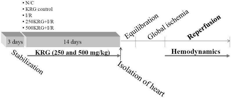
The animals were divided into five groups as shown. Animals in group 1 were health (non-I/R) guinea pigs and served as the normal control (N/C). Animals in group 2 were served as KRG control received with 500 mg/kg of KRG (non-I/R). Animals in group 3 were I/R-induced and KRG-untreated guinea pigs (served as I/R control). Group 4 (served as 250KRG+I/R) and group 5 (served as 500KRG+I/R) were treated orally with the KRG at doses of 250 and 500 mg/kg/day for 14 days, then ischemia was induced for 60 min and reperfusion for 120 min (n=8, each group) (Fig. 1). For treatment, KRG was dissolved in tap water at the doses of 250 and 500 mg/kg. At the end of the experiments, for biochemical and antioxidant analysis the coronary effluents were quickly frozen in -80℃ and cardiac tissues were fixed in 10% formalin, respectively.
Preparation of isolated heart
After pretreatment with KRG (250 and 500 mg/kg) for 14 days, the guinea pigs were anesthetized with pentobarbital (25~30 mg/kg, intraperitoneally). After median sternotomy, the heart was rapidly excised and then immersed in an ice-cold perfusion solution to prevent myocardial injury during the remainder of the procedure as described previously [16]. In brief, standard perfusion was carried out at 37℃ with a modified Krebs-Henseleit bicarbonate (KH) solution containing 118 mM NaCl, 4.7 mM KCl, 1.2 mM MgSO4, 1.2 mM KH2PO4, 25 mM NaHCO3, 10 mM glucose, 1.9 mM CaCl2, and 0.5 mM Na-EDTA., equilibrated with 94.4% O2 and 5.6% CO2 (pH 7.4). The veins entering the right atrium were ligated, so that coronary sinus effluent passed into the right ventricle and was ejected through the pulmonary artery. Coronary flow was continuously recorded with a flowmeter connected to the pulmonary artery [17].
Hemodynamic measurement
To study the effects of KRG, hemodynamic data after a 120 min reperfusion period were compared for changes in aortic flow, coronary flow, cardiac output and LVSP. Aortic flow was measured by the flow volume ejected from the aorta to the cannula located 100 cm above the heart. Coronary flow was also was measured by the timed collection of perfusate from the pulmonary trunk. Cardiac output was calculated by summing the aortic and coronary flows. LVSP was recorded by a transducer connected to the aortic cannula. In addition, the maximal rate of contraction (+dP/dtmax) and the maximal rate of relaxation (-dP/dtmax) are considered indices of ventricular contractility [18]. Therefore, in the study, the +dP/dtmax and -dP/dtmax values were recorded at 30 min intervals throughout the 120 min reperfusion period.
Preparation of ECG recording
As soon as the heart was attached to the isolated heart system, ECG recordings were taken from the epicardial surface. Two silver wire electrodes were placed on the epicardial surface. Signals from both electrodes were amplified by an electric amplifier (AB-621G, Nihon-Kohden, Tokyo), recorded on a personal computer (PC-9801VX, NEC, Tokyo) via an A/D converter (Analog-Pro Jr., Canopus Electric, Kobe), and analyzed with WAVE MASTER II and WM Read (Canopus Electric, Kobe) as described previously [19,20]. In the ECG recording, we measured the QRS interval or the "conduction interval," the QT interval which represents the "repolarization time," and the RR interval which signifies the "time between two consecutive R waves." In present study, if cardiac rhythm irregularities occurred during the stabilizatioin period, the heart was discarded.
Biochemical assays
The coronary effluent was collected throughout the 30 min stabilization and 120 min reperfusion separately, and stored at -80℃. Ischemic damage was assessed using LDH level [21], CK-MB activity [22] and cTnI level [23]. The LDH and CK-MB activities were determined with Hitachi 917 automated analyzer using commercial kits supplied from Roche Diagnostic (Mannheim, Germany). Troponin I levels were measured in ACS: 180 automated chemiluminescence system using commercial kits supplied from Bayer Diagnostics (Cedex, France). Also, oxidative stress was determined from cardiac tissue homogenates using malondialdehyde (MDA) and glutathione (GSH) content analysis. Briefly, following the 120 min reperfusion period, the hearts were rapidly arrested and stored at -80℃. The tissues were homogenized in 0.1 M phosphate-buffer (pH 7.4) with Ultra Turrax homogenizer (IKA T18 basic, Wilmington NC, USA). The homogenates were centrifuged at 5,000 rpm at 4℃ for 10 min, and the supernatants were removed and assayed for MDA and GSH levels. The levels of MDA [24] and GSH [25] content were measured using the methods referenced above.
Statistical analysis
All statistics were calculated using SigmaPlot for Windows version 12.0 (Systat Software, Inc., IL, USA). Data were subjected to one-way analysis of variance (ANOVA) and one-way repeated measures ANOVA. If statistical significance was established, the values from the control group and those from the other groups were compared using the Bonferroni t-test. For all studies, statistical significance was considered at p<0.05.
Go to :
RESULTS
UPLC analysis for ginsenosides contents in KRG
The contents of ginsenosides in KRG were composed of ginsenoside Rg1, 2.01 mg/g; Rb1, 8.27 mg/g; Rg3S, 1.04 mg/g; Re, 2.58 mg/g; Rc, 3.90 mg/g; Rb2, 3.22 mg/g; Rd, 1.09 mg/g; Rf, 1.61 mg/g; Rh1, 0.95 mg/g; Rg2S, 1.35 mg/g; and other minor ginsenosides and components.
Treatment of KRG improve the cardiac hemodynamic function
The effect of KRG was assessed by measuring cardiac function including aortic flow, coronary flow and cardiac output at 30 min intervals throughout the 120 min reperfusion. Aortic flow, coronary flow, and cardiac output were substantially decreased by I/R induction for 120 min to an average of 62.3±4.3%, 72.4±3.7% and 64.14±4.8% (compared to N/C as 100%), respectively. However, pretreatment with KRG (250 and 500 mg/kg) for 14 days increased aortic flow, coronary flow and cardiac output to an average of 71.5±4.7%, 81.6±4.9% and 73.2±5.1% using 250 mg/kg KRG, and to an average of 78.4±3.4%, 87.6±3.2% and 80.4±3.4% using 500 mg/kg KRG (compared to N/C as 100%), respectively (Fig. 2). Furthermore, I/R induction significantly decreased average LVSP values to 61.2±3.9 mmHg as compared to N/C (90.2±3.4 mmHg). In contrast, pretreatment with KRG significantly increased LVSP values to an average of 69.1±3.6 mmHg in 250 mg/kg of KRG and 75.3±3.6 mmHg in 500 mg/kg of KRG, respectively (Fig. 3). Meanwhile, compared to an average +dP/dtmax value of 1318.7±104.7 mmHg after 120 min reperfusion in N/C group, I/R induction resulted in a significant fall in average +dP/dtmax values to 696.2±95.4 mmHg, whereas pretreatment with KRG for 14 days significantly increased the +dP/dtmax values to an average of 919.3±98.0 mmHg in 250 mg/kg KRG and 979.0±94.2 mmHg in 500 mg/kg KRG, respectively (Fig. 4A). Under pretreatment of KRG for 14 days, the average -dP/dtmax values were 1211.2±86.1 mmHg in N/C group and 624.1±90.7 mmHg in I/R group. However, KRG significantly increased -dP/dtmax values to an average of 809.2±90.7 mmHg in 250 mg/kg KRG and 909.7±89.4 mmHg in 500 mg/kg KRG, respectively (Fig. 4B). As shown in Fig. 2, 3 and 4, there was no difference between hemodynamic parameters such as LVSP and ±dP/dtmax between N/C and KRG control groups. These results suggest that KRG per se did not influence cardiac function in the present study.
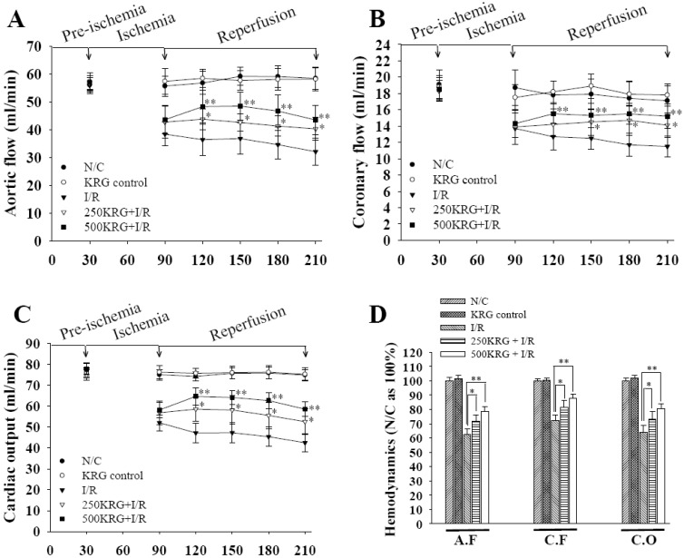 | Fig. 2Effects of 250 and 500 mg/kg KRG between aortic flow (A), coronary flow (B), cardiac output changes (C) and average percents for 120 min reperfusion on these hemodynamics (D). Each histogram represents the mean±SD (n=8). *p<0.05, **p<0.01 compared with the I/R group, respectively.
|
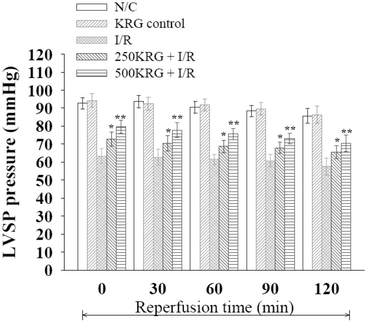 | Fig. 3Effects of 250 and 500 mg/kg KRG on left ventricular systolic pressure (LVSP). LVSP was estimated at 30 min intervals throughout the 120 min reperfusion period. Results were representative of eight independent experiments. Values are expressed as mean±SD. *p<0.05, **p<0.01 compared with the I/R.
|
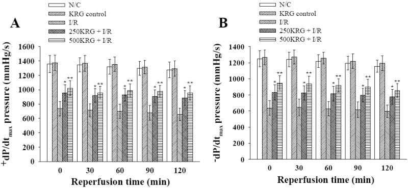 | Fig. 4Effects of 250 and 500 mg/kg KRG on the maximal rate of change in left ventricular contraction (+dP/dtmax) (A) and the maximal rate of change in left ventricular relaxation (-dP/dtmax) (B). Results were representative of eight independent experiments. Values are expressed as mean±SD. *p<0.05, **p<0.01 compared with I/R.
|
Treatment of KRG improves electrocardiographic parameters such as QRS, QT and RR intervals
As shown in Fig. 5A, the N/C group showed a normal ECG pattern. And, the KRG control group did not show any abnormal changes in ECG pattern compared with N/C. This indicates that ECG patterns were not affected by 500 mg/kg of KRG. Upon examination of the ECG patterns in I/R control, the QRS interval tend to be significantly prolonged compared to that of the N/C group (Fig. 5B). In the I/R group, the average QRS values were 138.7±4.51% during 120 min reperfusion (compared to 100% being the average of the N/C for 120 min). Whereas, the QRS interval was significantly shortened in the 250 and 500 mg/kg KRG groups (Fig. 5B). To be exact, the average values of the QRS interval were 129.7±3.8% in the 250 KRG mg/kg group and 118.6±4.3% in the 500 KRG mg/kg group. The QT interval showed a normal ECG pattern in the N/C. On the other hand, the QT interval in I/R control was significantly prolonged compared with the N/C group. However, the QT interval was significantly shortened by pretreatment with 250 and 500 mg/kg KRG. The average QT interval values were 119.4±2.4% in 250 mg/kg KRG and 112.6±2.7% in 500 mg/kg KRG compared with those of I/R control (i.e. 124.5±3.3%, N/C being as 100%). As shown in Fig. 5C, in terms of the QT interval during 120 min reperfusion, pretreatment with 500 mg/kg KRG was more effective than that seen with 250 mg/kg. In addition, the RR interval should be similar to those seen in N/C group (Fig. 5D). However, in I/R control, the RR interval was significantly prolonged compared to N/C. In I/R control, the average RR interval during the 120 min reperfusion was 118.1±3.4% compared to the average of 100% in N/C. In contrast, the RR interval after pretreatment with 250 and 500 mg/kg KRG for 14 days was significantly shorter than those seen in I/R control (Fig. 5D). Specifically, the average value of the RR interval was 110.9±3.3% in 250 mg/kg KRG and 107.3±2.2% in 500 mg/kg KRG. There were no significant differences between ECG parameters such as QRS, QT and RR intervals between N/C and KRG control (data not shown). These results suggest that KRG per se did not influence ECG function.
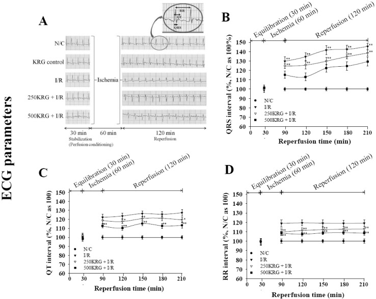 | Fig. 5Effects of 250 and 500 mg/kg KRG on representative electrocardiogram tracings. Enlarged ECG patterns such as QRS complex, QT and RR intervals are shown (the part shown in circle area on ECG of NC) (A). Namely, each group represents normal control (N/C), KRG only treated group (KRG control), I/R control (I/R), 250KRG+I/R, indicating ischemia and reperfusion treated 250 mg/kg KRG and 500KRG+I/R, indicating ischemia and reperfusion treated 500 mg/kg KRG. These pictures were representative ECG patterns in each group. Also, influence of 250 and 500 mg/kg KRG on QRS (B), QT (C) and RR intervals (D) was shown. Values are expressed as mean±SD for eight independent experiments in each group. *p<0.05, **p<0.01 compared with I/R.
|
Decreased cardiac tissue damage is observed in isolated heart when treated with KRG and KRG is required in antioxidant activities
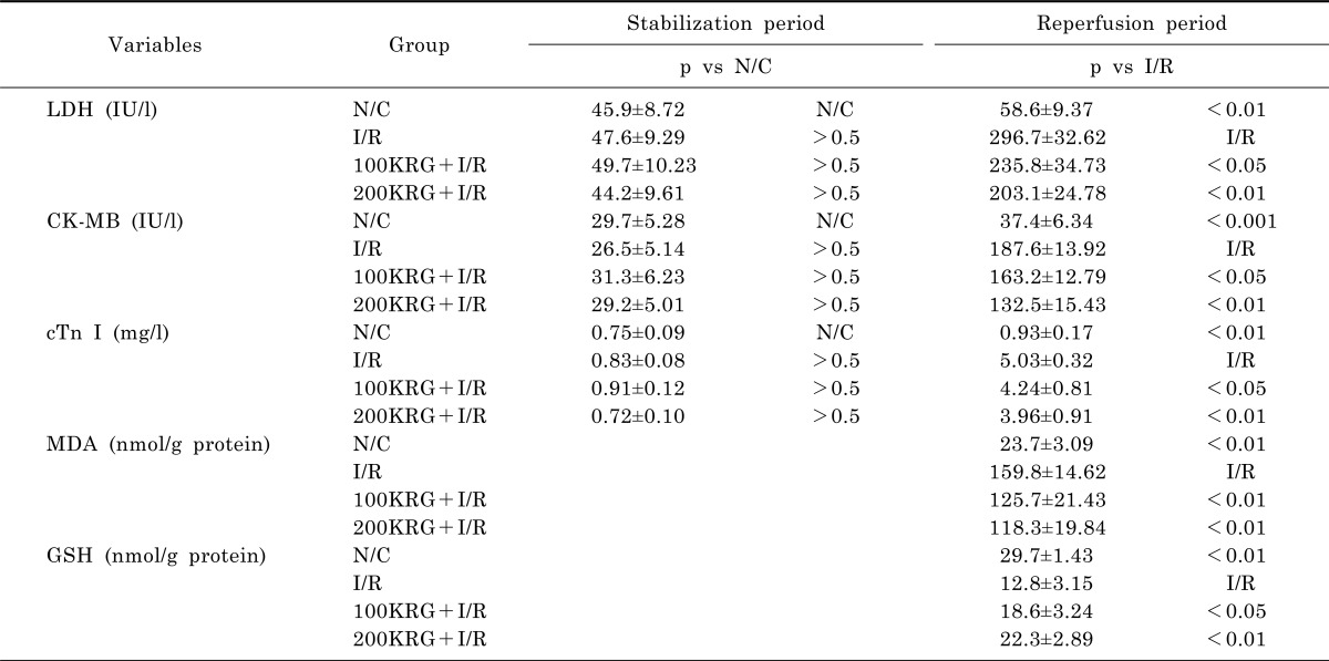
There were no significant differences in the biochemical parameters of coronary effluents such as LDH, CK-MB and cTnI levels among all groups following the stabilization period (Table 1, p>0.5). However, significant differences were observed during the 120 min reperfusion between each of the groups. LDH, CK-MB and cTnI levels during the reperfusion period in the group pretreated with 250 mg/kg KRG were significantly lower than those in I/R control group (p<0.05). Significantly less cardiac tissue damage was also found in the 500 mg/kg KRG throughout the 120 min reperfusion (p<0.01) (Table 1). Additionally, after reperfusion, the MDA levels in I/R group were significantly higher than N/C group, while pretreatment with 250 and 500 mg/kg KRG resulted in a significant decrease. Likewise, GSH level was significantly higher in the groups pretreated with 250 and 500 mg/kg KRG than in I/R control (Table 1). As shown in Table 1, pretreatment with 500 mg/kg KRG was more effective than 250 mg/kg with regard to the normalization of biochemical and oxidative stress indicators for GSH.
Go to :
DISCUSSION
In the present study, because of its free radical scavenging activities and enhancement of various hemodynamic factors, ginseng saponins showed the promising regulatory effects against cardiac I/R. Given its important antioxidant activity, together with the consistent regulatory effects on the hemodynamic parameters such as LVSP and ±dP/dtmax, these results suggest that the post-ischemic protective effects of ginseng saponins may be partly due to its antioxidative properties as well as by activation of antioxidative enzyme. Oxidative stress is well known as a primary contributing factor in the pathophysiology of cardiovascular disorders [26]. It is well reported that susceptibility to oxidative stress is higher in the cardiac tissue than in other tissue because of low levels of antioxidant enzymes [27]. Therefore, treatment of antioxidant agents can be an important therapeutic strategy to prevent cardiac ischemic damage by reactive oxygen species [28]. Also, it is reported that I/R damage involves oxygen radical formation [29]. Therefore, increased antioxidants could protect cardiac tissue from oxidative stress associated with I/R damage. In these experiments, we examined the influence of KRG on cardiac damage resulting from I/R on guinea pig and evaluated the cardioprotective effects of KRG. In previous studies, we reported that ginseng saponins have protective effects against reperfusion injury in the rat heart [15], but to the best of our knowledge, there has not been a report on these effects in guinea pig.
In the present study, the consequences of can be subdivided into three points. First, pretreatment of KRG resulted in an increase in coronary flow, aortic flow, cardiac output and LVSP. In this regard, the reason for increased coronary flow in the KRG-treated group may be due to the preconditioning-like role of KRG. Second, KRG preserved cardiac ventricular function as evidenced by the significant increases of -dP/dtmax indicating diastolic function as well as +dP/dtmax during reperfusion. Also, we found that I/R induce a changes in ECG parameters such as QRS complex, the QT and the RR interval. These results may be due to cardiac dysfunction induced by I/R, especially since the QT interval represents the periods of the ventricular depolarization and repolarization [30]. Therefore, our results revealed that cardiac ischemia can alter the repolarization period. It is known that these variations between the QT and RR intervals may have clinically important meaning [30]. Whereas, pretreatment with 250 and 500 mg/kg KRG inhibited the occurrence of pathological ECG patterns such as QRS complex, QT and RR interval, suggesting the protective effect of KRG. Third, KRG showed concentration-dependent cardioprotective effect by normalizing biochemical and oxidative parameters. The process of cardiac ischemia is a multiple process leading to the production of reactive oxygen species, which results in severe tissue injury [31] and finally lead to cardiac death [32]. Normally, ROS are eliminated by antioxidant systems [31]. Our present results suggested that the MDA, which is an index of the injury in lipid tissues by ROS, were found to be lower level in the KRG-treated groups. Similarly, GSH, which also indicates as a tissue defense system against ROS, were found to be higher level in the treated groups with 250 and 500 mg/kg KRG, indicating that KRG decreases oxidative stress. In addition, lower levels of biochemical parameter such as LDH, CK-MB and cTnI in KRG-treated groups indicate that there is lower I/R damage. Taken together, these effective results pointed towards an improved outcomes with the use of KRG. Also, these results may be due to the active concentration following treatment of KRG. In conclusion, in present study, KRG is suggested as a traditional medicine that provides beneficial effects against I/R-associated cardiac alterations and dysfunction in an ex vivo approach.
Go to :
 XML Download