

 PDF
PDF ePub
ePub Citation
 Print
Print


INTRODUCTION
Liver and kidney are both incorporated in the modulation of body homeostatic functions, metabolism and elimination of drugs and toxic products [1]. Any injury to either the liver or renal tissue may influence the other. Ischemia-reperfusion (IR) injury triggered acute renal damage is a frequent drawback in surgical procedures such as organ transplantation. The latest studies have established that acute kidney damage triggered by IR leads to dysfunction in liver [2]. Renal IR injury decreases antioxidant enzyme activities [1,3] and increases neutrophils and lymphocytes accumulation [4], oxidative stress and lipid peroxidation [1,5] in liver tissue.
Oxytocin (OT), a neurohypopysial nonapeptide generated in the paraventricular and the supraoptic nuclei in the hypothalamus, exhibits a wide spectrum of central and peripheral activities [6]. OT has reproduction related classical functions such as arousal of the uterine contractions at parturition and myoepithelial contractions in the mammary gland during suckling [7]. Besides its classical functions, OT exhibits a potent antistress [8], anti-inflammatory [6,9] and antioxidant effect [10,11]. In experimental IR models, OT represses neutrophil infiltration and controls the activation of proinflammatory mediators [10,11].
According to these data, this research was planned to explore the potential protective effect of OT against remote liver damage induced by renal IR in rats, by using biochemical and histological arguments.
Go to : 
METHODS
Animals
Twenty four Wistar albino male rats, each weighing between 200 and 240 g, were used in the present study. In the course of experiment, the rats were kept in constant laboratory conditions recommended by NIH, and were maintained with standard laboratory conditions. The study was initiated after obtaining approval from Dicle University Local Committee on Animal Research Ethics. We meticulously complied with the principles for the "Protection of Animal Rights" specified by the National Institutes of Health (NIH) during the entire course of the study.
Experimental method and procedure
The rats were put under anaesthesia with ketamine (75 mg/kg i.p.) and xylazine (8 mg/kg i.p.). Body temperature was preserved in every part of surgery at 37±1℃. All rats were submitted to surgical exposure of the left and right renal pedicles via midline incision. To create renal ischemia, both renal pedicles were blocked for 45 min with vascular clamps. After 45 min of blockage, the clamps were taken away, and kidneys were examined to endure reperfusion for 24 hours. The rats were randomly divided into four different groups (n=8). The groups were as follows: (1) Sham operated group; (2) Sham operated+OT group (3) Renal IR group; (4) Renal IR+OT group. OT (500µg/kg) (Sigma, St. Louis, MO, USA) was administered subcutaneously 12 and 24 hours before and immediately after ischemia. By the termination of experimental procedure, the rats were sacrificed. The excised liver tissue specimens for biochemical assay were weighed, and instantly stored at -70℃. Part of liver was fixed in 10% neutral formalin for histological examination.
Biochemical analyses
The liver tissues were washed with 1.15% ice-cold KCl, minced, then homogenized in five volumes (w/v) of the same solution. Measurements were performed on the supernatant of the homogenate that was prepared at 14.000 rpm for 30 min at +4℃. The protein density of the tissue was determined by the method of Lowry [12]. Lipid peroxidation level in the liver was represented as malondialdehyde (MDA). It was determined by the method of Ohkawa et al. [13]. Consistent with this method, samples were mixed with sodium dodecyl sulfate, acetic acid and 2-thiobarbituric acid. After vortexing, tissue samples were incubated for 1 h in 95℃ and butanol-pyridine 15:1 (v/v) was added. The mixture was shaken for 10 min and then centrifuged. Butanol-pyridine layer was measured fluorometrically (UV-1205 Shimadzu) at 552 nm. Data are shown as nmol/g protein. Nitric oxide (NO) levels were assessed with Griess' method [14]. Paraoxonase (PON-1) activity was evaluated spectrophotometrically by modified Eckerson [15] method.
The total antioxidant status (TAS) of supernatant fractions was evaluated by using a novel automated and colorimetric measurement method developed by Erel [16]. Hydroxyl radicals, the most potent biological radicals, are produced in this method. In the assay, the ferrous ion solution present in reagent 1 is mixed with hydrogen peroxide, which is present in reagent 2. The subsequently produced radicals, such as brown-colored dianisidine radical cations produced by the hydroxyl radicals, are also potent radicals. Using this method, the antioxidative effect of the sample is measured against the potent free radical reactions initiated by the produced hydroxyl radicals. The assay has excellent precision values lower than 3%. The assay is calibrated with Trolox (Vitamin E), and the TAS results are expressed as nmol Trolox equivalent/mg protein. The total oxidant status (TOS) of supernatant fractions was measured by using a novel automated and colorimetric measurement method [17]. Oxidants present in the sample oxidize the ferrous ion-o-dianisidine complex to ferric ion. The oxidation reaction is increased by glycerol molecules, which are abundantly present in the reaction medium. The ferric ion makes a colored complex with xylenol orange in an acidic medium. The color intensity, which can be measured spectrophotometrically, is related to the total amount of oxidant molecules present in the sample. The assay is calibrated with hydrogen peroxide, and the results are expressed in terms of nmol H2O2 equivalent/mg protein [18]. The TOS/TAS ratio was considered to be the oxidative stress index (OSI). The units of liver tissue TOS and TAS were µmole H2O2 Equiv./gram protein and mmole H2O2 Equiv./gram protein, respectively [19].
Histopathological assessment
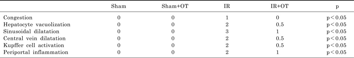
Liver injury was analyzed under light microscopy. All tissue specimens were taken from the central part of the right lobe of the liver and were fixed in 10% neutral buffered formalin solutions, refined for implanting in paraffin by routine protocols, and 4µm thick sections were then cut by microtome. The sections were stained with Hematoxylin-Eosin by using a routine protocol and examined with a photomicroscope (Eclipse 50i, Nikon). Congestion, hepatocyte vacuolization, sinusoidal dilatation, central vein dilatation, Kupffer cell activation and periportal inflammation were scored as 0 (no change), 1 (minimal change), 2 (moderate change), and 3 (severe change) and were assessed in a blinded manner.
Statistical analysis
Statistical analysis was performed by using GraphPad Prisma V3 program (GraphPad Software, Inc. San Diego, USA). Data were shown as means±standard deviation, and Kruskal-Wallis test was used for analysis. In the event of significant results, the Mann-Whitney U test was employed for comparisons of differences between the two independent groups. Histopathological results were expressed as median values and analyzed by Kruskal-Wallis and Mann-Whitney U test. A p value <0.05 was considered statistically significant.
Go to :
RESULTS
Biochemical results
The MDA, TOS, TAS, OSI and NO levels and PON-1 enzyme activities in the liver tissues are presented in Table 1. MDA, TOS and OSI levels were notably higher, and TAS, NO and PON-1 levels were notably lower in the IR as compared to the sham operated and sham+OT groups (p<0.01). However, TAS, NO and PON-1 levels were notably increased in oxytocin-treated group compared to the IR group (p<0.01). MDA levels were notably decreased in IR+OT group compared to the IR group (p<0.05).
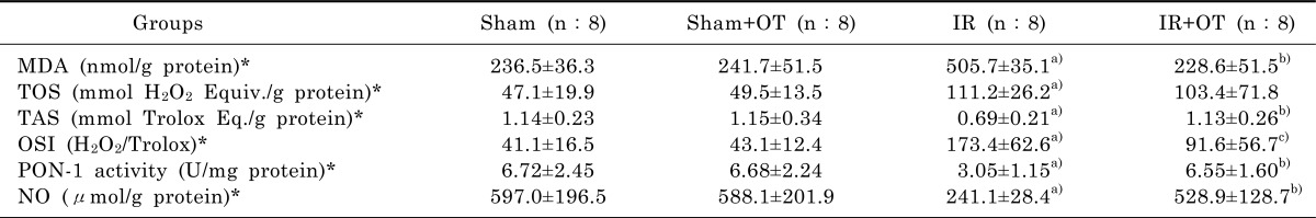
Table 1
Levels of MDA, TOS and TAS, OSI, PON-1 and NO in groups
Results are presented as means±standard deviation. *p<0.01 for Kruskal Wallis test. a)p<0.01 as compared to the sham operated and sham+OT groups, b)p<0.01 as compared to the IR group. c)p<0.05 as compared to the IR group. IR, ischemia-reperfusion; OT, oxytocin; MDA, malondialdehyde; TOS, total oxidant status; TAS, total antioxidant status; OSI, oxidative stress index; PON-1, paraoxonase activity; NO, nitric oxide.
![]()
Histopathological results
The histopathological findings are shown in Table 2. Light microscopic evaluation revealed that IR group resulted in widespread hepatic injury with marked congestion, hepatocyte vacuolization, sinusoidal dilatation, central vein dilatation, Kupffer cell activation and periportal inflammation (Fig. 1C). Histopathological alterations that were observed in IR group were significantly improved in IR+OT group (Fig. 1D). There were minimal changes in sham (Fig. 1A) and sham+OT (Fig. 1B) groups.
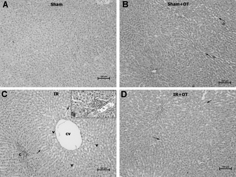 | Fig. 1Photomicrographs of Hematoxylin and Eosin stained sections from liver of rats. Minimal changes observed in sham (A) and sham+OT (B) groups. Congestion, hepatocyte vacuolization, sinusoidal dilatation, central vein dilatation, Kupffer cell activation and periportal inflammation are seen in IR group (C) sections. Histopathological alterations that were observed in IR group were significantly improved in IR+OT group (D). cv, central vein; v, vacuoles; kc, Kuppfer cells (inset); asterisk, inflammatory cells (inset); c, congestion; arrows, sinusoidal dilatation; IR, ischemia-reperfusion; OT, oxytocin.
|
Go to :
DISCUSSION
Acute renal failure appears in almost 5% of hospitalized patients and up to 30% of patients in intensive care units [20,21]. Acute renal ischemic damage persists to be linked with a high mortality rate and arises in many clinical situations, such as urinary tract surgery, septic shock cases, organ transplantation and trauma [22]. Numerous studies have shown that renal injury affects additional organs including the liver [1,5,22]. In this work, we have investigated the potential effect of OT on liver injury triggered by renal I/R. To that end, we measured the levels of liver MDA, TOS, TAS, NO and PON-1 activity.
IR of the tissue was determined to be connected with lipid peroxidation, which is an autocatalytic process causing oxidative demolition of the cellular membranes, and their breakdown can contribute to the generation of toxic, reactive products and cell death [5,23-25]. Lipid peroxidation, as a free radical-producing system, has been proposed to be tightly linked to IR-triggered tissue damage, and MDA is an important parameter of oxidative stress and a good pointer of lipid peroxidation [5,26,27]. In this research, in renal IR group, the level of MDA notably elevated in the liver tissue, while subcutaneously administered OT repressed MDA raising notably. This finding indicates that renal IR induced lipid peroxidation is likely to be improved with OT administration in liver tissue.
In our study, we evaluated oxidative status as TOS and TAS along with the measurement of OSI, a pointer of oxidative stress, which reveals the redox balance between oxidation and antioxidation [28]. Because of difficulties in measuring the levels of different oxidant molecules such as superoxide radical anion, hydrogen peroxide one by one, we measured TOS in serum as previously described by Erel [17]. Likewise, we appraised TAS, instead of measuring antioxidant molecules separately following the methods of Erel [29] and Cikrikcioglu et al. [30]. Lately, it has been widely documented that OSI may reveal the oxidative status more correctly than TOS or TAS level alone [31,32]. In the present study, the levels of TOS and OSI were notably augmented, and the level of TAS was notably reduced in IR group compared with sham operated group. On the other hand, in OT treated IR rats, TAS level was notably increased with respect to IR group. These results showed that renal IR leads to increased oxidative stress in liver tissue, and that the rising of oxidative stress was prevented by application of OT.
PON-1 is an enzyme associated with high-density lipoprotein that is mainly secreted by the liver. Although its physiological function has not been completely clarified, it seems that PON-1 is responsible for hydrolyzing lipid peroxides, also playing a major role in the antioxidant system [33,34]. PON-1 protects liver against inflammation, liver disease and fibrosis [35,36].
In this research, we revealed decreased PON-1 activity in the liver tissues of IR rats compared to sham operated rats. OT treatment reversed the reduced activity of PON-1 in IR+OT group. OT treatment prevented the reduced activity of PON-1 in liver tissue. These findings demonstrate that a decrease in PON-1 activity is related to oxidative damage in the liver tissues caused by renal IR and OT treatment reverses the decline of PON-1 activity in these tissues. Augmented PON-1 activity through OT treatment in these tissues may be associated with antioxidant and anti-inflammatory effect of OT.
NO has protective effects on cells during IR. NO has been demonstrated to inhibit oxidative stress, cytokine release, apoptosis, adhesion and aggregation of neutrophil leukocytes [24,37-39]. Several studies have shown a relationship between NO and IR injury [10,24,39,40]. A reduction of NO during IR is generally caused by endothelial dysfunction and reduction of endothelial nitric oxide synthase activity [41,42]. In this study, renal IR reduced the NO level, and OT treatment increased NO content in hepatic tissue. These findings showed that renal IR injury is associated with endothelial dysfunction and reduction of nitric oxide synthase activity. OT, which is revealed to be protective in IR induced remote liver injury, may exhibit its effect by increasing NO synthase activity and releasing NO from vascular endothelium.
In our research, we also investigated the histopathological consequences of exogenously administered OT in liver tissue. In IR group, intense hepatocyte vacuolization, evident sinusoidal dilatation, central vein dilation, Kupffer cell activation and periportal inflammation were observed. These alterations pointed out a severe damage in liver paranchyme, and the impairment of liver tissues in IR group was notably more severe than the IR+OT group.
These data show that renal ischemia provokes destructive alterations in liver histology, oxidative stress and induces a diminution in hepatic antioxidant capacity. OT ameliorates remote liver injury triggered by renal ischemia-reperfusion, and this preservation is related with suppression of inflammation and regulation of oxidant-antioxidant status.
Go to :
 XML Download