

 PDF
PDF ePub
ePub Citation
 Print
Print


ABBREVIATIONS
CMC
carboxy methyl cellulose
ECQ
extracts containing QGC
GSH
glutathione sulfhydryl
HETAB
hexadecyl trimethyl ammonium bromide
MDA
malondialdehyde
MPO
myeloperoxidase
QGC
quercetin-3-O-β-D-glucuronopyranoside
ROS
reactive oxygen species
TMB
tetra methylbenzidine
TBA
thiobarbituric acid
TBARS
thiobarbituric acid reactive substance
INTRODUCTION
Flavonoids, which are secondary metabolites in plants, are considered relatively non-toxic bioactive substances and play diverse biological effects, such as anti-inflammatory, anti-oxidant, anti-allergic, hepatoprotective, anti-thrombotic, anti-viral, and anti-carcinogenic activities [1-3]. The flavonoids are typical phenolic compounds and act as potent metal chelators and major ability of free radical scavengers [4]. Among the flavonoids, quercetin has gained special attention since it is antioxidant which scavenges highly reactive biological species such as peroxynitrite [5,6] and the hydroxyl radical [7] very efficiently.
Rumex Aquaticus is a family of Polygonaceae and this plant was used as a drug for disinfestations, diarrhea, anti-pyretic drug, edema, jaundice and constipation in the traditional oriental medicine. In the previous study performed in our research group, quercetin-3-β-D-glucuronopyranoside (QGC) was isolated from Rumex Aquaticus through several steps (Korean Intellectual Property Office, 10-0537432). QGC had more potent effect than quercetin on inhibition of experimental acute and chronic gastritis and ethanol-induced gastritis in Sprague-Dawley rats in vivo [8].
The Rumex Aquaticus Herba extracts containing quercetin-3-β-D-glucuronopyranoside (ECQ) was extracted from Rumex Aquaticus by ethanol. In our preliminary study, ECQ revealed protective effects on indomethacin or ethanol-induced gastric damage (not published yet).
Reflux esophagitis is one of the common gastrointestinal diseases that are increasingly recognized as a significant health problem. Abnormalities of the defense mechanisms in the esophagus are important in the pathophysiology of the disease. However, the degree of damage to esophageal mucosa also depends on the composition of the refluxed materials, the amount and duration of the reflux, and the defensive factors within the esophageal mucosa itself. Reflux esophagitis is caused by gastric juice gains access to the esophagus via imperfect lower esophageal sphincter relaxation [9], speed of esophageal clearance and mucosal resistance and other factors [10]. It has been believed that reflux of gastric juice causes ulceration and destruction of epithelium of esophagus. However, the exact pathophysiological mechanisms of esophageal cell damage during gastro-esophageal reflux are not fully explained by acid reflux alone.
Recently, experiments on rats and studies of esophageal biopsy samples from patients have shown that mucosal damage is mediated primarily by oxygen-derived free radicals accompanied by enhanced esophageal mucosal lipid peroxidation [11]. In addition, administration of various free-radical scavengers has been found to prevent esophageal mucosal damage induced by mixed reflux of gastro-duodenal contents, whereas acid suppression with ranitidine alone is not effective either in decreasing the degree of reflux esophagitis or in attenuating inflammation associated oxygen-derived free radical-mediated nuclear factor-κB activation [12].
Oxygen-derived free radicals have been known to play an important role in the pathogenesis of the injury of various tissues including the digestive system [13]. It has been shown that oxygen-derived free radicals lead to acute gastric and esophageal mucosal injury due to ischemia [14], non-steroidal anti-inflammatory drugs (NSAIDs) or ethanol [15]. Free radicals are also known to act as carcinogens, since they lead to DNA damage [16]. It has also been demonstrated that free radical damage to the gastric or esophageal mucosa [17] can be prevented by the administration of free radical scavengers.
Therapeutic medicines for reflux esophagitis are H2-receptor antagonists, prokinetic agents and proton pump inhibitors. H2-receptor antagonists and prokinetic agents promote symptomatic relief and esophageal healing in mild esophagitis, but are less effective in the treatment of moderate to severe esophagitis. For patients with moderate to severe esophagitis, rapid symptomatic relief and esophageal healing have been achieved with proton pump inhibitors through their profound and long-lasting anti-secretory activities [18].
This study was aimed at evaluating the protective effects of ECQ on i) the development of the reflux esophagitis induced by surgery procedure in rats, ii) gastric secretion, iii) myeloperoxidase (MPO) activity iv) lipid peroxidation which is a marker of oxidative stress, and v) GSH levels. Omeprazole was used as a reference drug for surgically induced reflux esophagitis in rats.
METHODS
Materials
ECQ was thankfully supplied by the Department of Pharmacognosy (Prof. Whang, Chung-Ang Univ., Seoul, Korea). MDA assay kits were purchased from BioRad (CELL BIOLABS, INC). Ether, omeprazole, carboxymethylcellulose (CMC), hexadecyl trimethyl ammonium bromide (HETAB), hydrogen peroxide and dimethylformamide were purchased from Sigma (St. Louis, MO, USA). Protein assay kits were purchased from BioRad (Richmond, CA, USA).
Animals
Male Sprague-Dawley rats with a body weight of about 200~220 g were used for the experiments. The rats were starved for 24 hours before the experiments, but were freely allowed to drink water. All animals were kept in raised mesh bottom cages to prevent coprophagy. All animal experiments were approved by the Institutional Animal Care and Use Committee of Chung-Ang University, in accordance with the guidelines for the Care and Use of Laboratory Animals in Seoul, Korea.
Generation of reflux esophagitis in rats
Experiments were carried out under general anesthesia. The abdomen was incised along the midline and then the pylorus and the limiting ridge which is a transitional region between the forestomach and corpus were simultaneously ligated [9]. A longitudinal cardiomyotomy of about 1 cm length across the gastroesophageal junction was performed to enhance reflux. The vagus nerves were left intact. Thirty five rats were divided into seven groups of 5 rats each. For a normal group, a midline laparotomy alone without further surgical or medical interventions was performed. In a control group, a surgical procedure was done to induce reflux esophagitis, but ECQ was not treated. But, other 5 groups with reflux esophagitis induced by the surgical procedure were treated with ECQ (1, 3, 10, and 30 mg/kg) and omeprazole (30 mg/kg). The test drugs were administered immediately after the ligation. All seven groups of rats were starved of food for 24 hours after surgery, but had free access to water. After 24 hours, the rats were sacrificed under deep ether anesthesia and the esophageal portion of the digestive tract was excised. The esophageal mucosa was stripped off from muscle layer, and stored at -80℃ for the following biochemical assays including MPO activity, MDA level, and reduced glutathione level.
Evaluation of esophagus lesions
After 24 hour experiments, the animals were sacrificed and the esophagus was excised, opened along the greater curvature and spread out with pins on cork board. The area (mm2) of mucosal erosive lesions was measured under a dissecting microscope with a squared grid (X10; Olympus, Tokyo, Japan). The number of animals used in each experiment is indicated in the respective figure legends.
Evaluation of gastric secretion
Twenty four hours after pylorus ligation, rats were sacrificed by cervical dislocation and the esophagus clamped. Samples of gastric juice were collected in graduated conical centrifuge tubes and centrifuged at 3,000 g for 10 min at 4℃. After centrifugation, the supernatant was measured for volume (ml/rat), pH (Toledo 320, Mettler, Swiss) and acidity (mEq/l). Total acidity was determined by titration of the gastric juice against 0.1 N NaOH to pH 7.0. Acid ouput was expressed as µEq/hr [19].
Biochemical investigation of esophagus tissues
MPO activities and MDA levels in rat esophagus tissues were determined. To prepare the tissue homogenates, esophagus tissues were cut with iris scissors. The grinded tissues were then treated with 2.0 ml of phosphate or tris buffer. The mixtures were homogenized on ice using a homogenizer (TMZ-20DN, TAEMIN, Korea) for 30 sec. Homogenates were centrifuged at 600 g for 10 min at 4℃ and then supernatant was sonicated for 45 sec, and recentrifuged at 12,000 g for 15 min at 4℃ by using a refrigerated centrifuge to obtain a mitochondrial fraction. These supernatants were used for assays.
TBARS assay
Lipid peroxidation was determined according to the method of Buege and Aust by measuring spectrophotometrically the formation of thiobarbituric acid reactive substances (TBARS). Esophageal mucosa was harvested, sonicated in 1ml of Tris-HCl buffer (pH 7.0). After centrifugation at 12,000 g for 15 min at 4℃ (Micro 17 TR, Hanil, Korea), 0.9 ml of 8℃ trichloroacetic acid (TCA) was added to 0.3 ml of supernatant. After centrifugation at 10,000 g for 5 min at 4℃, 0.25 ml of TBA (1%) was added to 1 ml of supernatant and the resulting solution was heated at 100℃ for 20 min. The tubes were cooled, 2 ml of n-butanol was added and each tubes was vortexed for 90 sec. After centrifugation at 3,000 g for 5 min at 4℃, 1 ml of butanol phase was utilized for TBARS asay at 532 nm (UV-160A, Shimadzu, Japan) against malondialdehyde (MDA) standards. Results were expressed as nmol/mg protein. Protein assay was determined according to the Bradford method (1976) using bovine serum albumin as a standard.
Measurement of MPO activity
MPO assay was performed as previously described [20] and partly modified. One milliliter of the leukocyte suspension was centrifuged at 600 g at 4℃ for 7 min. The precipitate was suspended in 1 ml of 80 mM sodium phosphate buffer, pH 5.4, containing 0.5% Hexadecyl trimethyl ammonium bromide (0.5% HETAB solution), sonicated and centrifuged at 12,000 g at 4℃ for 15 min. Duplicate 30 µl samples of resulting supernatant were poured into 96 well microtiter plates. For assay, 200 µl of a mixture containing 100 µl phosphate buffered saline, 85 µl 0.22 M sodium phosphate buffer, pH 5.4, and 15 µl of 0.017% hydrogen peroxide were added to the wells. The reaction was started by the addition of 20 µl of 18.4 mM TMB•HCl in 8% aqueous dimethylformamide. Plates were stirred and incubated at 37℃ for 3 min and then placed on ice where the reaction was stopped by addition to each well of 30 µl of 1.46 M sodium acetate, pH 3.0. The MPO value was evaluated by measuring the absorbance of samples at 620 nm (OD value) using MPO standard.
GSH assay
GSH concentration of esophagus tissue was determined according to Beutler and partially modified method was used [21]. Obtained mitochondrial fraction was added to metaphophoric acid for protein precipitation and stand for 5 min. And phosphate buffer and 5'5-dithiobis-2-nitro-benzoic acid were added for color development. Then GSH was determined by spectrophotometrically at 415 nm using GSH (Sigma) standard.
RESULTS
The effect of ECQ on esophagus lesions in reflux esophagitis model
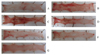
The effects of surgical procedure and agents were shown in Fig. 1. A. Normal. B. Control. C. Treatment with ECQ 1 mg/kg. D. Treatment with ECQ 3 mg/kg. E. Treatment with ECQ 10 mg/kg. F. Treatment with ECQ 30 mg/kg. G. Treatment with omeprazole 30 mg/kg. In the reflux esophagitis models, severe ulcerations were observed in the esophagus of control group which was not treated with any drug. In the control group with reflux esophagitis induced by the surgical procedure, esophagus lesions were significantly observed. But, ECQ and omeprazole treated groups were decreased the development of reflux esophagitis significantly and dose-dependently. This data suggest that ECQ and omeprazole have similar pharmacological activities.
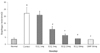
Measurements of the esophagus lesions induced by the surgical procedure were shown in Fig. 2. In the control group, the surgical procedure significantly affected esophagus lesion generation. The control group lesions were about 22.25±4.15 mm2. Treatment with ECQ of 1 mg/kg decreased the esophagus lesions gradually but was not significantly. Treatment with ECQ of 3 mg/kg decreased esophagus lesions by 52% at p<0.05. And 70% of inhibition of esophagus lesions was revealed at treatment with ECQ of 10 mg/kg at p<0.05. ECQ of 30 mg/kg significantly decreased by 80% and omeprazole of 30 mg/kg significantly decreased by 81% of esophagus lesions compared with those of the control group. The result indicated that the dose of ECQ for inhibition of esophagus lesions was 3 mg/kg.
The effect of ECQ on gastric secretion in reflux esophagitis
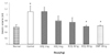
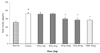
The gastric volume in the control group was significantly increased compared to that in the normal group by 48% at p<0.01 (Fig. 3). ECQ administered by intraduodenal (i.d.) routes gradually decreased the gastric volume in a dose-dependent manner and ECQ of 30 mg/kg significantly decreased by 52%.
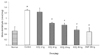
The pH in the control group with reflux esophagitis was decreased, compared to that in the normal group (Fig. 4). After the administration of ECQ, the pH was significantly increased by 25% at p<0.05 and omeprazole also increased the pH by 39% at p<0.05. ECQ and omeprazole administered by i.d. decreased acid output effectively (Fig. 5). Total acidity in the control group was significantly increased by 41% at p<0.05. However, total acidities in ECQ 10 mg and 30 mg treated groups were decreased by 18% at p<0.05 and by 21% at p<0.05, respectively. Similarly, total acidity in the omeprazole treated group was decreased by 22% at p<0.05.
The effect of ECQ on MPO activity in reflux esophagitis
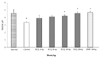
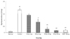
During the initial inflammatory stage, tissue damage caused by ROS or other factors induces infiltration of neutrophil, which can be assessed by myeloperoxidase (MPO) activity. In the control group, the surgical procedure significantly increased MPO activity after 24 hr (p<0.01) (Fig. 6). Treatment with ECQ decreased MPO activity significantly and dose dependently. At the doses of 1 and 3 mg/kg ECQ, the MPO activities were decreased by 23% and 56%, respectively, at p<0.05 compared with that in the control group. And treatment with ECQ 10 mg/kg and 30 mg/kg decreased MPO activity to 85% and 87%, respectively, at p<0.01. The treatment with 30 mg/kg of omeprazole also decreased MPO activity to 87% at p<0.01 compared with the control.
The effect of ECQ on MDA levels in reflux esophagitis models
Because of reactivity of ROS with lipids, MDA levels were significantly increased when the tissue was damaged by surgical procedure which can cause generation of ROS in tissue. As an index of lipid peroxidation, esophagus tissue MDA levels were shown in Fig. 7. The control group induced by surgical procedure markedly enhanced the tissue MDA levels (1.29±0.03 nmol/mg protein, p<0.05) after 24 hours. The treatment with ECQ prevented increasing of MDA levels dose-dependently. When the rats were treated with 3 mg/kg and 10 mg/kg of ECQ, the tissue MDA levels were significantly decreased to 0.80±0.04 nmol/mg protein and 0.74±0.07 nmol/mg protein, resepectively, at p<0.01. In the 30 mg/kg of ECQ treated group, the increased levels of MDA induced by the surgical procedure were decreased to 0.55±0.12 nmol/mg protein at p<0.01. The treatment of omeprazole (30 mg/kg) also decreased MDA level to 0.63±0.30 nmol/mg protein.
The effect of ECQ on GSH levels in reflux esophagitis models
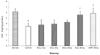
Reflux gastric acid-induced esophagus damage caused esophagus lesions and increased MDA levels. Also it can cause GSH depletion since the reflux gastric acid-induced esophagus damage can result in generation of ROS and intracellular redox disequilibrium, Consequently, GSH is oxidized with toxic lipid or H2O2 by glutathione S-transferase (GST). As expected, the GSH levels of the control group were significantly decreased to 40% compared to the normal group (p<0.05) (Fig. 8). In the reflux esophagitis rats treated with ECQ and omeprazole, GSH levels were restored. At dose of 10 mg/kg and 30 mg/kg ECQ, GSH levels were restored to 24% and 42%, respectively. The treatment with omeprazole 30 mg/kg also restored GSH levels to 42%, compared with those of the control group.
DISCUSSION
Gastroesophageal reflux is a common condition that affects children and adults, and if untreated, may result in chronic esophagitis, aspiration pneumonia, esophageal strictures and Barrett's esophagus, a premalignant condition [22]. Reflux esophagitis is a multifactorial disease that may depend on transient LES relaxation, speed of esophageal clearance, mucosal resistance and other factors, and is often associated with LES pressure [10].
Flavonoids are potent bioactive molecules proposed to exert beneficial effects including neuroprotective, cardioprotective, chemoprotective actions through not only antioxidative activity but also modulatory actions in cells. And many of the biological actions of flavonoids have been attributed to their antioxidant properties, either through reducing capacities or through their possible influences on redox status [23]. Quercetin is one of the most frequently researched natural flavonoid compounds with various bioactivities. In the previous studies, it has been shown to scavenge superoxide anions [24,25] or to chelate iron ions [26]. Also it has been reported that quercetin prevented the gastric mucosal lesions produced by ethanol [27] and cold-restraint stress. Quercetin was found to be an active anti-inflammatory [28], anti-thrombotic [29], anti-bacterial and anti-tumoral [30] effects. Quercetin has been investigated for its abilities to express antiproliferative effect [31] and induce cell death predominantly by an apoptotic mechanism in cancer cell lines [32]. Quercetin has been applied for clinical therapy because of its multiple pharmacological activities [33]. QGC, quercetin derivatives, is extracted from Rumex Aquaticus and isolated to purify QGC through several steps. In our previous study, it has been shown that QGC has protective effects on reflux esophagitis and gastritis in rats and the oral administration of QGC significantly reduced gastric content dose-dependently [8]. Also, in feline esophageal epithelial cell, QGC had protective effects against ethanol-induced cell damage by inhibition of ROS generation, downstream activation of the ERK and interaction with downstream signal transduction induced by interleukin-1 beta [34].
ECQ is extracted from Rumex Aquaticus by simple ethanol-extracting method and it was analyzed by HPLC. ECQ contains QGC by 10.78% per gram of extracts. It was useful to determine the dose of ECQ. In our previous research, the effective dose of QGC was between 0.1 mg/kg and 3 mg/kg [8]. Therefore, we planned the doses of ECQ between 1 mg/kg and 30 mg/kg so that those are containing QGC by 10.78%.
Flavonoids have anti-gastric secretory activity [10,35]. It has been reported that the pH-dependent pattern of mucosal injury is best explained by the very low activity of gastric pepsin over pH 4.0 [36]. It has also been reported that the effect of antisecretory therapy on reflux esophagitis can be predicted from the duration of suppression of intragastric acidity above pH 4.0 achieved by each drug regimen [10].
In our results, ECQ inhibited the gastric acid output dose-dependently, suggesting that ECQ compounds may be useful as much as omeprazole for the inhibition of acid output. Generally, the gastric pH in normal rat is between 2 and 3. But, in our study, to evaluate the gastric pH, it was essential to incise duodenal. Therefore, the pH was higher than that in the common theory due to intestinal juices. It has been shown that acid output inhibition is enough to prevent the development of esophagitis [37], and gastric acid is considered essential to esophagus damage [38]. The results of this study suggest that ECQ show efficacy on the development of reflux esophagitis by the inhibition of gastric secretion.
Flavonoids appeared to have anti-ulcerogenic properties in rats and guinea pigs; such properties appeared to be of interest with respect to the adverse effect of esophagus ulceration, which develops commonly in subjects taking NSAIDs [39]. It has been shown that apigenin blocked cytokine-induced expression of intercellular adhesion molecule-1 [40,41], vascular cell adhesion molecule-1 [40], and E-selectin on human endothelial cells [40].
We evaluated previously protective effects of ECQ on indomethacin-induced chronic gastritis and ethanol-induced gastric damage (not published yet). In the present study, MDA level, the end product of lipid peroxidation, was significantly increased in the esophageal mucosa after the induction of reflux esophagitis. Increases in antioxidant levels such as GSH levels and decreases in MDA in esophagus tissues of ECQ treated groups can be explained by the antioxidant effect and free radical scavenging activity.
It has been shown that reflux esophagitis in rats is mediated by oxygen-derived free radicals, and that superoxide anions appear to be a main source of free-radical damage in reflux esophagitis of rats [42]. Recently, one study shows that the production of free radical and lipid peroxidation increased with the degree of esophagitis and was the highest in patients with Barrett's esophagus, a premalignant condition [43]. In the present experiments, we compared the inhibitory effects of ECQ and omeprazole on esophagitis and lipid peroxidation. These compounds significantly prevented the lipid peroxidation. It has been reported that omeprazole attenuates oxygen-derived free radical production [44]. Esophagus injury and MPO activity induced by reflux esophagitis were increased after surgical procedure. The stimulated MPO activity may indicate the contribution of granulocytes to the inflammatory response. ECQ ameliorated the esophagus mucosal injury and reduced MPO activity increased by reflux esophagitis. MPO uses the generated superoxides, leading to the formation of HOCl and other reactive oxidants that mediate generation of lipid peroxides and worsen esophagus mucosal injury [45]. The present study suggests that ECQ has a protective effect against reflux esophagitis in rats.
Our results demonstrated that esophagus mucosal GSH concentrations were depleted in reflux esophagitis. GSH acts as a scavenger of free radicals and toxic substances ingested with foods or produced directly in the gastrointestine [46] and a major non-protein thiol in living organisms, and plays a critical role in coordinating the body's antioxidant defense process [47]. Administration of ECQ normalized mucosal GSH. It was shown that TBARS was found to be increased in reflux esophagitis rat models. The decrease in TBARS and the increase in antioxidant level (GSH level) in esophagus tissues of ECQ treated groups can be explained by the antioxidant properties of ECQ.
In conclusion, ECQ inhibited experimental reflux esophagitis induced by a surgical procedure showing the reductions of esophagus lesions, acid output, MPO activity and MDA levels. Also it recovered pH and GSH level. Therapeutic effect of ECQ (30 mg/kg) was similar to that of omeprazole (30 mg/kg) on esophagus lesions, pH, acid ouput, MPO activity, MDA level and GSH level.
Taken together, the results indicate that the therapeutic effects of ECQ in reflux esophagitis may be attributed to its anti-inflammatory, anti-acid secretory and antioxidant properties. This ECQ was also proven to be safe in our recent study which investigated the acute toxicity and the general pharmacological effects of the ECQ on general behavior, central nervous system, digestive system, smooth muscles, cardiovascular and respiratory systems to search for any side effects in rats, mice, guinea pigs, and cats [48]. Therefore, we suggest that ECQ could be a promising, safe and effective drug for the prevention and treatment of reflux esophagitis.
 XML Download