

 PDF
PDF ePub
ePub Citation
 Print
Print


ABBREVIATIONS
CAT
catalase
CMC
carboxy methyl cellulose
ECQ
extracts containing QGC
GAPDH
glyceraldehyde-3-phosphate dehydrogenase
GSH
glutathione sulfhydryl
HETAB
hexadecyl trimethyl ammonium bromide
IND
indomethacin
MDA
malondialdehyde
MPO
myeloperoxidase
QGC
quercetin-3-O-β-D-glucuronopyranoside
ROS
reactive oxygen species
SOD
superoxide dismutase
TMB
tetra methylbenzidine
TBA
thiobarbituric acid
TBARS
thiobarbituric acid reactive substances
INTRODUCTION
Non-steroidal anti-inflammatory drugs (NSAIDs) are widely used in the treatment of fever, pain and inflammation. However, these drugs have some side effects, especially on the gastrointestinal tract such as gastric mucosal erosions, ulcerations, bleeding, and perforations [1,2].
Infection by Helicobacter pylori is a leading cause of peptic ulcer disease without associations with NSAIDs. H. pylori colonizes the deep mucosal layers coated in the gastric mucosa and presumably disrupts their protective properties. H. pylori is thought to infect clinically all patients with chronic active gastritis. NSAIDs such as aspirin also interfere with the protective mucus layer by inhibiting mucosal cyclooxygenase activity and reducing levels of mucosal prostaglandins. Many people with H. pylori colonization or taking NSAIDs do not suffer from gastritis or peptic ulcer disease, which indicates that other important causative factors can be involved [3].
Reactive oxygen species (ROS) which cause lipid peroxidation, have been known to play a critical role in the development of pathogenesis in acute gastric damage induced by stress, ethanol and NSAIDs [4,5]. Thus, much attention has been attracted on ROS which includes oxygen ions, free radicals, and peroxides. When ROS attack to cells, the cells defend themselves using radical scavenging enzymes such as superoxide dismutase (SOD) and catalase (CAT). Therefore their antioxidative activity is very important for protection of gastric damage [6].
SOD catalyzes the dismutation of superoxide into oxygen and hydrogen peroxide. SOD is an important to defend cells from toxic reactive oxygen species [7]. Previous researches demonstrated that antioxidative ability, as determined SOD activity, was decreased in indomethacin-induced gastric ulcer rat [8]. In addition, CAT catalyzes the decomposition of hydrogen peroxide to water and oxygen molecules. Therefore, the enzymes should quickly less dangerous substances to prevent cell damage [9,10].
Flavonoids, secondary metabolites in the plants, have been recognized to possess anti-inflammatory, anti-oxidant, anti-allergic, hepatoprotective, anti-thrombotic, anti-viral, and anti-carcinogenic activities [11,12]. The flavonoids are typical phenolic compounds acting as potent metal chelators and free radical scavengers [13].
The major component in the extract used in this study is quercetin-3-O-β-D-glucuronopyranoside (QGC). QGC is a flavonoid glycoside extracted from a Rumex Aquaticus Herba through a series of steps. Rumex Aquaticus is a family of Polygonaceae. In the traditional oriental medicine, this plant has been used as a drug for disinfestation, diarrhea, antipyretic drug, edema, jaundice and constipation. In the previous study performed in our research group, QGC is more potent than a quercetin on inhibition of experimental reflux esophagitis and indomethacin-induced gastritis in Sprague-Dawley rats, in vivo [14]. The efficacies of QGC were caused by decreasing ulcer index, gastric volume, total acidity and thiobarbituric acid reactive substance (TBARS). Our previous study indicated that QGC induces HO-1 known as one of the anitoxidative enzymes in cultured feline esophageal epithelial cells [15].
The aim of the present study is (i) to evaluate the gastroprotective effect of extract containing quercetin-3-O-β-D-glucuronopyranoside (ECQ) on indomethacin-induced gastric lesions in rat; (ii) to determine the effect of ECQ on the activity or the expression of antioxidative enzymes, such as SOD and CAT; and (iii) to study its effects on the activity of myeloperoxidase (MPO) as a marker of neutrophil infiltration and the TBARS level as an index of lipid peroxidation in indomethacin-induced gastritis model.
Go to : 
METHODS
Materials
ECQ was thankfully supplied by Pharmacal Botany Resources Laboratory (Prof. Whang, Chung-Ang Univ., Seoul, Korea). It was extracted from Rumex Aquaticus with ethanol and subsequently partitioned between chloroform and water to provide a chloroform-soluble fraction and a water-soluble fraction. ECQ is a water-soluble fraction and we used ECQ containing 8~10% QGC. The chemical structure of QGC was shown in our previous study [15]. SOD assay kit was from Sigma Chemical Co. (St Louis, MO, USA), CAT assay kit was an Express EIA kit from Cayman chemical (Ann arbor, MI, USA), and MDA assay kits were purchased from BioRad (Cell Biolabs, Inc.). Trichloroacetic acid (TCA), indomethacin, carboxymethylcellulose (CMC), thiobarbituric acid (TBA), n-butanol, hexadecyltrimethlyammonium bromide (HETAB), hydrogen peroxide and dimethylformamide were purchased form Sigma (St. Louis, MO, USA). Superoxide dismutase-2 and GAPDH antibody were purchased from Santa Cruz Biotechnology (Santa Cruz, CA, USA). Protein assay kits were purchased from BioRad (Richmond, CA, USA).
Animals
Male Sprague-Dawley rats with a body weight of about 200~220 g were used for the experiments. The animals were group-housed in cages with wire-net floors to prevent coprophagy in a room controlled for temperature (24~25℃) and humidity (70~75%) and were fed a normal laboratory diet (Samtako Bio). The rats were starved for 24 hours before the experiments, but were freely allowed to drink water. For experiments, animals were randomized into groups of n=5. All animal experiments were approved by the Institutional Animal Care and Use Committee of Chung-Ang University, in accordance with the guide for the Care and Use of Laboratory Animals in Seoul, Korea.
Evaluation of indomethacin-induced gastritis
Gastric mucosal injury was produced by intragastric gavage of 40 mg/kg of indomethacin, suspended in 0.5% carboxymethylcellulose (CMC). ECQ was given orally one hour before indomethacin administration. The volume of the extract or vehicle was 2 ml/kg of body weight. Six hours after the indomethacin administration, the animals were sacrificed with an overdose of ether and their stomachs were excised, opened along the greater curvature and spread out with pins on a board. The area (mm2) of mucosal erosive lesions was measured under a dissecting microscope with a squared grid (X10; Olympus, Tokyo, Japan).
Biochemical investigation of stomach tissues
After the macroscopic analyses, SOD and CAT activities, SOD expression, MPO and MDA levels in rat stomach tissues were determined. To prepare the tissue homogenates, stomach tissues were cut with iris scissors. The grinded tissues were then treated with 2.0 ml of appropriate buffer. The mixtures were homogenized on ice using a homogenizer (TMZ-20DN, TAEMIN, Korea) for 30 sec, 3 times. Homogenates were filtered and centrifuged by using a refrigerated centrifuge at 4℃. Then, these supernatants were used for the determination of the enzymatic activities. All assays were carried out at room temperature in duplicate.
TBARS assay (MDA levels)
Lipid peroxidation was determined according to the method of Buege and Aust by measuring spectrophotometrically the formation of thiobarbituric acid reactive substances (TBARS) [16]. Stomach tissue was homogenized on ice for 30 sec, 3 times (TME-20DN, TAEMIN, Korea), sonicated in 1 ml of Tris-HCl buffer (pH 7.0). After centrifugation at 12,000 g for 20 min at 4℃ (Micro17TR, Hanil, Korea), 0.9 ml of 8% trichloroacetic acid (TCA) was added to 0.3 ml of supernatant. The sample was again centrifuged at 10,000 g at 4℃ for 5 min. Then 0.25 ml of TBA (1%) was added to 1 ml aliquot of supernatant and the resulting solution was heated at 100℃ for 20 min. The tubes were cooled, 2 ml of n-butanol was added and each tube was vortexed for 90 sec. After centrifugation at 3,000 g for 5 min at 4℃, 1 ml of the n-butanol phase was utilized for TBARS assay at 532 nm (UV-160A, Shimadzu, Japan) against malondialdehyde (MDA) standards. Results were expressed as ng/mg protein.
Measurement of SOD-2 and catalase activity
Stomach tissue was homogenized on ice in 5 ml of 50 mM potassium phosphate buffer (pH 7.4, containing 1 mM EDTA) per gram tissue. After centrifugation at 10,000 g for 15 min at 4℃, the supernatant was removed for assaying SOD and catalase using each assay kit. Assays were performed according to the manufacturer's instruction.
Measurements of mucosal MPO levels
The MPO assay was modified from previously described method [17]. One milliliter of the leukocyte suspension was centrifuged at 620×g at 4℃ for 2 min. The precipitate was suspended in 1 ml of 80 mM sodium phosphate buffer, pH 5.4, containing 0.5% Hexadecyltrimethlyammonium bromide (0.5% HETAB solution), freeze-thawed 3 times and centrifuged at 1,400×g at 4℃ for 5 min. Duplicate 30 µl samples of resulting supernatant were poured into a 96-well microtiter plate. For the assay, 200 µl of a mixture containing 100 µl phosphate buffered saline, 85 µl of 0.22 M sodium phosphate buffer, pH 5.4, and 15 µl of 0.017% hydrogen peroxide was added to each well. The reaction was started by the addition of 20 µl of 18.4 mM TMB·HCl in 8% aqueous dimethylformamide. Plates were stirred and incubated at 37℃ for 3 min and then placed on ice where the reaction was stopped by addition of 30 µl of 1.46 M sodium acetate, pH 3.0 to each well. The MPO value was calculated by measuring the absorbance of samples at 620 nm (OD value) and comparing to a MPO standard.
Protein determination
The protein concentration of the supernatant in each reaction vial was measured spectrophotometrically using the Bio-Rad assay kit (Bio-Rad Chemical Devision, Richmond, CA). Absorption was monitored at 595 nm.
Western blot analysis
Equal amounts of protein from each sample were subjected to electrophoresis on a 12% SDS-polyacrylamide gel and transferred to a nitrocellulose (NC) membrane, using power supply, Power Pac 1000 (Bio-Rad, Melville, NY). To block nonspecific binding, the NC membrane was incubated in 5% nonfat dry milk in PBS for 60 min followed by three rinses in milk-free PBS. The membranes were incubated for 1 hour with shaking with primary antibodies raised against SOD-2 followed by three washes with PBS containing 0.05% Tween 20. This was followed by 60 min incubation in horseradish peroxidase-conjugated secondary antibody. Detection was performed with an enhanced chemiluminescence agent. Molecular masses were estimated by comparison with a prestained molecular mass. To confirm uniformity of protein loading, the same blots was subsequently stripped with western blot stripping buffer and reproved with GAPDH antibody. Developed films were scanned and analyzed densitometrically using Scion Image. Percent of SOD-2 expression was calculated as the ratios of SOD-2 to GAPDH.
Data analysis
The data are expressed as the means±S.E.M. of n separate experiments and the statistical differences between means were determined by Student's t-test (two-tailed), with p<0.05 considered as significant.
Go to :
RESULTS
Effect of ECQ on indomethacin-induced gastric hemorrhagic lesions
Six hours after oral administration of indomethacin 40 mg/kg, gross linear hemorrhagic mucosal lesions were observed in the gastric mucosa, and pretreatment with ECQ markedly reduced the number and severity of these lesions (Fig. 1A). Quantitative measurement for the gastric lesions is shown in Fig. 1B. Pretreatment with ECQ reduced the areas of gastric lesions induced by indomethacin in a dose-dependent manner. In particular, ECQ at dose of 10 mg/kg significantly prevented the gastric damage by 75% (p<0.001)compared to indomethacin group, while pretreatment of ECQ at dose of 0.3 mg/kg decreased the gastric damage by 25% compared to indomethacin group. Pretreatment with ECQ at dose of 1 mg/kg and 3 mg/kg also significantly reduced the gastric damage induced by indomethacin (p<0.01).
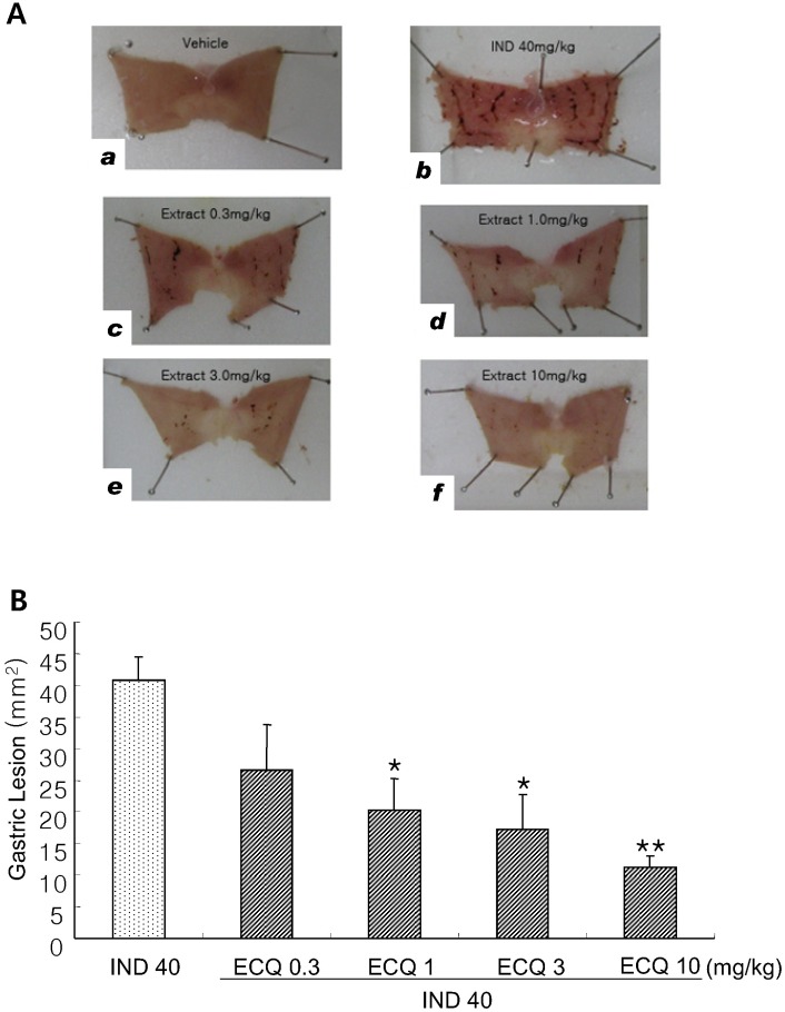 | Fig. 1Protective Effect of ECQ on gastric lesions induced by indomethacin (IND) in rats. (A) Representative macroscopic findings of the gastric lesions induced by IND 40 mg/kg in rats: (a) Vehicle group, (b) IND group, (c~f) One hour before oral administration of IND 40 mg/kg, animals were orally pretreated with ECQ at dose of 0.3, 1.0, 3.0, or 10 mg/kg, respectively. Six hours later, the animals were then sacrificed by cervical dislocation and the stomach was harvested. (B) Macroscopic damage was quantified by measuring the extent of the gastric mucosal lesions. Each value represents the mean±S.E.M. (n=5 per group). *p<0.01 and **p<0.001 vs. IND group.
|
Effect of ECQ on SOD or CAT activities in indomethacin-induced gastric damage
Oral administration of indomethacin 40 mg/kg induced a marked decrease in SOD activity compared to vehicle group. However, pretreatment with ECQ seemed to show increase in the activity of SOD in a dose-dependent manner. In particular, ECQ at dose of 10 mg/kg almost completely restored the loss of SOD activity induced by indomethacin in gastric mucosa (Fig. 2).
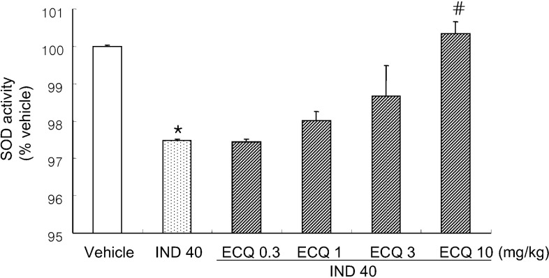 | Fig. 2Effect of ECQ on indomethacin (IND)-induced gastric mucosal SOD activity in rats. ECQ (0.3, 1.0, 3.0, or 10 mg/kg) were pretreated one hour before administration of IND, and then SOD activity was estimated 6 hours after the IND administration. Each value represents the mean±S.E.M. (n=5 per group). *p<0.001 vs. vehicle group, #p<0.05 vs. IND group.
|
Indomethacin administration also showed a reduction in CAT activity compared to vehicle group, and pretreatment with ECQ tended to induce an increase in the activity of CAT in a dose-dependent manner. Pretreatment with ECQ at dose of 10 mg/kg significantly increased CAT activity compared to indomethacin group (Fig. 3).
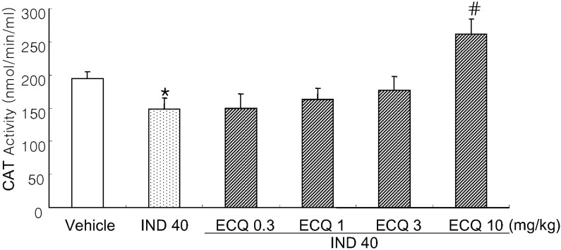 | Fig. 3Effect of ECQ on indomethacin (IND)-induced gastric mucosal CAT activity in rats. ECQ (0.3, 1.0, 3.0, or 10 mg/kg) were given one hour before administration of IND, and then CAT activity was estimated 6 hours after the IND administration. Each value represents the mean±S.E.M. (n=5 per group). *p<0.05 vs. vehicle group, #p<0.01 vs. IND group.
|
Effect of ECQ on expression of SOD-2 in indomethacin-induced gastric damage
SOD-2 expression was significantly decreased by 20% in the group treated with indomethacin alone compared to vehicle group. However, pretreatment with ECQ seemed to induce an increase in the SOD-2/GAPDH ratio compared to indomethacin alone group. Interestingly, ECQ at dose of 10 mg/kg significantly increased the SOD-2/GAPDH ratio compared to indomethacin group in gastric mucosa (Fig. 4).
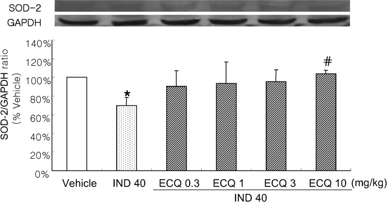 | Fig. 4The expression of SOD-2 in indomethacin (IND) treated groups with or without ECQ. ECQ (0.3, 1.0, 3.0, or 10 mg/kg) were pretreated one hour before administration of IND, and then SOD-2 expression was detected by Western blot analysis 6 hours after the IND administration (The representative blot is shown). Each value represents the mean±S.E.M. (n=5 per group). *p<0.05 vs. vehicle group, #p<0.05 vs. IND group.
|
Effect of ECQ on MPO levels in indomethacin-induced gastric damage
To estimate the degree of mucosal infiltration by neutrophils elicited by oral administration of indomethacin 40 mg/kg, the MPO levels were measured with or without pretreatment of ECQ in the gastric mucosa. In the group of rats that received indomethacin alone, MPO activity was significantly increased by 189% compared to vehicle group. However, pretreatment of ECQ tended to induce a reduction in the MPO activity in a dose-dependent manner. Especially, pretreatment of ECQ at the dose of 10 mg/kg significantly reduced the MPO levels by 75.3% compared to indomethacin group (Fig. 5).
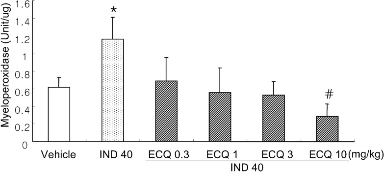 | Fig. 5Effect of ECQ on indomethacin (IND)-induced gastric MPO activity in rats. ECQ (0.3, 1.0, 3.0, or 10 mg/kg) were pretreated one hour before administration of IND, and then MPO activity was estimated 6 hours after the IND administration. Each value represents the mean±S.E.M. (n=5 per group). *p<0.05 vs. vehicle group, #p<0.05 vs. IND group.
|
Effect of ECQ on MDA levels in indomethacin-induced gastric damage
As shown in the Fig. 6, MDA levels used as an index of lipid peroxidation in the gastric tissue, were significantly higher in the indomethacin alone group than in the vehicle group. On the other hand, pretreatment of ECQ at dose of 1, 3 and 10 mg/kg significantly prevented the increase of the MDA levels induced by indomethacin.
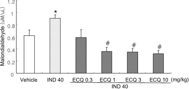 | Fig. 6Effect of ECQ on indomethacin (IND)-induced increases in gastric MDA level in rats. ECQ (0.3, 1.0, 3.0, or 10 mg/kg) were pretreated one hour before IND administration, and then MDA level was estimated 6 hours after the IND administration. Each value represents the mean±S.E.M. (n=5 per group). *p<0.05 vs. vehicle group, #p<0.01 vs. IND group.
|
Go to :
DISCUSSION
Flavonoids are naturally occurring plant polyphenols found in abundance in the fruit, vegetables and plant-derive beverages such as green tea. It is well known that flavonoids have anti-inflammation and anti-oxidative effects [18]. Quercetin, a flavonoid, has been shown to chelate iron ions [19] or to scavenge superoxide anions [20,21]. It has been reported to exert anti-inflammatory [22], anti-thrombotic [23], anti-bacterial and anti-tumoral [24] effects: it prevents gastric mucosal lesions produced by ethanol [25], cold-restraint stress, and the HCl plus ethanol [26]. Interestingly, our previous study revealed that QGC is more potent than a quercetin on inhibition of experimental reflux esophagitis and indomethacin-induced gastritis in Sprague-Dawley rats: Oral administration of QGC decreased ulcer index, injury area, gastric volume and acid output, and increased gastric pH. The elevation of MDA levels in reflux esophagitis was aslo inhibited by QGC, but not by quercetin [14]. Therefore, we have continued to focus on investigating the effects of QGC and its mechanism on upper gastrointestinal damage: QGC protection from indomethacin-induced damage is associated with increased gastric mucus secretion, inhibition of free radical production by activated neutrophils via ICAM-1, and the downregulation of pro-inflammatory cytokine [27]; the cytoprotective effect of QGC against ethanol-induced cell damage may involve inhibition of ROS generation and downstream activation of the ERK 1/2 in cultured feline esophageal epithelial cells [28]. Our previous study also revealed that QGC could result in enhanced antioxidant enzyme defense system via HO-1 expression and Nrf2 translocation involving both the ERK and PI3K-Akt pathways as well as partly PKC pathways in cultured feline esophageal epithelial cells [15].
In the present study, we investigated the protective effects of extract containing 8~10% QGC against gastric damage induced by oral administration of indomethacin. The result suggested that the extract at 10 mg/kg has a protective effect and an anti-oxidative effect on indomethacin-induced gastric damage. The pretreatment with ECQ by oral administration significantly reduced the indomethacin-induced gastric damage with an increase in the activities of antioxidative enzymes such as SOD and catalase, and significantly increased SOD-2 expression. Some authors have demonstrated that oxygen free radicals were directly implicated in the pathogenesis of indomethacin-induced mucosal damage [29]. Free radicals can also alter the cellular antioxidant defense system including SOD and CAT [30]. This might lead to aggravated tissue damage during stomach ulceration [31].
In the present study, when the MPO levels were measured in the gastric mucosa in order to estimate the degree of mucosal infiltration by neutrophils elicited by treatment with indomethacin, the pretreatment with ECQ by oral administration attenuated MPO activity. The circulating neutrophils play a critical role in the pathogenesis of mucosal lesions provoked by NSAIDs [1], acetic acid [32], and ethanol [33]. These leukocytes might contribute to gastric ulceration by various mechanisms, such as production of reactive oxygen metabolites or release of proteases and lipid mediators such as leukotrienes and PAF which affect vascular tone and permeability, exacerbating tissues ischemia [34]. Leukotrienes which are potent vasoconstrictors, have been identified in the gastric mucosa, particularly after exposure to ethanol. They exert various biological actions including vasoconstrictive effect, that could contribute to their role as mediators of ischemic and tissue damage.
It is well known that induction of gastric lesions by indomethacin is the oxidative damage with its dual events of reactive oxygen species (ROS) generation and lipid peroxidation [35,36]. Indomethacin activates the polymorphonuclear leucocytes in peripheral blood and enhances the release of ROS from these cells [37]. ROS induce lipid peroxidation which is believed to be an important cause of destruction and damage of the cellular membranes. In the present study, the involvement of extensive lipid peroxidation in indomethacin-induced gastric damage was evidenced by the accumulation of MDA level, as an index of lipid peroxidation, and MPO activity, as a marker of neutrophil aggregation, which was probably substantial enough to contribute to the gastritis. However, the pretreatment with ECQ also reversed the elevation of MDA levels induced by indomethacin. Therefore, the inhibitory activity of ECQ may result from its ability to scavenge ROS produced by indomethacin administration which initiate lipid peroxidation.
Taken together, the pretreatment with ECQ was proven to protect against gastric damage, which is susceptible to NSAIDs in rats. The underlying mechanism of gastroprotective effects of the ECQ on gastric damage induced by indomethacin could be related to its anti-inflammatory actions and its antioxidant properties, which reduce MDA levels and MPO activity and increase SOD and CAT activities and SOD-2 expression. This ECQ was also proven to be safe in our recent study which examined the acute toxicity and the general pharmacological action of the ECQ to search for any side effects in rats, mice, guinea pigs, and cats [38]. Therefore, the results suggest that ECQ could be a promising new drug for the prevention of NSAIDs-induced gastric damage.
Go to :
 XML Download