Journal List > Korean J Physiol Pharmacol > v.16(6) > 1025829

Chung, Bey, and Jiang: Synaptic Plasticity in Mouse Models of Autism Spectrum Disorders

Abstract

Analysis of synaptic plasticity together with behavioral and molecular studies have become a popular approach to model autism spectrum disorders in order to gain insight into the pathosphysiological mechanisms and to find therapeutic targets. Abnormalities of specific types of synaptic plasticity have been revealed in numerous genetically modified mice that have molecular construct validity to human autism spectrum disorders. Constrained by the feasibility of technique, the common regions analyzed in most studies are hippocampus and visual cortex. The relevance of the synaptic defects in these regions to the behavioral abnormalities of autistic like behaviors is still a subject of debate. Because the exact regions or circuits responsible for the core features of autistic behaviors in humans are still poorly understood, investigation using region-specific conditional mutant mice may help to provide the insight into the neuroanatomical basis of autism in the future.

ABBREVIATIONS

AS

Angelman syndrome

ASD

autism spectrum disorders

CaMKII

Ca2+/calmodulin-dependent protein kinase II

DHPG

dihydroxyphenylglycine

E-LTP

early phase LTP

FMRP

fragile X mental retardation protein

FXS

Fragile X syndrome

HFS

high frequency stimulation

ID

intellectual disability

KO

knock-out

LFS-LTD

low frequecy stimulation LTD

L-LTP

late phase LTP

LTD

long term depression

LTP

long term potentiation

mEPSC

miniature excitatory postsynaptic current

mGluR

metabotropic glutamate receptor

mGluR-LTD

mGluR mediated LTD

NMDA

N-methyl-D-aspartate

PP-LFS

paired-pulse low frequency stimulation

PSD

postsynaptic density

RTT

Rett syndrome

TSC

tuberous sclerosis complex

INTRODUCTION

Synaptic plasticity is frequently measured by analysis of long term potentiation (LTP) and long term depression (LTD) of synapses in different brain regions [1-3]. Historically, the best studied region for synaptic plasticity is the Schaffer collateral pathway in hippocampal CA1 region. The physiological and biochemical mechanisms underlying LTP and LTD have been extensively investigated [3,4]. Over the last decade, analysis of synaptic plasticity has become a popular technique to characterize animal models of neurodevelopmental disorders including autism spectrum disorders (ASD) [5,6]. Various abnormal findings in synaptic plasticity from different brain regions have been reported in mouse models with targeted mutations in genes implicated in ASD (Table 1). However, identifying and interpreting the defects in synaptic plasticity relevant to behavioral manifestations and disease pathophysiology of ASD remain a significant challenge. In this review, we will focus on reviewing the studies of synaptic plasticity in several prominent mouse models for neurodevelopmental disorders with pronounced autistic features and discussing the challenges and future directions in the field.
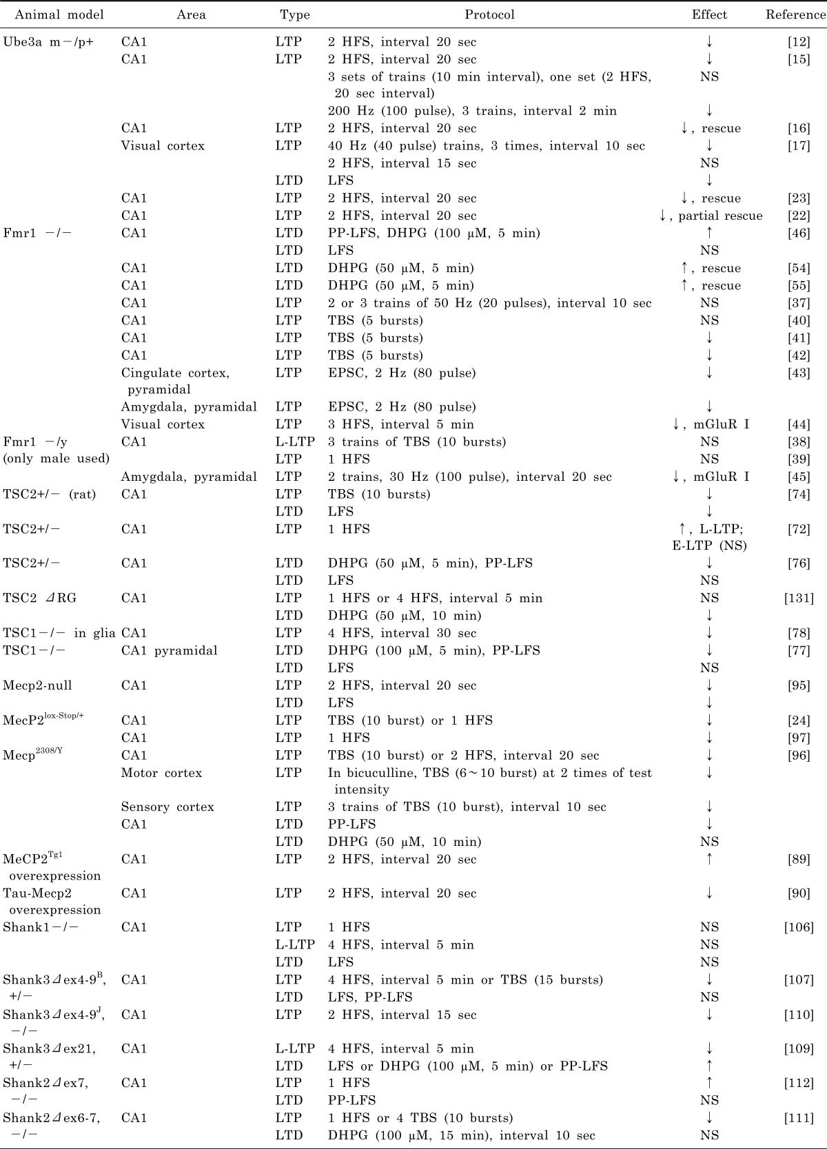
ANGELMAN SYNDROME

Angelman syndrome (AS) is characterized by profound intellectual disability (ID), movement disorders, absence of speech, epilepsy, and autistic behaviors [7,8]. The molecular defects causing AS include maternal microdeletions on chromosome15q11-q13 (60% of cases), point mutations in the maternal copy of the UBE3A gene (20%), paternal uniparental disomy (5%), and imprinting center defects (1%) [9]. Despite the presence of different molecular defects, it is a well-supported fact that the deficiency of maternal expression of the UBE3A gene in the brain is responsible for the key clinical features of AS [10,11]. To model human AS in mice, the first knock-out (KO) mouse that targeted exon 2 of Ube3a was reported in 1998 and recapitulated the major features of AS in maternal deficiency mice (Ube3a m-/p+) [12]. Subsequently, Ube3a mutant mouse with a mutation in the last coding exon encoding the ubiquitin ligase domain and was reported [13]. In addition, mutant mice with a 1.6 Mb deletion from Ube3a to Gabrb3 that is more similar to AS deletion patients were also reported [14]. However, the Ube3a exon 2 deletion mutant mice have been used more widely by investigators in the research community over the last 15 years.
Synaptic plasticity has been studied extensively in Ube3a m-/p+ mice in different brain regions using different protocols (Table 1). In CA1, LTP is reduced in Ube3a m-/p+ mice using an induction protocol of two trains of high frequency stimulation (HFS) of 100 pulses at 100 Hz (Table 1) [12]. Interestingly, the reduced LTP in CA1 of Ube3a m-/p+ mice could be rescued if a stronger stimulation protocol, three sets of two trains of HFS, was applied [15]. This indicates that the role of Ube3a in synaptic plasticity is probably as a modulator for the expression of LTP and less likely to play an essential role in LTP induction. Unfortunately, LTD at the same synapses has not been investigated so far. In the same study, reduced Ca2+/calmodulin-dependent protein kinase II (CaMKII) activity due to increased inhibitory autophosphorylation was observed in Ube3a m-/p+ mice [15]. In the subsequent rescue experiment, genetic reduction of inhibitory autophosphorylation of CaMKII rescued the LTP deficit as well as hippocampal-dependent learning as assessed by Morris water maze and fear conditioning in Ube3a m-/p+ mice [16]. This in vivo rescue indicates that a biochemical mechanism mediated by CaMKII activity underlies the impaired synaptic plasticity in Ube3a-deficient synapses. However, it remains unclear what is the molecular mechanism through which the deficiency of Ube3a contributes to the altered activity of CaMKII.
Synaptic plasticity was also investigated in visual cortex in Ube3a m-/p+ mice [17,18]. Both LTP and LTD are reduced in visual cortex in Ube3a m-/p+ mice (Table 1) [17]. Low frequency stimulation was used for N-methyl-D-aspartate (NMDA) receptor-dependent LTD (LFS-LTD) induction but group I metabotropic glutamate receptor (mGluR) mediated LTD (mGluR-LTD) was not analyzed. Interestingly, in vitro synaptic plasticity was affected by the in vivo visual experience of the mice. Deprivation of visual input achieved by a dark rearing environment could actually restore the impaired LTP and LTD in Ube3a m-/p+ mice [17]. This observation is intriguing because it indicates that the defective synaptic plasticity due to deficiency of Ube3a can be easily reversed in animals. On the other hand, it would be interesting to learn whether dark rearing may actually reverse abnormal behavioral phenotypes in Ube3a m-/p+ mice.
As the UBE3A gene is a brain-specific imprinted gene that is expressed primarily from the maternal chromosome, the UBE3A gene on the paternal chromosome is structurally intact but transcriptionally repressed [9,19]. Through a small molecule screening program, Huang et al. discovered that the silenced Ube3a from the paternal chromosome can be activated by topotecan, a toposiomerase inhibitor, in vitro and in vivo in mice [20]. This discovery opens an exciting research avenue to explore the treatment of AS using pharmacological interventions and presumably through epigenetic modification [21]. The immediate question is whether postnatal treatment with toposomeriase inhibitors can rescue the synaptic plasticity and ultimately behavioral defects in Ube3a m-/p+ mice. Another approach of treatment is to use a virus-mediated gene delivery method in hippocampus in vivo. Restoring the expression of Ube3a could rescue the early phase of LTP impairment and cognitive deficits in Ube3a m-/p+ mice [22]. In yet another group, use of an ErbB inhibitor could rescue LTP in Ube3a m-/p+ [23]. These observations together suggest that impaired synaptic plasticity and behavioral abnormalities in Ube3a m-/p+ mice is reversible even during late development. A similar finding was also reported in Mecp2 mutant mice [24,25]. However, it remains a subject of debate if the same phenomenon may exist in the treatment of human neurodevelopmental disorders.
A subset of AS patients meet the diagnostic criteria for ASD [26]. However, the behavioral studies of Ube3a m-/p+ mice have not revealed significant impairments in ASD-like behaviors although these mice have recapitulated the most salient features of intellectual disability and movement disorder seen in AS [27]. This result may indicate that other genes in the 15q11-q13 region may also contribute to the ASD in human AS. Alternatively, more extensive behavioral tests and comparisons between mutant mice with mutation only in Ube3a and a deletion from Ube3a to Gabrb3 may be warranted [14].

FRAGILE X SYNDROME

Fragile X syndrome (FXS) is one of the best studied disorders with intellectual disability ID and ASD both in humans and mouse models [28]. In addition to ID and ASD, FXS patients are characterized by seizures, macroorchidism, and dysmorphic facial features [29]. Molecularly, FXS is caused by the CGG triplet expansion in the 5' untranslated region of the FMR1 gene on the X chromosome [30,31]. The expanded CGG repeat results in promoter methylation that represses the transcription of FMR1 [32]. The FMR1 gene encodes the fragile X mental retardation protein (FMRP), an RNA-binding protein which inhibits local protein translation stimulated by group I mGluR signaling [28,33,34]. To understand the pathogenesis of fragile X syndrome, Fmr1 KO mutant mice were first developed in 1994 [35]. For almost two decades, Fmr1 mutant mice have been extensively studied from many angles by numerous investigators [36]. The full review of the findings from studying Fmr1 mutant mice is beyond the scope of this review. Instead, we will focus on synaptic plasticity in Fmr1 mutant mice.
The initial studies of synaptic plasticity in hippocampal CA1 region in Fmr1 mutant mice did not reveal any impairment in LTP by a standard LTP induction protocol (Table 1) [37-40]. However when the stimulation for LTP induction was reduced to near the induction threshold level in subsequent studies, LTP in CA1 was found to be reduced in Fmr1 KO [41,42]. Interestingly, LTP in brain regions including somatosensory cortex, anterior cingular cortex, anterior piriform cortex and lateral amygdala was also found to be decreased [39,40,43]. Impaired LTP was also observed in visual cortex and basolateral amygdala, but notably they were mGluR dependent [44,45]. These observations indicate different region- or synapse-specific defects in Fmrp deficient mice. However, the most important finding from synaptic plasticity studies is the observation of enhanced mGluR-LTD in hippocampal CA1 region [46]. A similar phenomenon of enhanced LTD was observed in cerebellum [47]. The observation of enhanced LTD in hippocampal CA1 region led to a theory of aberrant mGluR signaling underlying the pathophysiology of FXS [48]. The central hypothesis of the mGluR theory is that loss of FMRP in the synapse leads to the up-regulation of the mGluR-mediated signaling pathway. The mGluR theory and the molecular mechanism underlying the enhanced mGluR-LTD have been tested extensively since it was proposed and these studies have validated the central hypothesis [46,49-51]. However, alterations of many signaling pathways and a long list of potential protein targets in synapses have been revealed in Fmr1 mutant mice [28,52]. It is not entirely clear how the disruption of these different pathways can be integrated into a unifying mechanism responsible for the pathophysiology of FXS. Recent reports indicate an involvement of Homer proteins in the dysregulated mGluR signaling pathway [53]. Genetic reduction of mGluR5 or pharmacological inhibition of mGluR5 could rescue the abnormal behaviors in Fmr1 mutant mice [54,55]. Similarly, genetic reduction of Homer1a in Fmr1 KO could also improve behaviors, though this did not rescue mGluR-LTD in hippocampus [56]. These rescue experiments raise an interesting possibility for potential reversal of neurological impairments in human fragile X syndrome. The various synaptic defects found in different brain regions in Fmr1 mutant mice raise an immediate question about the correlation between the defective synaptic plasticity and the abnormal behaviors for future investigation. For example, social behaviors are impaired in Fmr1 KO mice which is consistent with autistic behaviors frequently seen in human fragile X syndrome patients [57-60]. These studies support Fmr1 KO mice as a good model to dissect the pathophysiology and explore treatment strategies for ASD [28].

TUBEROUS SCLEROSIS COMPLEX

Tuberous sclerosis complex (TSC) is a neurocutaneous condition with prominent neurobehavioral manifestations including seizures, ID, and autistic behaviors [61,62]. The neurobehavioral features are quite variable and range from mild to severe presentations in TSC patients [61]. TSC is caused by mutations in TSC1 or TSC2 genes that show a dominant inheritance pattern [63,64]. The proteins, hamartin encoded by TSC1 and tuberin encoded by TSC2 genes, form a heterodimeric complex that functions as a negative regulator for the mTOR pathway [65-67]. Therefore, it has been hypothesized that loss of function mutations in TSC1 or TSC2 disinhibit mTOR signaling and lead to the up-regulation of the signaling pathway downstream of mTOR which promotes cell growth and proliferation [67,68].
Both homozygous Tsc1 or Tsc2 KO mice are embryonic lethal [69-71]. Heterozygotes of Tsc1 or Tsc2 mutation exhibit cognitive impairment and synaptic dysfunction in the absence of apparent neuroanatomical defects or seizures [72-75]. In Tsc2+/- rats (Eker rat), LTP and LFS-LTD was decreased in CA1 [74]. In Tsc2+/- mice, early phase LTP (E-LTP) in hippocampal CA1 is not affected but late phase LTP (L-LTP) was enhanced [72]. In Tsc2+/-, mGluR-LTD was decreased but LFS-LTD was intact [76]. The reduced mGluR-LTD in Tsc2+/- is opposite to what is seen in Fmr1 KO mice although the mGluR-LTDs from both were insensitive to protein synthesis inhibitors [76]. As in Tsc2+/-, Tsc1+/- mutant mice showed a similar impairment in synaptic plasticity. In hippocampal CA1 pyramidal neurons with conditionally deleted Tsc1, mGluR-LTD was reduced but LFS-LTD was intact [77]. Interestingly, synaptic plasticity is also impaired when Tsc1 was knocked out in non-neuronal cells. For instance, the E-LTP was reduced in Tsc1 glia-specific conditional KOs [78]. A recent study on Tsc1 deleted specifically in cerebellar Purkinje cells showed impaired social interaction, enhanced repetitive behaviors and abnormal ultrasonic vocalizations [79]. However, synaptic plasticity was not tested in cerebellum in this mouse model [79]. This observation raises a provocative question regarding the brain regions and circuits that are important for the pathophysiology of autistic behaviors because social interaction was significantly reduced both in Tsc1+/- and Tsc2+/- [73,79,80]. The advantage of TSC models over other ASD mouse models is that the signaling pathway involving dysregulation of mTOR is well defined in both Tsc1 and Tsc2 mutant mice.

RETT SYNDROME

Rett syndrome (RTT) is a neurological disorder that primarily affects females and is caused by mutations in the MeCP2 gene [81,82]. The clinical presentations of RTT are characterized by normal early neurodevelopment for the first 12~18 months followed by developmental regression [83]. The major symptoms of RTT include movement disorders, absence of speech, and repetitive hand movements [83]. MeCP2 protein generally is considered to suppress transcription by binding to methylated CpG DNA [84]. However, recent evidence suggests a role of bidirectional regulation with both repression and activation of transcription mediated by MeCP2 [85]. Several Mecp2 mutant mice carrying slightly different mutations have been produced and characterized [24,86-88]. In addition, mutant mice with overexpression of Mecp2 was also reported [89,90]. These mice are valuable models to ASD research because RTT is a prototype for syndromic ASD and because impairments in social behaviors were observed in both whole brain- and region specific Mecp2 mutant mice [91-94].
In general, mice lacking the functional copy of Mecp2 recapitulate the major features of RTT. In Mecp2-null mouse, synaptic plasticity was analyzed at two different ages because of the age-dependent regression in human RTT [86,95]. In male mice at a presymptomatic age (3~5 weeks old), no difference in LTP at hippocampal CA1 region was found. However, at a symptomatic age (6~10 weeks old) LTP and LFS-LTD in CA1 was reduced. This indicates that the trajectory of impaired synaptic plasticity correlates well with the developmental phenotype changes as suggested in humans. In a model where Mecp2 was truncated as in some human patients (Mecp2308/Y), LTP and paired-pulse low frequency stimulation (PP-LFS) (Table 1) but not the group I mGluR agonist dihydroxyphenylglycine (DHPG)-LTD was reduced in hippocampal CA1 from male mice [87,96]. Similarly, LTP in primary motor cortex and sensory cortex was reduced [96]. More interestingly, the impaired LTP in CA1 and neurological phenotypes in Mecp2lox-Stop/+ mice could be rescued by reintroduction of Mecp2 by genetic manipulation in male or female mice [24,97]. At least in three different RTT models, decreased CA1 LTP was consistent.
Synaptic plasticity was also investigated in mice with overexpressed MeCP2 via BAC mediated transgenics (MeCP2Tg1) [89]. As predicted from the finding of reduced LTP in Mecp2 deficiency mice, LTP in CA1 was increased in MeCP2Tg1 [89]. However LTP in CA1 was decreased in a different animal model with the overexpression of Mecp2 driven by Tau promoter in neurons (Tau-Mecp2) [90]. The explanation for this discrepancy is not apparent and additional investigation is warranted.

SHANK FAMILY GENE CAUSING ASD

SHANK family proteins, SHANK1, SHANK2, and SHANK3, are scaffolding proteins enriched at the postsynaptic density (PSD) of excitatory synapses [98]. SHANK proteins share a similar protein domain structure that mediates protein-protein interaction at the PSD for synaptic function [98,99]. Molecular defects in SHANK3 were first found in patients with ASD and ID [100]. Subsequently, genetic defects of SHANK1 and SHANK2 were also reported in ASD and ID [101-103]. Because of the existing knowledge of the function of SHANK family proteins at synapses, the discovery of mutations in SHANK family genes provide direct support for the notion that the pathogenesis of ASD may reside in the dysfunction of synapses [104,105]. Mutant mice for all Shank family genes have been reported [106-112]. Shank1 mutant mice were first reported [106]. Surprisingly, the phenotype of Shank1 deficiency mice was unexpectedly mild. No synaptic plasticity changes were detected in LTP and LFS-LTD in the Shank1 KO even though mEPSC frequency and synaptic strength were decreased [106]. Because of the findings of both microdeletions of and point mutations in the SHANK3 gene in human ASD [100,113], the interest to model Shank3 mutations in mice has been intensified recently. This led to the simultaneous generation of multiple Shank3 mutant mice by disrupting different portions of Shank3 exons [107-110]. These mutations include deletion of exons 4-7 (Δex4-7) [108], exons 4-9 (Δex4-9J) [110] and (Δex4-9B) [107], exon 11 (Δex11) [112], exons 13-16 (Δex13-16) [108] and exon 21 (Δex21) [109]. We recently discovered that Shank3 has an array of protein isoforms resulting from the combination of multiple intragenic promoters and extensive alternative splicing of coding exons [110]. Therefore, we concluded that different mutations in different exons resulted in the disruption of different Shank3 isoforms but none of these mutant mice were Shank3 complete knockouts. Shank proteins regulate the abundance and signaling of ionotropic and metabotropic glutamate receptors at excitatory synapses [105,114]. Accordingly, synaptic transmission and plasticity were examined in different brain regions in all Shank3 mutant mice. Measurements of miniature excitatory postsynaptic current (mEPSC) frequency and amplitude, paired pulse ratio, input/output (I/O) curves, fiber volley, and population spikes indicated that synaptic transmission was reduced at hippocampal CA1 synapses of Δex4-9B+/- mice [107], but not in mice bearing Δex4-9J-/- [110], Δex13-16-/- [108], or Δex21+/- mutations [109]. The explanation for the difference between Δex4-9B+/- and Δex4-9J-/- is not immediately clear.
In striatum, the frequency of mEPSCs and amplitude of population spikes were significantly decreased in Δex13-16-/- mice, but only mildly affected in Δex4-7-/- mice [108]. Presynaptic responses measured by paired pulse ratio and input/output curves were not altered at corticostriatal synapses in Δex13-16-/- or Δex4-7-/- mice [108]. The different degree of synaptic transmission defects in mice with specific Shank3 mutations supports the notion of an isoform-specific contribution to synaptic function. Moreover, the reduced NMDA receptor-mediated responses at cortical synapses of Δex21+/- [109] but not in the corticostriatal synapses of Δex13-16-/- mice [108] indicate distinct functions of Shank3 at different synapses.
In terms of plasticity, hippocampal LTP was reduced at CA1 synapses of Δex4-9J-/-, Δex4-9B+/-, and ex21+/- mice [107,109,110]. In contrast, LFS-LTD was reduced in CA1 of Δex21+/- mice [109] but not in Δex4-9B+/- mice [107], suggesting an alteration in the set-point for bidirectional Hebbian synaptic plasticity [115]. mGluR-LTD induced by DHPG or PP-LFS was enhanced in CA1 hippocampus of Δex21+/- mice. However, a similar enhancement of mGluR-LTD was not evident in the Δex4-9B+/- mice induced by PP-LFS [107]. In addition, mGluR1/5 protein levels were not altered in Δex21+/- mice [109].
Collectively, these data support synaptic defects mediated by glutamate receptors in Shank3 mutant mice that appear to be both synapse- and mutation-specific. It is not yet clear whether there are common core circuit defects in the various mutant mice, but the phenotypic heterogeneity itself appears consistent with the clinical heterogeneity of patients harboring SHANK3 mutations. Since different mutations affect different isoforms of Shank3, some of the observed phenotypes may arise from isoform-specific effects on synaptic transmission.
Two mutant models for Shank2 were reported recently [111,112]. Schmeisser et al. reported Shank2 exon 7 deletion mutant mice (Shank2 Δex7) in which LTP in hippocampal CA1 was increased but no change in LTD with PP-LFS was observed [112]. Reduced social interaction, increased stereotypical behavior, hyperactivity, and altered ultrasonic vocalization pattern were found in Shank2 Δex7 -/- mice. Won et al. generated Shank2 mutant mice where exons 6-7 were deleted (Shank2 Δex6-7) [111]. Both exon 7 and exon 6-7 deletion resulted in a frame shift mutation shortly after exon 7. Intriguingly, LTP in hipppocampal CA1 was reduced in Shank2 Δex6-7 mice and this is opposite to Shank2 Δex7 mice [111]. In addition, the NMDA current and LFS-induced LTD were reduced in hippocampal CA1 region in Shank2 Δex7 mice but DHPG-induced LTD was not affected. The behavioral profile of Shank2 Δex6-7 mice is very similar to Shank2 Δex7. Interestingly, treatment with NMDA agonist current mediated signaling could rescue social interaction deficits [111]. The explanation for the apparent discrepancy in synaptic plasticity but similar behavioral profile in mice with two very similar mutations is not immediately clear and further investigation is warranted. However, available data strongly support that both Shank2 and Shank3 mutant mice are valid ASD models to dissect the pathophysiology.

FUTURE DIRECTIONS AND CHALLENGES

It is clear that abnormalities in synaptic plasticity vary significantly among different animal models of ASD. For instance, Tsc2 mutations in rat and mice have opposite effects on LTP [72,74]. Shank1 mutant mice do not show plasticity defects using standard protocols unlike Shank2 and Shank3 mutants (Table 1). In Shank2 mutants, LTP impairment is in opposite directions in two different lines of mutant mice despite similar mutations and behavioral profiles [111,112]. However, as an example of convergence, LTP in three different lines of Shank3 mutants from three different groups was decreased consistently but the LTD defects are significantly different [107,109,110]. On the other hand, it is difficult to correlate the abnormal plasticity with the corresponding behavioral manifestations in each model.
Currently, most synaptic plasticity experiments were performed in the hippocampus while the deficiency of targeted genes was in the whole brain. Therefore, it is difficult to establish causality between brain region and abnormal behaviors studied in the models. Because the neuroanatomical basis for autism is still poorly understood, an unbiased survey for synaptic plasticity in other different brain areas may provide more informative data about the pathophysiology of autism.
In human brain, the superior temporal sulcus region, the fusiform gyrus and amygdala are considered important for social interaction and gaze behaviors [116]. However, gaze behavior in mice, which are nocturnal, is difficult to monitor technically. A neural circuit involving amygdala could be important region to study in ASD mouse model. The study of amygdala and related circuits such as medial prefrontal cortex in autism mouse models is within reach [117,118].
For stereotypical behaviors, cortical-striatal circuits are hypothesized to be important in ASD [119-121]. Significant repetitive behaviors measured by increase in self-grooming and inflexibility in the reversal phase of the Morris water maze are frequently reported in ASD mouse models [107,108]. The synaptic plasticity in cortical-striatal circuit activity is less well characterized in these most ASD models [122].
For the aspect of communication/language impairment in humans, ultrasonic vocalization (USV) recording in mice has become a popular approach despite ongoing debate about the value of the USV relevant to human communication [123,124]. Abnormal USV measurements have been reported in numerous ASD mouse models [14,108-112,125-128]. These observations support the value of USV recording because of easy quantification and detailed numerical analysis. However, several challenges remain. First, what is the ethological meaning of USV in mice? Second, what is the circuit in rodent brain responsible for USVs [129,130]. More investigation are clearly warranted in future.
The studies of synaptic plasticity and behaviors in these high profile mouse models with defined genetic defects have produced many interesting findings but also raise numerous challenging questions. First, what is the implication of variable, or opposite in some cases, synaptic plasticity related to understanding the pathophysiology of ASD and other comorbidities? Second, what is the molecular mechanism underlying the different synaptic plasticity between different brain regions? Third, can impaired synaptic plasticity in a particular brain region predict abnormal behaviors? Fourth, which is a more reliable biomarker, the synaptic plasticity or behavioral defects, to use for future drug screening? Future investigations may focus on 1) generation of brain region-, cell type-, or circuit-specific targeted gene KO, 2) in vivo physiology or circuit analysis, and 3) development of new and sensitive behavioral tests. Despite these challenges, we have reasonable confidence that studying these and other new ASD models will lead to better understanding the pathophysiology of ASD and ultimately lead to the development of new treatments.

ABBREVIATIONS

AS

Angelman syndrome

ASD

autism spectrum disorders

CaMKII

Ca2+/calmodulin-dependent protein kinase II

DHPG

dihydroxyphenylglycine

E-LTP

early phase LTP

FMRP

fragile X mental retardation protein

FXS

Fragile X syndrome

HFS

high frequency stimulation

ID

intellectual disability

KO

knock-out

LFS-LTD

low frequecy stimulation LTD

L-LTP

late phase LTP

LTD

long term depression

LTP

long term potentiation

mEPSC

miniature excitatory postsynaptic current

mGluR

metabotropic glutamate receptor

mGluR-LTD

mGluR mediated LTD

NMDA

N-methyl-D-aspartate

PP-LFS

paired-pulse low frequency stimulation

PSD

postsynaptic density

RTT

Rett syndrome

TSC

tuberous sclerosis complex

References

1. Bliss TV, Collingridge GL. A synaptic model of memory: long-term potentiation in the hippocampus. Nature. 1993; 361:31–39. PMID: 8421494.
crossref
2. Kandel ER. The molecular biology of memory storage: a dialogue between genes and synapses. Science. 2001; 294:1030–1038. PMID: 11691980.
crossref
3. Collingridge GL, Peineau S, Howland JG, Wang YT. Long-term depression in the CNS. Nat Rev Neurosci. 2010; 11:459–473. PMID: 20559335.
crossref
4. Nicoll RA. Expression mechanisms underlying long-term potentiation: a postsynaptic view. Philos Trans R Soc Lond B Biol Sci. 2003; 358:721–726. PMID: 12740118.
crossref
5. Zoghbi HY, Bear MF. Synaptic dysfunction in neurodevelopmental disorders associated with autism and intellectual disabilities. Cold Spring Harb Perspect Biol. 2012; 4:pii: a009886.
crossref
6. Ting JT, Peça J, Feng G. Functional consequences of mutations in postsynaptic scaffolding proteins and relevance to psychiatric disorders. Annu Rev Neurosci. 2012; 35:49–71. PMID: 22540979.
crossref
7. Dagli A, Buiting K, Williams CA. Molecular and Clinical Aspects of Angelman Syndrome. Mol Syndromol. 2012; 2:100–112. PMID: 22670133.
crossref
8. Williams CA, Beaudet AL, Clayton-Smith J, Knoll JH, Kyllerman M, Laan LA, Magenis RE, Moncla A, Schinzel AA, Summers JA, Wagstaff J. Angelman syndrome 2005: updated consensus for diagnostic criteria. Am J Med Genet A. 2006; 140:413–418. PMID: 16470747.
crossref
9. Jiang Y, Lev-Lehman E, Bressler J, Tsai TF, Beaudet AL. Genetics of Angelman syndrome. Am J Hum Genet. 1999; 65:1–6. PMID: 10364509.
crossref
10. Kishino T, Lalande M, Wagstaff J. UBE3A/E6-AP mutations cause Angelman syndrome. Nat Genet. 1997; 15:70–73. PMID: 8988171.
crossref
11. Matsuura T, Sutcliffe JS, Fang P, Galjaard RJ, Jiang YH, Benton CS, Rommens JM, Beaudet AL. De novo truncating mutations in E6-AP ubiquitin-protein ligase gene (UBE3A) in Angelman syndrome. Nat Genet. 1997; 15:74–77. PMID: 8988172.
crossref
12. Jiang YH, Armstrong D, Albrecht U, Atkins CM, Noebels JL, Eichele G, Sweatt JD, Beaudet AL. Mutation of the Angelman ubiquitin ligase in mice causes increased cytoplasmic p53 and deficits of contextual learning and long-term potentiation. Neuron. 1998; 21:799–811. PMID: 9808466.
crossref
13. Miura K, Kishino T, Li E, Webber H, Dikkes P, Holmes GL, Wagstaff J. Neurobehavioral and electroencephalographic abnormalities in Ube3a maternal-deficient mice. Neurobiol Dis. 2002; 9:149–159. PMID: 11895368.
14. Jiang YH, Pan Y, Zhu L, Landa L, Yoo J, Spencer C, Lorenzo I, Brilliant M, Noebels J, Beaudet AL. Altered ultrasonic vocalization and impaired learning and memory in Angelman syndrome mouse model with a large maternal deletion from Ube3a to Gabrb3. PLoS One. 2010; 5:e12278. PMID: 20808828.
crossref
15. Weeber EJ, Jiang YH, Elgersma Y, Varga AW, Carrasquillo Y, Brown SE, Christian JM, Mirnikjoo B, Silva A, Beaudet AL, Sweatt JD. Derangements of hippocampal calcium/calmodulin-dependent protein kinase II in a mouse model for Angelman mental retardation syndrome. J Neurosci. 2003; 23:2634–2644. PMID: 12684449.
crossref
16. van Woerden GM, Harris KD, Hojjati MR, Gustin RM, Qiu S, de Avila Freire R, Jiang YH, Elgersma Y, Weeber EJ. Rescue of neurological deficits in a mouse model for Angelman syndrome by reduction of alphaCaMKII inhibitory phosphorylation. Nat Neurosci. 2007; 10:280–282. PMID: 17259980.
17. Yashiro K, Riday TT, Condon KH, Roberts AC, Bernardo DR, Prakash R, Weinberg RJ, Ehlers MD, Philpot BD. Ube3a is required for experience-dependent maturation of the neocortex. Nat Neurosci. 2009; 12:777–783. PMID: 19430469.
crossref
18. Sato M, Stryker MP. Genomic imprinting of experience-dependent cortical plasticity by the ubiquitin ligase gene Ube3a. Proc Natl Acad Sci USA. 2010; 107:5611–5616. PMID: 20212164.
crossref
19. Mabb AM, Judson MC, Zylka MJ, Philpot BD. Angelman syndrome: insights into genomic imprinting and neurodevelopmental phenotypes. Trends Neurosci. 2011; 34:293–303. PMID: 21592595.
crossref
20. Huang HS, Allen JA, Mabb AM, King IF, Miriyala J, Taylor-Blake B, Sciaky N, Dutton JW Jr, Lee HM, Chen X, Jin J, Bridges AS, Zylka MJ, Roth BL, Philpot BD. Topoisomerase inhibitors unsilence the dormant allele of Ube3a in neurons. Nature. 2011; 481:185–189. PMID: 22190039.
crossref
21. Beaudet AL. Angelman syndrome: Drugs to awaken a paternal gene. Nature. 2011; 481:150–152. PMID: 22190038.
22. Daily JL, Nash K, Jinwal U, Golde T, Rogers J, Peters MM, Burdine RD, Dickey C, Banko JL, Weeber EJ. Adeno-associated virus-mediated rescue of the cognitive defects in a mouse model for Angelman syndrome. PLoS One. 2011; 6:e27221. PMID: 22174738.
crossref
23. Kaphzan H, Hernandez P, Jung JI, Cowansage KK, Deinhardt K, Chao MV, Abel T, Klann E. Reversal of impaired hippocampal long-term potentiation and contextual fear memory deficits in Angelman syndrome model mice by ErbB inhibitors. Biol Psychiatry. 2012; 72:182–190. PMID: 22381732.
crossref
24. Guy J, Gan J, Selfridge J, Cobb S, Bird A. Reversal of neurological defects in a mouse model of Rett syndrome. Science. 2007; 315:1143–1147. PMID: 17289941.
crossref
25. Gadalla KK, Bailey ME, Cobb SR. MeCP2 and Rett syndrome: reversibility and potential avenues for therapy. Biochem J. 2011; 439:1–14. PMID: 21916843.
crossref
26. Williams CA. The behavioral phenotype of the Angelman syndrome. Am J Med Genet C Semin Med Genet. 2010; 154C:432–437. PMID: 20981772.
crossref
27. Allensworth M, Saha A, Reiter LT, Heck DH. Normal social seeking behavior, hypoactivity and reduced exploratory range in a mouse model of Angelman syndrome. BMC Genet. 2011; 12:7. PMID: 21235769.
crossref
28. Bhakar AL, Dölen G, Bear MF. The pathophysiology of fragile X (and what it teaches us about synapses). Annu Rev Neurosci. 2012; 35:417–443. PMID: 22483044.
crossref
29. Martin JP, Bell J. A pedigree of mental defect showing sex-linkage. J Neurol Psychiatry. 1943; 6:154–157. PMID: 21611430.
crossref
30. Pieretti M, Zhang FP, Fu YH, Warren ST, Oostra BA, Caskey CT, Nelson DL. Absence of expression of the FMR-1 gene in fragile X syndrome. Cell. 1991; 66:817–822. PMID: 1878973.
crossref
31. Verkerk AJ, Pieretti M, Sutcliffe JS, Fu YH, Kuhl DP, Pizzuti A, Reiner O, Richards S, Victoria MF, Zhang FP, et al. Identification of a gene (FMR-1) containing a CGG repeat coincident with a breakpoint cluster region exhibiting length variation in fragile X syndrome. Cell. 1991; 65:905–914. PMID: 1710175.
crossref
32. Sutcliffe JS, Nelson DL, Zhang F, Pieretti M, Caskey CT, Saxe D, Warren ST. DNA methylation represses FMR-1 transcription in fragile X syndrome. Hum Mol Genet. 1992; 1:397–400. PMID: 1301913.
crossref
33. Laggerbauer B, Ostareck D, Keidel EM, Ostareck-Lederer A, Fischer U. Evidence that fragile X mental retardation protein is a negative regulator of translation. Hum Mol Genet. 2001; 10:329–338. PMID: 11157796.
crossref
34. Li Z, Zhang Y, Ku L, Wilkinson KD, Warren ST, Feng Y. The fragile X mental retardation protein inhibits translation via interacting with mRNA. Nucleic Acids Res. 2001; 29:2276–2283. PMID: 11376146.
crossref
35. The Dutch-Belgian Fragile X Consortium. Fmr1 knockout mice: a model to study fragile X mental retardation. Cell. 1994; 78:23–33. PMID: 8033209.
36. Santoro MR, Bray SM, Warren ST. Molecular mechanisms of fragile X syndrome: a twenty-year perspective. Annu Rev Pathol. 2012; 7:219–245. PMID: 22017584.
crossref
37. Godfraind JM, Reyniers E, De Boulle K, D'Hooge R, De Deyn PP, Bakker CE, Oostra BA, Kooy RF, Willems PJ. Long-term potentiation in the hippocampus of fragile X knockout mice. Am J Med Genet. 1996; 64:246–251. PMID: 8844057.
crossref
38. Paradee W, Melikian HE, Rasmussen DL, Kenneson A, Conn PJ, Warren ST. Fragile X mouse: strain effects of knockout phenotype and evidence suggesting deficient amygdala function. Neuroscience. 1999; 94:185–192. PMID: 10613508.
crossref
39. Li J, Pelletier MR, Perez Velazquez JL, Carlen PL. Reduced cortical synaptic plasticity and GluR1 expression associated with fragile X mental retardation protein deficiency. Mol Cell Neurosci. 2002; 19:138–151. PMID: 11860268.
crossref
40. Larson J, Jessen RE, Kim D, Fine AK, du Hoffmann J. Age-dependent and selective impairment of long-term potentiation in the anterior piriform cortex of mice lacking the fragile X mental retardation protein. J Neurosci. 2005; 25:9460–9469. PMID: 16221856.
crossref
41. Lauterborn JC, Rex CS, Kramár E, Chen LY, Pandyarajan V, Lynch G, Gall CM. Brain-derived neurotrophic factor rescues synaptic plasticity in a mouse model of fragile X syndrome. J Neurosci. 2007; 27:10685–10694. PMID: 17913902.
crossref
42. Lee HY, Ge WP, Huang W, He Y, Wang GX, Rowson-Baldwin A, Smith SJ, Jan YN, Jan LY. Bidirectional regulation of dendritic voltage-gated potassium channels by the fragile X mental retardation protein. Neuron. 2011; 72:630–642. PMID: 22099464.
crossref
43. Zhao MG, Toyoda H, Ko SW, Ding HK, Wu LJ, Zhuo M. Deficits in trace fear memory and long-term potentiation in a mouse model for fragile X syndrome. J Neurosci. 2005; 25:7385–7392. PMID: 16093389.
crossref
44. Wilson BM, Cox CL. Absence of metabotropic glutamate receptor-mediated plasticity in the neocortex of fragile X mice. Proc Natl Acad Sci USA. 2007; 104:2454–2459. PMID: 17287348.
crossref
45. Suvrathan A, Hoeffer CA, Wong H, Klann E, Chattarji S. Characterization and reversal of synaptic defects in the amygdala in a mouse model of fragile X syndrome. Proc Natl Acad Sci USA. 2010; 107:11591–11596. PMID: 20534533.
crossref
46. Huber KM, Gallagher SM, Warren ST, Bear MF. Altered synaptic plasticity in a mouse model of fragile X mental retardation. Proc Natl Acad Sci USA. 2002; 99:7746–7750. PMID: 12032354.
crossref
47. Koekkoek SK, Yamaguchi K, Milojkovic BA, Dortland BR, Ruigrok TJ, Maex R, De Graaf W, Smit AE, VanderWerf F, Bakker CE, Willemsen R, Ikeda T, Kakizawa S, Onodera K, Nelson DL, Mientjes E, Joosten M, De Schutter E, Oostra BA, Ito M, De Zeeuw CI. Deletion of FMR1 in Purkinje cells enhances parallel fiber LTD, enlarges spines, and attenuates cerebellar eyelid conditioning in Fragile X syndrome. Neuron. 2005; 47:339–352. PMID: 16055059.
crossref
48. Bear MF, Huber KM, Warren ST. The mGluR theory of fragile X mental retardation. Trends Neurosci. 2004; 27:370–377. PMID: 15219735.
crossref
49. Gallagher SM, Daly CA, Bear MF, Huber KM. Extracellular signal-regulated protein kinase activation is required for metabotropic glutamate receptor-dependent long-term depression in hippocampal area CA1. J Neurosci. 2004; 24:4859–4864. PMID: 15152046.
crossref
50. Hou L, Klann E. Activation of the phosphoinositide 3-kinase-Akt-mammalian target of rapamycin signaling pathway is required for metabotropic glutamate receptor-dependent long-term depression. J Neurosci. 2004; 24:6352–6361. PMID: 15254091.
crossref
51. Osterweil EK, Krueger DD, Reinhold K, Bear MF. Hypersensitivity to mGluR5 and ERK1/2 leads to excessive protein synthesis in the hippocampus of a mouse model of fragile X syndrome. J Neurosci. 2010; 30:15616–15627. PMID: 21084617.
crossref
52. Bassell GJ, Warren ST. Fragile X syndrome: loss of local mRNA regulation alters synaptic development and function. Neuron. 2008; 60:201–214. PMID: 18957214.
crossref
53. Ronesi JA, Huber KM. Homer interactions are necessary for metabotropic glutamate receptor-induced long-term depression and translational activation. J Neurosci. 2008; 28:543–547. PMID: 18184796.
crossref
54. Dölen G, Osterweil E, Rao BS, Smith GB, Auerbach BD, Chattarji S, Bear MF. Correction of fragile X syndrome in mice. Neuron. 2007; 56:955–962. PMID: 18093519.
crossref
55. Michalon A, Sidorov M, Ballard TM, Ozmen L, Spooren W, Wettstein JG, Jaeschke G, Bear MF, Lindemann L. Chronic pharmacological mGlu5 inhibition corrects fragile X in adult mice. Neuron. 2012; 74:49–56. PMID: 22500629.
crossref
56. Ronesi JA, Collins KA, Hays SA, Tsai NP, Guo W, Birnbaum SG, Hu JH, Worley PF, Gibson JR, Huber KM. Disrupted Homer scaffolds mediate abnormal mGluR5 function in a mouse model of fragile X syndrome. Nat Neurosci. 2012; 15:431–440. PMID: 22267161.
crossref
57. Spencer CM, Alekseyenko O, Serysheva E, Yuva-Paylor LA, Paylor R. Altered anxiety-related and social behaviors in the Fmr1 knockout mouse model of fragile X syndrome. Genes Brain Behav. 2005; 4:420–430. PMID: 16176388.
crossref
58. McNaughton CH, Moon J, Strawderman MS, Maclean KN, Evans J, Strupp BJ. Evidence for social anxiety and impaired social cognition in a mouse model of fragile X syndrome. Behav Neurosci. 2008; 122:293–300. PMID: 18410169.
crossref
59. Thomas AM, Bui N, Graham D, Perkins JR, Yuva-Paylor LA, Paylor R. Genetic reduction of group 1 metabotropic glutamate receptors alters select behaviors in a mouse model for fragile X syndrome. Behav Brain Res. 2011; 223:310–321. PMID: 21571007.
crossref
60. Bhattacharya A, Kaphzan H, Alvarez-Dieppa AC, Murphy JP, Pierre P, Klann E. Genetic Removal of p70 S6 Kinase 1 Corrects Molecular, Synaptic, and Behavioral Phenotypes in Fragile X Syndrome Mice. Neuron. 2012; 76:325–337. PMID: 23083736.
crossref
61. Crino PB, Nathanson KL, Henske EP. The tuberous sclerosis complex. N Engl J Med. 2006; 355:1345–1356. PMID: 17005952.
crossref
62. Curatolo P, Bombardieri R, Jozwiak S. Tuberous sclerosis. Lancet. 2008; 372:657–668. PMID: 18722871.
crossref
63. Kandt RS, Haines JL, Smith M, Northrup H, Gardner RJ, Short MP, Dumars K, Roach ES, Steingold S, Wall S, et al. Linkage of an important gene locus for tuberous sclerosis to a chromosome 16 marker for polycystic kidney disease. Nat Genet. 1992; 2:37–41. PMID: 1303246.
crossref
64. van Slegtenhorst M, de Hoogt R, Hermans C, Nellist M, Janssen B, Verhoef S, Lindhout D, van den Ouweland A, Halley D, Young J, Burley M, Jeremiah S, Woodward K, Nahmias J, Fox M, Ekong R, Osborne J, Wolfe J, Povey S, Snell RG, Cheadle JP, Jones AC, Tachataki M, Ravine D, Sampson JR, Reeve MP, Richardson P, Wilmer F, Munro C, Hawkins TL, Sepp T, Ali JB, Ward S, Green AJ, Yates JR, Kwiatkowska J, Henske EP, Short MP, Haines JH, Jozwiak S, Kwiatkowski DJ. Identification of the tuberous sclerosis gene TSC1 on chromosome 9q34. Science. 1997; 277:805–808. PMID: 9242607.
65. Plank TL, Yeung RS, Henske EP. Hamartin, the product of the tuberous sclerosis 1 (TSC1) gene, interacts with tuberin and appears to be localized to cytoplasmic vesicles. Cancer Res. 1998; 58:4766–4770. PMID: 9809973.
66. van Slegtenhorst M, Nellist M, Nagelkerken B, Cheadle J, Snell R, van den Ouweland A, Reuser A, Sampson J, Halley D, van der Sluijs P. Interaction between hamartin and tuberin, the TSC1 and TSC2 gene products. Hum Mol Genet. 1998; 7:1053–1057. PMID: 9580671.
crossref
67. Tee AR, Manning BD, Roux PP, Cantley LC, Blenis J. Tuberous sclerosis complex gene products, Tuberin and Hamartin, control mTOR signaling by acting as a GTPase-activating protein complex toward Rheb. Curr Biol. 2003; 13:1259–1268. PMID: 12906785.
crossref
68. de Vries PJ, Howe CJ. The tuberous sclerosis complex proteins--a GRIPP on cognition and neurodevelopment. Trends Mol Med. 2007; 13:319–326. PMID: 17632034.
69. Onda H, Lueck A, Marks PW, Warren HB, Kwiatkowski DJ. Tsc2(+/-) mice develop tumors in multiple sites that express gelsolin and are influenced by genetic background. J Clin Invest. 1999; 104:687–695. PMID: 10491404.
crossref
70. Kobayashi T, Minowa O, Kuno J, Mitani H, Hino O, Noda T. Renal carcinogenesis, hepatic hemangiomatosis, and embryonic lethality caused by a germ-line Tsc2 mutation in mice. Cancer Res. 1999; 59:1206–1211. PMID: 10096549.
71. Kobayashi T, Minowa O, Sugitani Y, Takai S, Mitani H, Kobayashi E, Noda T, Hino O. A germ-line Tsc1 mutation causes tumor development and embryonic lethality that are similar, but not identical to, those caused by Tsc2 mutation in mice. Proc Natl Acad Sci USA. 2001; 98:8762–8767. PMID: 11438694.
crossref
72. Ehninger D, Han S, Shilyansky C, Zhou Y, Li W, Kwiatkowski DJ, Ramesh V, Silva AJ. Reversal of learning deficits in a Tsc2+/- mouse model of tuberous sclerosis. Nat Med. 2008; 14:843–848. PMID: 18568033.
crossref
73. Goorden SM, van Woerden GM, van der Weerd L, Cheadle JP, Elgersma Y. Cognitive deficits in Tsc1+/- mice in the absence of cerebral lesions and seizures. Ann Neurol. 2007; 62:648–655. PMID: 18067135.
74. von der Brelie C, Waltereit R, Zhang L, Beck H, Kirschstein T. Impaired synaptic plasticity in a rat model of tuberous sclerosis. Eur J Neurosci. 2006; 23:686–692. PMID: 16487150.
crossref
75. Nie D, Di Nardo A, Han JM, Baharanyi H, Kramvis I, Huynh T, Dabora S, Codeluppi S, Pandolfi PP, Pasquale EB, Sahin M. Tsc2-Rheb signaling regulates EphA-mediated axon guidance. Nat Neurosci. 2010; 13:163–172. PMID: 20062052.
crossref
76. Auerbach BD, Osterweil EK, Bear MF. Mutations causing syndromic autism define an axis of synaptic pathophysiology. Nature. 2011; 480:63–68. PMID: 22113615.
crossref
77. Bateup HS, Takasaki KT, Saulnier JL, Denefrio CL, Sabatini BL. Loss of Tsc1 in vivo impairs hippocampal mGluR-LTD and increases excitatory synaptic function. J Neurosci. 2011; 31:8862–8869. PMID: 21677170.
crossref
78. Zeng LH, Ouyang Y, Gazit V, Cirrito JR, Jansen LA, Ess KC, Yamada KA, Wozniak DF, Holtzman DM, Gutmann DH, Wong M. Abnormal glutamate homeostasis and impaired synaptic plasticity and learning in a mouse model of tuberous sclerosis complex. Neurobiol Dis. 2007; 28:184–196. PMID: 17714952.
crossref
79. Tsai PT, Hull C, Chu Y, Greene-Colozzi E, Sadowski AR, Leech JM, Steinberg J, Crawley JN, Regehr WG, Sahin M. Autistic-like behaviour and cerebellar dysfunction in Purkinje cell Tsc1 mutant mice. Nature. 2012; 488:647–651. PMID: 22763451.
crossref
80. Chévere-Torres I, Maki JM, Santini E, Klann E. Impaired social interactions and motor learning skills in tuberous sclerosis complex model mice expressing a dominant/negative form of tuberin. Neurobiol Dis. 2012; 45:156–164. PMID: 21827857.
crossref
81. Amir RE, Van den Veyver IB, Wan M, Tran CQ, Francke U, Zoghbi HY. Rett syndrome is caused by mutations in X-linked MECP2, encoding methyl-CpG-binding protein 2. Nat Genet. 1999; 23:185–188. PMID: 10508514.
crossref
82. Chahrour M, Zoghbi HY. The story of Rett syndrome: from clinic to neurobiology. Neuron. 2007; 56:422–437. PMID: 17988628.
crossref
83. Smeets EE, Pelc K, Dan B. Rett Syndrome. Mol Syndromol. 2012; 2:113–127. PMID: 22670134.
crossref
84. Nan X, Campoy FJ, Bird A. MeCP2 is a transcriptional repressor with abundant binding sites in genomic chromatin. Cell. 1997; 88:471–481. PMID: 9038338.
crossref
85. Chahrour M, Jung SY, Shaw C, Zhou X, Wong ST, Qin J, Zoghbi HY. MeCP2, a key contributor to neurological disease, activates and represses transcription. Science. 2008; 320:1224–1229. PMID: 18511691.
crossref
86. Chen RZ, Akbarian S, Tudor M, Jaenisch R. Deficiency of methyl-CpG binding protein-2 in CNS neurons results in a Rett-like phenotype in mice. Nat Genet. 2001; 27:327–331. PMID: 11242118.
crossref
87. Shahbazian M, Young J, Yuva-Paylor L, Spencer C, Antalffy B, Noebels J, Armstrong D, Paylor R, Zoghbi H. Mice with truncated MeCP2 recapitulate many Rett syndrome features and display hyperacetylation of histone H3. Neuron. 2002; 35:243–254. PMID: 12160743.
crossref
88. Na ES, Nelson ED, Kavalali ET, Monteggia LM. The Impact of MeCP2 Loss- or Gain-of-Function on Synaptic Plasticity. Neuropsychopharmacology. 2012; [Epub ahead of print].
crossref
89. Collins AL, Levenson JM, Vilaythong AP, Richman R, Armstrong DL, Noebels JL, David Sweatt J, Zoghbi HY. Mild overexpression of MeCP2 causes a progressive neurological disorder in mice. Hum Mol Genet. 2004; 13:2679–2689. PMID: 15351775.
crossref
90. Na ES, Nelson ED, Adachi M, Autry AE, Mahgoub MA, Kavalali ET, Monteggia LM. A mouse model for MeCP2 duplication syndrome: MeCP2 overexpression impairs learning and memory and synaptic transmission. J Neurosci. 2012; 32:3109–3117. PMID: 22378884.
crossref
91. Moretti P, Bouwknecht JA, Teague R, Paylor R, Zoghbi HY. Abnormalities of social interactions and home-cage behavior in a mouse model of Rett syndrome. Hum Mol Genet. 2005; 14:205–220. PMID: 15548546.
crossref
92. Samaco RC, Mandel-Brehm C, McGraw CM, Shaw CA, McGill BE, Zoghbi HY. Crh and Oprm1 mediate anxiety-related behavior and social approach in a mouse model of MECP2 duplication syndrome. Nat Genet. 2012; 44:206–211. PMID: 22231481.
crossref
93. Gemelli T, Berton O, Nelson ED, Perrotti LI, Jaenisch R, Monteggia LM. Postnatal loss of methyl-CpG binding protein 2 in the forebrain is sufficient to mediate behavioral aspects of Rett syndrome in mice. Biol Psychiatry. 2006; 59:468–476. PMID: 16199017.
crossref
94. Chao HT, Chen H, Samaco RC, Xue M, Chahrour M, Yoo J, Neul JL, Gong S, Lu HC, Heintz N, Ekker M, Rubenstein JL, Noebels JL, Rosenmund C, Zoghbi HY. Dysfunction in GABA signalling mediates autism-like stereotypies and Rett syndrome phenotypes. Nature. 2010; 468:263–269. PMID: 21068835.
crossref
95. Asaka Y, Jugloff DG, Zhang L, Eubanks JH, Fitzsimonds RM. Hippocampal synaptic plasticity is impaired in the Mecp2-null mouse model of Rett syndrome. Neurobiol Dis. 2006; 21:217–227. PMID: 16087343.
crossref
96. Moretti P, Levenson JM, Battaglia F, Atkinson R, Teague R, Antalffy B, Armstrong D, Arancio O, Sweatt JD, Zoghbi HY. Learning and memory and synaptic plasticity are impaired in a mouse model of Rett syndrome. J Neurosci. 2006; 26:319–327. PMID: 16399702.
crossref
97. Weng SM, McLeod F, Bailey ME, Cobb SR. Synaptic plasticity deficits in an experimental model of rett syndrome: long-term potentiation saturation and its pharmacological reversal. Neuroscience. 2011; 180:314–321. PMID: 21296130.
crossref
98. Grabrucker AM, Schmeisser MJ, Schoen M, Boeckers TM. Postsynaptic ProSAP/Shank scaffolds in the cross-hair of synaptopathies. Trends Cell Biol. 2011; 21:594–603. PMID: 21840719.
crossref
99. Boeckers TM, Bockmann J, Kreutz MR, Gundelfinger ED. ProSAP/Shank proteins - a family of higher order organizing molecules of the postsynaptic density with an emerging role in human neurological disease. J Neurochem. 2002; 81:903–910. PMID: 12065602.
crossref
100. Durand CM, Betancur C, Boeckers TM, Bockmann J, Chaste P, Fauchereau F, Nygren G, Rastam M, Gillberg IC, Anckarsäter H, Sponheim E, Goubran-Botros H, Delorme R, Chabane N, Mouren-Simeoni MC, de Mas P, Bieth E, Rogé B, Héron D, Burglen L, Gillberg C, Leboyer M, Bourgeron T. Mutations in the gene encoding the synaptic scaffolding protein SHANK3 are associated with autism spectrum disorders. Nat Genet. 2007; 39:25–27. PMID: 17173049.
crossref
101. Berkel S, Marshall CR, Weiss B, Howe J, Roeth R, Moog U, Endris V, Roberts W, Szatmari P, Pinto D, Bonin M, Riess A, Engels H, Sprengel R, Scherer SW, Rappold GA. Mutations in the SHANK2 synaptic scaffolding gene in autism spectrum disorder and mental retardation. Nat Genet. 2010; 42:489–491. PMID: 20473310.
crossref
102. Pinto D, Pagnamenta AT, Klei L, Anney R, Merico D, Regan R, Conroy J, Magalhaes TR, Correia C, Abrahams BS, Almeida J, Bacchelli E, Bader GD, Bailey AJ, Baird G, Battaglia A, Berney T, Bolshakova N, Bölte S, Bolton PF, Bourgeron T, Brennan S, Brian J, Bryson SE, Carson AR, Casallo G, Casey J, Chung BH, Cochrane L, Corsello C, Crawford EL, Crossett A, Cytrynbaum C, Dawson G, de Jonge M, Delorme R, Drmic I, Duketis E, Duque F, Estes A, Farrar P, Fernandez BA, Folstein SE, Fombonne E, Freitag CM, Gilbert J, Gillberg C, Glessner JT, Goldberg J, Green A, Green J, Guter SJ, Hakonarson H, Heron EA, Hill M, Holt R, Howe JL, Hughes G, Hus V, Igliozzi R, Kim C, Klauck SM, Kolevzon A, Korvatska O, Kustanovich V, Lajonchere CM, Lamb JA, Laskawiec M, Leboyer M, Le Couteur A, Leventhal BL, Lionel AC, Liu XQ, Lord C, Lotspeich L, Lund SC, Maestrini E, Mahoney W, Mantoulan C, Marshall CR, McConachie H, McDougle CJ, McGrath J, McMahon WM, Merikangas A, Migita O, Minshew NJ, Mirza GK, Munson J, Nelson SF, Noakes C, Noor A, Nygren G, Oliveira G, Papanikolaou K, Parr JR, Parrini B, Paton T, Pickles A, Pilorge M, Piven J, Ponting CP, Posey DJ, Poustka A, Poustka F, Prasad A, Ragoussis J, Renshaw K, Rickaby J, Roberts W, Roeder K, Roge B, Rutter ML, Bierut LJ, Rice JP, Salt J, Sansom K, Sato D, Segurado R, Sequeira AF, Senman L, Shah N, Sheffield VC, Soorya L, Sousa I, Stein O, Sykes N, Stoppioni V, Strawbridge C, Tancredi R, Tansey K, Thiruvahindrapduram B, Thompson AP, Thomson S, Tryfon A, Tsiantis J, Van Engeland H, Vincent JB, Volkmar F, Wallace S, Wang K, Wang Z, Wassink TH, Webber C, Weksberg R, Wing K, Wittemeyer K, Wood S, Wu J, Yaspan BL, Zurawiecki D, Zwaigenbaum L, Buxbaum JD, Cantor RM, Cook EH, Coon H, Cuccaro ML, Devlin B, Ennis S, Gallagher L, Geschwind DH, Gill M, Haines JL, Hallmayer J, Miller J, Monaco AP, Nurnberger JI Jr, Paterson AD, Pericak-Vance MA, Schellenberg GD, Szatmari P, Vicente AM, Vieland VJ, Wijsman EM, Scherer SW, Sutcliffe JS, Betancur C. Functional impact of global rare copy number variation in autism spectrum disorders. Nature. 2010; 466:368–372. PMID: 20531469.
crossref
103. Sato D, Lionel AC, Leblond CS, Prasad A, Pinto D, Walker S, O'Connor I, Russell C, Drmic IE, Hamdan FF, Michaud JL, Endris V, Roeth R, Delorme R, Huguet G, Leboyer M, Rastam M, Gillberg C, Lathrop M, Stavropoulos DJ, Anagnostou E, Weksberg R, Fombonne E, Zwaigenbaum L, Fernandez BA, Roberts W, Rappold GA, Marshall CR, Bourgeron T, Szatmari P, Scherer SW. SHANK1 Deletions in Males with Autism Spectrum Disorder. Am J Hum Genet. 2012; 90:879–887. PMID: 22503632.
crossref
104. Naisbitt S, Kim E, Tu JC, Xiao B, Sala C, Valtschanoff J, Weinberg RJ, Worley PF, Sheng M. Shank, a novel family of postsynaptic density proteins that binds to the NMDA receptor/PSD-95/GKAP complex and cortactin. Neuron. 1999; 23:569–582. PMID: 10433268.
crossref
105. Tu JC, Xiao B, Naisbitt S, Yuan JP, Petralia RS, Brakeman P, Doan A, Aakalu VK, Lanahan AA, Sheng M, Worley PF. Coupling of mGluR/Homer and PSD-95 complexes by the Shank family of postsynaptic density proteins. Neuron. 1999; 23:583–592. PMID: 10433269.
crossref
106. Hung AY, Futai K, Sala C, Valtschanoff JG, Ryu J, Woodworth MA, Kidd FL, Sung CC, Miyakawa T, Bear MF, Weinberg RJ, Sheng M. Smaller dendritic spines, weaker synaptic transmission, but enhanced spatial learning in mice lacking Shank1. J Neurosci. 2008; 28:1697–1708. PMID: 18272690.
crossref
107. Bozdagi O, Sakurai T, Papapetrou D, Wang X, Dickstein DL, Takahashi N, Kajiwara Y, Yang M, Katz AM, Scattoni ML, Harris MJ, Saxena R, Silverman JL, Crawley JN, Zhou Q, Hof PR, Buxbaum JD. Haploinsufficiency of the autism-associated Shank3 gene leads to deficits in synaptic function, social interaction, and social communication. Mol Autism. 2010; 1:15. PMID: 21167025.
crossref
108. Peça J, Feliciano C, Ting JT, Wang W, Wells MF, Venkatraman TN, Lascola CD, Fu Z, Feng G. Shank3 mutant mice display autistic-like behaviours and striatal dysfunction. Nature. 2011; 472:437–442. PMID: 21423165.
crossref
109. Bangash MA, Park JM, Melnikova T, Wang D, Jeon SK, Lee D, Syeda S, Kim J, Kouser M, Schwartz J, Cui Y, Zhao X, Speed HE, Kee SE, Tu JC, Hu JH, Petralia RS, Linden DJ, Powell CM, Savonenko A, Xiao B, Worley PF. Enhanced polyubiquitination of Shank3 and NMDA receptor in a mouse model of autism. Cell. 2011; 145:758–772. PMID: 21565394.
110. Wang X, McCoy PA, Rodriguiz RM, Pan Y, Je HS, Roberts AC, Kim CJ, Berrios J, Colvin JS, Bousquet-Moore D, Lorenzo I, Wu G, Weinberg RJ, Ehlers MD, Philpot BD, Beaudet AL, Wetsel WC, Jiang YH. Synaptic dysfunction and abnormal behaviors in mice lacking major isoforms of Shank3. Hum Mol Genet. 2011; 20:3093–3108. PMID: 21558424.
crossref
111. Won H, Lee HR, Gee HY, Mah W, Kim JI, Lee J, Ha S, Chung C, Jung ES, Cho YS, Park SG, Lee JS, Lee K, Kim D, Bae YC, Kaang BK, Lee MG, Kim E. Autistic-like social behaviour in Shank2-mutant mice improved by restoring NMDA receptor function. Nature. 2012; 486:261–265. PMID: 22699620.
crossref
112. Schmeisser MJ, Ey E, Wegener S, Bockmann J, Stempel AV, Kuebler A, Janssen AL, Udvardi PT, Shiban E, Spilker C, Balschun D, Skryabin BV, Dieck St, Smalla KH, Montag D, Leblond CS, Faure P, Torquet N, Le Sourd AM, Toro R, Grabrucker AM, Shoichet SA, Schmitz D, Kreutz MR, Bourgeron T, Gundelfinger ED, Boeckers TM. Autistic-like behaviours and hyperactivity in mice lacking ProSAP1/Shank2. Nature. 2012; 486:256–260. PMID: 22699619.
crossref
113. Moessner R, Marshall CR, Sutcliffe JS, Skaug J, Pinto D, Vincent J, Zwaigenbaum L, Fernandez B, Roberts W, Szatmari P, Scherer SW. Contribution of SHANK3 mutations to autism spectrum disorder. Am J Hum Genet. 2007; 81:1289–1297. PMID: 17999366.
crossref
114. Uchino S, Wada H, Honda S, Nakamura Y, Ondo Y, Uchiyama T, Tsutsumi M, Suzuki E, Hirasawa T, Kohsaka S. Direct interaction of post-synaptic density-95/Dlg/ZO-1 domain-containing synaptic molecule Shank3 with GluR1 alpha-amino-3-hydroxy-5-methyl-4-isoxazole propionic acid receptor. J Neurochem. 2006; 97:1203–1214. PMID: 16606358.
crossref
115. Cho KK, Bear MF. Promoting neurological recovery of function via metaplasticity. Future Neurol. 2010; 5:21–26. PMID: 20209094.
crossref
116. Belger A, Carpenter KL, Yucel GH, Cleary KM, Donkers FC. The neural circuitry of autism. Neurotox Res. 2011; 20:201–214. PMID: 21213096.
crossref
117. Pare D, Duvarci S. Amygdala microcircuits mediating fear expression and extinction. Curr Opin Neurobiol. 2012; 22:717–723. PMID: 22424846.
crossref
118. Sotres-Bayon F, Quirk GJ. Prefrontal control of fear: more than just extinction. Curr Opin Neurobiol. 2010; 20:231–235. PMID: 20303254.
crossref
119. Langen M, Durston S, Kas MJ, van Engeland H, Staal WG. The neurobiology of repetitive behavior: ... and men. Neurosci Biobehav Rev. 2011; 35:356–365. PMID: 20153769.
120. Langen M, Kas MJ, Staal WG, van Engeland H, Durston S. The neurobiology of repetitive behavior: of mice... Neurosci Biobehav Rev. 2011; 35:345–355. PMID: 20156480.
crossref
121. Kreitzer AC, Malenka RC. Striatal plasticity and basal ganglia circuit function. Neuron. 2008; 60:543–554. PMID: 19038213.
crossref
122. Lüscher C, Huber KM. Group 1 mGluR-dependent synaptic long-term depression: mechanisms and implications for circuitry and disease. Neuron. 2010; 65:445–459. PMID: 20188650.
crossref
123. Scattoni ML. Special interest section on mouse ultrasonic vocalizations. Genes Brain Behav. 2011; 10:1–3. PMID: 21251214.
crossref
124. Fischer J, Hammerschmidt K. Ultrasonic vocalizations in mouse models for speech and socio-cognitive disorders: insights into the evolution of vocal communication. Genes Brain Behav. 2011; 10:17–27. PMID: 20579107.
crossref
125. Rotschafer SE, Trujillo MS, Dansie LE, Ethell IM, Razak KA. Minocycline treatment reverses ultrasonic vocalization production deficit in a mouse model of Fragile X Syndrome. Brain Res. 2012; 1439:7–14. PMID: 22265702.
crossref
126. Young DM, Schenk AK, Yang SB, Jan YN, Jan LY. Altered ultrasonic vocalizations in a tuberous sclerosis mouse model of autism. Proc Natl Acad Sci USA. 2010; 107:11074–11079. PMID: 20534473.
crossref
127. De Filippis B, Ricceri L, Laviola G. Early postnatal behavioral changes in the Mecp2-308 truncation mouse model of Rett syndrome. Genes Brain Behav. 2010; 9:213–223. PMID: 19958389.
128. Wöhr M, Roullet FI, Hung AY, Sheng M, Crawley JN. Communication impairments in mice lacking Shank1: reduced levels of ultrasonic vocalizations and scent marking behavior. PLoS One. 2011; 6:e20631. PMID: 21695253.
crossref
129. Van Daele DJ, Cassell MD. Multiple forebrain systems converge on motor neurons innervating the thyroarytenoid muscle. Neuroscience. 2009; 162:501–524. PMID: 19426785.
crossref
130. Van Daele DJ, Fazan VP, Agassandian K, Cassell MD. Amygdala connections with jaw, tongue and laryngo-pharyngeal premotor neurons. Neuroscience. 2011; 177:93–113. PMID: 21211549.
crossref
Table 1
Summary of synaptic plasticities with protocols
kjpp-16-369-i001

↓, decrease; ↑, increase; HFS, 100 Hz, 100 pulses; LFS, low frequency stimulation, 1 Hz, 900 pulse; L-LTP, late-LTP; LTD, long-term depression; LTP, long-term potentiation; mGluR I, group I metabotropic glutamate receptor; NS, no significant difference; PP-LFS, paired pulse LFS, paired pulse (50 ms interval), 1 Hz, 900 or 1200 pulse); TBS, theta burst stimulation, 5 to 10 bursts (100 Hz, 4~5 pulse) interval 200 ms. All animals are mice except a note of "rat".

TOOLS
Similar articles