

 PDF
PDF ePub
ePub Citation
 Print
Print


ABBREVIATIONS
IEC
ion exchange chromatography
RP-HPLC
reversed-phase high-performance liquid chromatography
PITC
phenylisothiocyanateor
OPA
o-phthalaldehyde
AQC
6-aminoquinolyl-N-hydroxysuccinimidyl carbamate
AMQ
6-aminoquinoline
UV
ultraviolet
Glu
glutamate
Gly
glycine
Ala
alanine
BWI
body weight index
QC
quality control
RE
relative error
RSD
relative standard deviations
INTRODUCTION
Several excitatory amino acids, such as endogenous glutamate, glycine, and alanine, which are involved in glutamate neurotransmission via NMDA receptors, are being investigated as potential indicators of health status and disease risk. Increased glutamate is closely related with neuronal damage by cerebral ischemia and plasma glutamate concentration is the most powerful predictor biomarker of lesion enlargement in the acute phase of ischemic stroke [1,2]. In some cases, the peripheral blood levels of glutamate, glycine, and alanine could be diagnostic, therapeutic or symptomatic biological markers [3-6]. Thus determination of these amino acids plays an important role in diagnosis and therapy of some psychiatric and neurological disorders. Moreover, the preparations of amino acids are increasingly used to treat a wide array of diseases in recent years. In china, glutamate, glycine, and alanine tablet or capsule is wildly used in the treatment of chronic prostatitis [7]. Simultaneous analysis of glutamate, glycine, and alanine may provide help for the study of the pharmacokinetics of these preparations.
The method commonly employed for the analysis of free plasma amino acids is automated ion exchange chromatography (IEC) in combination with postcolumn ninhydrin detection. This method provides simplicity in sample preparation and high reproducibility [8,9]. However, the long analytical run time is the main drawback [8,10]. Reversed-phase high-performance liquid chromatography (RP-HPLC) is used as an alternative due to its higher sensitivity and faster analysis. The most common RP-HPLC methods involve precolumn derivatization with phenylisothiocyanate (PITC) or o-phthalaldehyde (OPA) [11-14]. However, each reagent has its disadvantages or drawbacks: PITC needs a long derivatization time (20 min) and it is necessary to remove any excess reagent by drying, and OPA does not react with secondary amino acids, and glycine derivatives, for instance, are unable [15,16]. Precolumn derivatization utilizing 6-aminoquinolyl-N-hydroxysuccinimidyl carbamate (AQC) might constitute a feasible alternative, as to method characteristics that overcome these major drawbacks. In this procedure, primary and secondary amines react with the AQC reagent, yielding stable compounds. Excess reagent is hydrolyzed to yield 6-aminoquinoline (AMQ), carbon dioxide and N-hydroxysuccinimide [17]. The advantages of this method include a very simple derivatization procedure, stable derivatives, excellent separation, detection by either absorbance or fluorescence, and commercial availability of reagents. Disadvantages include relatively long chromatography, high solvent consumption and no possibility of on-line derivatization, which seems limited compared to the defects of other methods.
In the present study, we describe a sensitive and rapid RP-HPLC method, using AQC derivatization and ultraviolet (UV) detection. The objective is to obtain a simple and rapid simultaneous measurement of glutamate, glycine, and alanine in human plasma.
METHODS
Chemicals
AccQ.Fluor reagent kit (consisting of AQC reagent, acetonitrile and 0.2 mM sodium borate buffer, pH 8.8) as well as glass reaction tubes (6×50 mm) were provided by Waters (Milford, MA, USA). Amino acid standards (glutamate: Glu, glycine: Gly, alanine: Ala) were obtained from Sigma (St Louis, MO, USA). HPLC grade acetonitrile and triethylamine were from Merck (Darmstadt, Germany). Perchloric acid and sodium acetate were of analytical reagent grade and purchased from J.T. Baker (Phillipsburg, NJ, USA). Ultrapure water was generated using a Milli-Q system purchased from Millipore (Bedford, MA, USA).
Chromatographic instruments and conditions
The HPLC system used was a Waters Alliance consisting of a 2,690 separation model, a thermostat-controlled column oven, a system interface module, and a 2,487 Dual Wavelength UV Detector (all Waters components, Millipore, Milford, MA, USA). A Millennium Chromatography Manager workstation running Millennium software controls system operation and results management. Separation was carried out using a 20×3.9 mm Sentry guard column (Nova-Pak C18 bonded silica) connected to a 4 µm AccQ-Tag C18 column (3.9×150 mm I.D.; both from Waters).
Working eluents
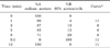
Eluent A was 0.14 M sodium acetate with 0.017 M triethanolamine (pH 4.95); eluent B was 60% (v/v) acetonitrile aqueous solution. The gradient program is shown in Table 1.
The AccQ-Tag column was thermostated at 37℃ and operated at a flow-rate of 1 ml/min. Detection was accomplished by UV detector and the wavelength was set at 248 nm. For column regeneration, 3 min with 60% acetonitrile and 40% water was sufficient to wash out the column and before the next injection, 100% sodium acetate for 6 min was needed to equilibrate the system. Injections were made every 18 min (injection-to-injection), using an injection volume of 10 µl.
Standard solutions
Stock solutions of Glu, Gly, and Ala needed in the method were prepared by dissolving appropriate amount of the compounds in 0.1 M hydrochloric acid and diluting to the appropriate volumes. These solutions were kept at 4℃ for several days without noticeable change. The working solutions were prepared by appropriate dilutions with water as needed, and processed without delay.
Plasma preparation
Twenty healthy male volunteers were recruited for this study. They are 19~25 years old with BWI (body weight index) between 19~24 kg/m2. The study was performed in accordance with the Helsinki Declaration and the design and execution of the experiment were explained thoroughly to the participants, and informed consent was obtained.
Blood was collected, following an overnight fast, into evacuated lithium heparin gel tubes, immediately placed on ice, and centrifuged at 3,000 rpm for 15 min at 4℃. Plasma was then used for the determination of Glu, Gly, and Ala without delay or stored at -70℃. Prior to analysis, a 100 µl portion of the plasma sample was added to 100 µl of 5% perchloric acid, and the solution was mixed with a vortex-mixer. After vortex-mixing, the precipitate was removed by centrifugation at 15,000 rpm for 10 min at 4℃ and the supernatant was used for the analysis.
Quality control (QC) samples preparation
For validation of the method, QC samples were made by adding the appropriate amount of working standard solution of Glu, Gly, and Ala to the plasma samples from an apparently healthy donor. The nominal concentrations of low, medium, and high QC samples were 40, 80, 160 µM for Glu; 160, 400, 640 µM for Gly and 200, 500, 800 µM for Ala, respectively.
Derivatization
In a typical analysis, 10 µl of standards or samples were mixed with 70 µl buffer solution (0.2 M borate buffer) and afterwards 20 µl derivatization reagent (2 mg/ml AQC) were added. After ten minutes response at 55℃, the derivatized solution was directly injected to separate the amino acids by HPLC.
Calibration and linearity
The retention time of each amino acid was studied using dilutions from stock solutions. Linearity was assured by calculating the regression coefficients for the calibration curves. Range of the analysis was determined by assuring that all the points on the calibration curve were characterised by acceptable precision and accuracy. For each curve, the peak area were calculated and plotted against concentration of the corresponding amino acid.
Precision and accuracy
Precision and accuracy were determined by the analysis of QC samples spiked at three concentrations listed above. The intra-assay was determined by replicate analysis of a blank (unspiked) plasma sample (n=6) and a QC sample at each concentration (n=6) in a single run. The inter-assay was determined by replicate analysis of the same QC sample stored at -70℃ and analyzed 6 separate times over a period of 2 months. The calculated mean concentration relative to the nominal concentration (relative error: RE%) was used to express accuracy and relative standard deviations (RSD%) calculated from the blank sample and QC values was used to estimate the inter- and intra-day precision.
Recovery and stability
Recovery of Glu, Gly, and Ala from plasma was assessed by analyzing pooled human plasma obtained from a healthy volunteer spiked at the three QC concentration levels listed above. Six replicate 'control' (unspiked) plasma samples and six spiked samples of each QC level were analyzed and recovery was determined by comparing the nominal amino acid concentration. The recovery of each amino acid was defined as 100%. Means, standard deviations (SD) and RSD% were calculated.
The stability of plasma samples stored at -70℃ within 2 months was assessed. Also, QC samples were subjected to three freeze-thaw cycles (-70℃ to room temperature) to evaluate sample stability. In addition, stability of processed samples was assessed. Triplicate low and high QC samples were processed and analyzed, then re-injected and analyzed up to 24 h post-processing.
RESULTS
Chromatographic separation
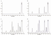
Representative chromatograms of the derivatized blank, standard solution, blank (unspiked) plasma sample and QC plasma sample are depicted in Fig. 1. The method demonstrated good chromatographic separation of all the amino acids studied, and showed an excellent signal-to-noise ratio, allowing easy quantification. Retention times for Glu, Gly, and Ala were approximately 6.3, 7.9, and 9.3 min, respectively. The peaks of interest were well separated and there was no interference from other amino acids and endogenous compounds in plasma.
Linearity, precision and accuracy
Linear calibration curves were obtained for Glu, Gly, and Ala over the concentration ranges of 5~320 µM, 18.75~1,200 µM, and 21.9~1400 µM, respectively. The mean regression equations were y=1,414.2x+1791.3 for Glu, y=1,590.7x+6,982.6 for Gly, and y=1,626.6x+7289.2 for Ala, with correlation coefficients (r)>0.999 for all three amino acids. The lower limit of quantitation (LOQ) for each amino acid demonstrated acceptable precision.
The intra- and inter-day RSD% were between 1.8% and 9.5% and most RSD% values calculated from the high QC samples of each amino acid were smaller than those from low and medium QC samples especially pronounced in Ala group. The accuracy, expressed as RE%, was within ±10% (Table 2).
Stability and recovery
The plasma samples were stable for up to 2 months at -70℃. Three freeze-thaw cycles had no effect on the stability of Glu, Gly, and Ala. Additionally, amino acid derivatives are stable at room temperature for 24 hours. The mean recoveries of all analytes from plasma at all concentrations ranged from 95% to 103%. At the same time, the RSD% values were less than 5% which showed significant inverse relationship with the concentration of the analyte (Table 3).
Analysis of human plasma samples
The method described in this paper was applied for amino acid analysis in plasma samples from twenty male volunteers (Table 4). Free plasma amino acids exhibit wide variability between subjects. This probably reflects differences in metabolic control since the amino acids belong to endogenous substances which can be influenced by physiologic conditions of different volunteers as well as diet. Plasma concentrations of Glu, Gly, and Ala were 51.7±16.5, 289.7±46.5, and 350.1±59.9 µM, respectively.
DISCUSSION
The increasing significance of plasma concentrations of Glu, Gly, and Ala in clinical fields, such as being used as diagnostic or therapeutic biomarkers, has prompted the study of these amino acids analyses. In this study, we intended to develop a method of simultaneous determination of Glu, Gly, and Ala in plasma which would be highly accurate, but at the same time not so time-consuming. This method is based on the use of precolumn AQC derivatization as well as the optimized mobile phase and gradient for the separation. In addition, the sensitivity, linearity and detection limits of these amino acids by using UV detector were assessed strictly. The second step is method validation, which verifies the performance of the entire analytical process. Finally, plasma Glu, Gly, and Ala were determined using this method, and the concentrations of these amino acids were generally consistent with those reported in other studies [18,19].
As developed by the Waters Co., Ltd, the mobile phase used in the traditional AQC method [17] has been modified by increasing the proportion of eluent B (60% acetonitrile) while decrease the gradient time. The optimized gradient (Table 1) accelerated the separation of Glu, Gly, and Ala, which therefore shortened the time required for assay and the total run time is only about 18 min.
The other critical step in the whole procedure is sample preparation including the sample collection, storage and deproteinization. When deprotenization of the plasma samples cannot be done immediately after centrifugation, it is recommended to store the samples at temperatures lower than -18℃ to prevent further hydrolysis of proteins. Previous studies demonstrated that the Glu increased markedly when the storage temperature higher than -68℃, so the plasma samples were frozen immediately and stored at -70℃ until analyzed in this study, and all the studied amino acids remained stable within 8 weeks. Another point regarding deproteinization should be emphasized as well. The most wildly used methods for deproteinization of plasma samples include precipitation with acids or organic solvents. In our experiment, we tried acetonitrile, 5-sulfosalicylic acid, and perchloric acid for the deproteinization of plasma samples and compared the efficacy of them. We found that the concentrations of Glu and Gly using acetonitril, were lower than those measured with 5-sulfosalicylic acid and perchloric acid. Also, we observed that perchloric acid protein precipitation was superior to 5-sulfosalicylic acid because 5-sulfosalicylic acid interfered with the chromatography of Glu peaks, which was consistent with a previous report [20]. For this reason, perchloric acid was used as the deproteinizing reagent in this analysis.
Our modified method has been successfully validated. The mobile phase conditions produce excellent separation of Glu, Gly, and Ala in plasma (Fig. 1) with good sensitivity, i.e. lower LOQ for all analytes. After deproteinization with 5% perchloric acid, perfect recovery (95% to 103%; Table 3) of all analytes from plasma was achieved. Another important characteristic of the method is high precision. The experimental results abstained from the intra- and inter-day precision and accuracy (Table 2) showed that RSD% values of these three amino acids were below 10%. Meanwhile, most RSD% values of high QC samples were smaller than those of low and medium QC samples, and similar results are also shown in Table 3. Precision relates to the random error of a measurement system and the obvious relationship between the precision of an analytical method and the concentration of the analyte was usually observed in many studies. The results of our study also demonstrated that smaller RSD%, which indicates higher precision, was achieved when the high QC samples were analyzed.
In summary, the method described here, using a precolumn derivatization with AQC, is a rapid, sensitive, and selective technique for simultaneously analyzing Glu, Gly, and Ala in human plasma. The chromatographic conditions allow for the separation and quantification of amino acids using a simplified gradient. This assay shows good precision and reproducibility, and it is a relatively simple and fast method. Finally, this method was used to determine Glu, Gly, and Ala in the plasma of twenty healthy human volunteers.


 XML Download