INTRODUCTION
METHODS
Reagents
Cell culture
Nuclear extraction
Immunoblotting
Collagen assay
Statistical analysis
RESULTS
Effect of high glucose on type I collagen in human gingival cells
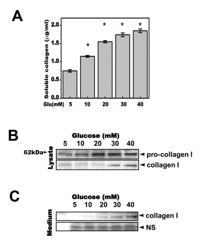
 | Fig. 1Glucose increases the secretion of soluble collagen. Cells were exposed to the indicated concentrations of glucose for 1, 2, 3 or 4 days. (A) Cells were exposed to the indicated concentrations of glucose for 3 days. Soluble collagen was then analyzed in the media. (B) Cell lysates were used for immunoblotting with anti-pro-collagen 1 or collagen 1 antibody. (C) Immunoblotting using the medium from cells cultured with 40 mM glucose for the indicated periods. Anti-collagen 1 antibody immunoblot (upper) and a separate Coomassie blue-stained gel (lower) are shown. Data represent means±S.E. (n=3). *p<0.05, significantly different from 5 mM glucose-treated cells; NS, non-specific. |
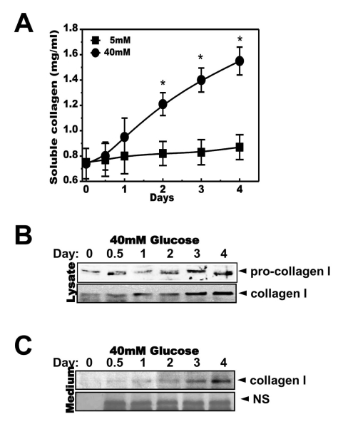
 | Fig. 2High concentrations of glucose increased secretion of soluble collagen in a time-dependent manner. Cells were exposed to the indicated concentrations of glucose for 1, 2, 3 or 4 days. Soluble collagen was analyzed in the media (A). Cells were exposed to a concentration of 40 mM glucose for the same period. Cell lysates were then used for immunoblotting with anti-pro-collagen 1 or collagen 1 antibody (B). Immunoblotting using the medium from cells cultured with 40 mM glucose for the indicated periods with anti-collagen I antibody (C) and separate Coomassie blue staining (C, lower). Data represent means±S.E. (n=4). *p<0.05, significantly different from 5 mM glucose-exposed cells during each same period; NS, non-specific. |
High glucose induces an ER stress response in human gingival cells
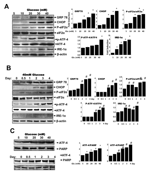
 | Fig. 3Glucose induces the ER stress response. Cells were exposed to the indicated concentrations of glucose for 3 days. Immunoblotting was performed with anti-GRP78, CHOP, peIF-2α, eIF-2α, IRE-1α or β-actin antibodies (A). Quantification analysis was performed (right). Cells were exposed to 40 mM glucose for the indicated periods. Immunoblotting was performed with anti-GRP78, CHOP, p-eIF-2α, eIF-2α, IRE-1α or β-actin antibodies (B). Quantification was performed (right). After cells were exposed to the indicated concentrations of glucose for 3 days (C, upper) and separately cells were exposed to 40 mM glucose for the indicated periods (C, lower), nuclear fractions were obtained and immunoblotting was performed with anti-ATF-4 or PARP antibodies. Quantification was performed (right). Data represent means±S.E. (n=3). *p<0.05, significantly different from 5 mM glucose-exposed cells during each same period. #p<0.05, significantly different from 40 mM glucosetreated condition during 0 day. |
The ER stress regulator 4-PBA inhibits collagen secretion in human gingival cells
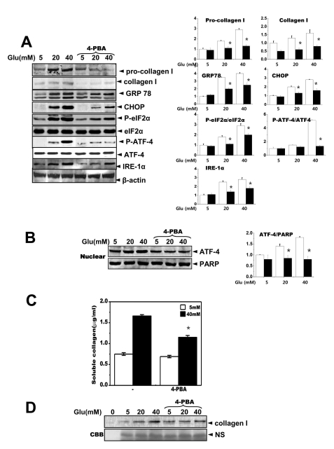
 | Fig. 4The chemical chaperone, 4-PBA inhibits the ER stress response as well as collagen synthesis and secretion. Cells were exposed to the indicated concentrations of glucose with or without 4-PBA for 3 days. Immunoblotting was performed with anti-GRP78, CHOP, p-eIF-2α, eIF-2α, IRE-1α, pro-collagen I, collagen I or β-actin antibodies (A). Quantification was performed (right). Separately, cells were exposed to the indicated concentrations of glucose with or without 4-PBA for 3 days and nuclear fractions were obtained and immunoblotting was performed with either anti-ATF-4 or PARP antibodies (B). Quantification was performed (right). Soluble collagen was analyzed in media from the cells cultured with 5 or 40 mM glucose for 3 days (C). Immunoblotting was performed with the medium from the cells cultured with 40 mM glucose for 3 days, using anti-collagen I antibody (D) and separately stained with Coomassive blue dye (lower). Data represent means±S.E. (n=5). *p<0.05, significantly different from each indicated concentration of glucose-treated cells without 4-PBA. CBB; Coomassive blue staining. |


 PDF
PDF ePub
ePub Citation
 Print
Print


 XML Download