

 PDF
PDF ePub
ePub Citation
 Print
Print


INTRODUCTION
Smooth muscle cells are the contractile component of body passages, including blood vessels, airways, gastrointestinal tract, and others [1]. Two types of smooth muscle have been described based on their electromechanical properties [2]. Phasic smooth muscle contract and relax rapidly, and their spontaneous, rhythmic activities are advantageous to the physiological functions, as found in digestive tract, ileum, uterus and bladder. Tonic smooth muscle contract slowly and they can maintain force with very low energy consumption, as found in most vessels and airways.
Contractions of both phasic and tonic smooth muscles are regulated by the phosphorylation of myosin regulatory light chain (MRLC) and the muscle shortening velocity is closely correlated with the extent of the phosphorylation although there is considerable debate about the in vivo role of additional mechanisms that might modulate crossbridge interactions [3,4]. In slow tonic smooth muscles MRLC phosphorylation is not a simple switch enabling crossbridge attachment and cycling. Sustained contraction (tone) is associated with reductions in cell Ca2+, MRLC phosphorylation, and parameters reflecting crossbridge cycling rates such as shortening velocity and ATP consumption [5]. This phenomenon termed "latch" is advantageous for a muscle type that has an important skeletal role in stabilizing organ dimensions against imposed loads. A simple scheme in which MRLC phosphorylation is the sole regulatory mechanism can predict the dependence of velocity and force on phosphorylation in the tonic swine carotid media and the time course of phosphorylation and contraction in the bovine trachealis stimulated by neurotransmitters, hormones, or depolarization [6-9].
If 4-state model is sufficient to explain crossbridge interactions in tonic smooth muscle, then it should also quantitatively account for crossbridge interactions in phasic smooth muscle. However, the 4-state crossbridge model does not explain many observations of permeabilized smooth muscle or intact fast, phasic tissues [9,10]. The contractile system of phasic smooth muscle is poorly understood. Phasic smooth muscles usually exhibit spontaneous, rhythmic contractile activity at 37℃. In vitro studies are typically conducted at room temperature to block this activity. Lower temperatures are also employed with skinned preparations to enhance their stability [11,12]. It has been reported that an isolated myosin light chain phosphatase (MLCP) has unusually high temperature dependence with Q10=5.2 [13]. Thus, lowering temperature should favor the fast crossbridge cycle over the slow cycle for a given myoplasmic [Ca2+] if 4-state model is correct. The rabbit bladder is a phasic smooth muscle that is normally exhibit spontaneous, rhythmic contractile activity at 37℃ in vivo. The axial alignment of cells makes it suitable for a biological analysis of contractile system function.
The purpose of the present study was to employ a phasic smooth muscle preparation that could be activated by KCl and carbachol stimulation and determine the relationship between myoplasmic [Ca2+], crossbridge phosphorylation, crossbridge cycling rates, and force generation. Our goals was also to evaluate that the 4-state model that predicts latch and other properties of tonic smooth muscle is also applicable to phasic smooth muscle to predict the maintenance of the latch state during a phasic smooth muscle contraction.
METHODS
Tissue preparations
Male New Zealand white rabbits weighing approximately 2.5~3 kg were killed by inhalation of halothane and urinary bladder was isolated promptly, and placed in a cold (4℃) physiological salt solution (PSS) of the following composition (mM): 117.8 mM NaCl, 6.0 KCl, 24.3 NaHCO3, 1.2 NaH2PO4, 0.026 Na2-EDTA, 1.6 CaCl2, 1.2 MgSO4 and 5.6 glucose at 37℃ gassed with 95% O2 and 5% CO2 which result in a pH 7.4. Strips were dissected from the ridges formed by the bladders contraction in the cold PSS on the abdominal external surface of the bladder. Histological examination showed that all the cells are aligned in the longitudinal axis of the preparation in which length and force are measured. 70 mM KCl-depolarization was accomplished by substituting KCl stoichiometrically for NaCl in PSS. Lengths and weights of rabbit bladder were measured in all experiments. Tissue wet weights were calculated from dry weights using 0.15 for the dry-to-wet weight ratio. Stress (in N/m2) was calculated from force development and tissue cross-sectional area at Lo, which was calculated from tissue length, wet weight, and density of 1.05 kg/l.
Isometric measurements
Muscle strips were mounted in jacketed baths and attached to Grass FT.03 (Quincy, Massachusetts) force transducers. After 1 hr equilibration period the length for maximal active force development was determined by increasing the length of each muscle strip by 1-mm increments until the maximal active contractile responses to 70 mM KCl was achieved. Force was measured in the isometric mode and was collected by a personal computer.
Estimation of isometric step-shortening velocity
After determination of a partial length-tension curve, the strips were set to their optimum length for force development, Lo [14]. A rapid micrometer shortening from Lo to 0.9 Lo reduced the force to the passive value. The rate of stress redevelopment, expressed as the time required for half maximal force redevelopment after shortening ("half-time") is an index of average crossbridge cycling rates. Its reciprocal, 1/half-time, is proportional to changes in shortening velocity.
Measurement of Myoplasmic [Ca2+]
Myoplasmic [Ca2+] was estimated using the photoprotein aequorin [15]. Aequorin was loaded intracellularly by iso-osmotic hyperpermeabilization. This procedure involves incubation of free-floating tissues in a series of Ca2+-free solutions at 2℃. Solution 1 chelates extracellular Ca2+ with high EGTA (ethylene glycol-bis-(β-aminoethylether)-N,N,N',N'-tetraacetic acid), solution 2 contains aequorin, solution 3 contains higher [Mg2+] which may help to reseal the membrane, and solution 4 is Ca2+-free PSS with high MgCl2. The tissues were mounted isometrically, stretched to a length that produced a stress of approximately 0.5×10⁵ N/m2, and warmed to 22℃. Extracellular CaCl2 was slowly increased to 1.6 mM over 30 min, and the tissues were equilibrated overnight at 37℃. The next morning, the tissues were stretched to within 5% of their optimal length for stress development. The loading procedure did not affect maximal stress development. Light measurements were made in a light-tight photomultiplier tube that allowed simultaneous measurement of aequorin-emitted light and stress measurement [15]. Force was measured with a servo (model 300H, Cambridge Technology, Cambridge, USA) in the isometric mode and was collected by a personal computer in synchronization with the light signal. Aequorin light signals are presented as log L/Lmax, where L is the photon counter (in counters per second) and Lmax is a measure of the total undischarged aequorin present in the tissue. Lmax was calculated at each time point to correct for aequorin consumption. Log L/Lmax is a function of [Ca2+], light signals are reported as a change log L/Lmax in which the resting log L/Lmax is subtracted from all subsequent log L/Lmax values. This normalization markedly decreased interexperimental variability and provided enhanced sensitivity to small changes in [Ca2+]. Aequorin light signals were calibrated in Ca2+-EGTA buffers at 37℃ with [Mg2+]=0.5 mM.
Measurement of MRLC phosphorylation
Phosphorylation of the 20,000-Dalton myosin light chains was determined under isometric conditions. Muscle strips were rapidly frozen in a acetone/dry ice slurry (-78℃) in the unstimulated state and at various times during contraction, stored overnight in dry ice and then allowed to thaw slowly to room temperature. The acetone-dried tissues were then removed, the tissues air dried and weight. Tissues were then homogenized at 4℃ in homogenization buffer containing 1% sodium dodecyl sulfate (SDS), 10% glycerol, and 20 mM dithiothreitol. Tissue homogenates were stored at -20℃ until ready for two-dimensional polyacrylamide gel electrophoresis. Each 35 µl of homogenate was applied to an isoelectric focusing polyacrylamide tube gel (1.5 mm in inner diameter and 6 mm in outer diameter) with 8 M Urea and 5% ampholytes pH 4~6.5. After isoelectric focusing overnight the appropriate portion of tube gel was placed on a slab gel consisting of a stacking gel of 5% acrylamide and a separating gel of 14% acrylamide. The separating gel was removed, fixed and stained overnight with shaking in a solution containing 25% isopropanol, 10% acetic acid, and then slowly destained (3 hrs) in 10% acetic acid until background was clear. The myosin light chains were quantified by densitometric scanning of gels using a densitometer (Helena). Myosin phosphorylation in mol phosphate per total light chain was the ratio of monophosphorylated light chain divided by the sum of unphosphorylated and monophosphorylated light chains. In rabbit bladder, there is also approximately 5~10% of the monophosphorylated light chain is diphosphorylated. This amount was not counted in the reported phosphorylation values. Nonmuscle light chains were ignored.
Statistics
The results of the experiments are expressed as mean±S.E.M. Unpaired Student's t-test was used for statistical analysis of the results and the number of preparations taken from separate animals was indicated by n. Significant tests were performed by Student's paired or unpaired t test. p values of less than 0.05 were considered significant.
RESULTS
Contraction produced by CCh and KCl
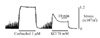
Unstimulated bladder preparations occasionally showed spontaneous, rhythmic contractile activity at 37℃ in vivo. And K+ depolarization also elicited transient contractions diagnostic of a phasic smooth muscle. Fig. 1 shows representative recordings of changes in tension induced by 1 µM CCh and 70 mM KCl depolarization. The application of 1 µM CCh induced sustained contraction. When exposed to 70 mM KCl, the level of tension increased rapidly and then slowly decreased to achieve sustained level. The tension during activation by 1 µM CCh was significantly greater than that during activation by 70 mM KCl.
Dose-dependent effects of KCl and CCh on myoplasmic [Ca2+], MRLC phosphorylation and stress
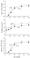
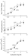
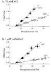
The relationship between aequorin-estimated myoplasmic [Ca2+], MRLC phosphorylation and active stress was examined by comparing dose-dependent relationships obtained from KCl (Fig. 2) and CCh (Fig. 3) at both 37℃ and 22℃. Fig. 2 shows that KCl (15, 25, 40, 70 and 100 mM) induced concentration-dependent increases in myoplasmic [Ca2+], MRLC phosphorylation and steady-state active stress. As shown in Fig. 3, CCh (0.01, 0.03, 0.1, 0.3, 1, and 3 µM) increased myoplasmic [Ca2+], MRLC phosphorylation and steady-state active stress in a dose-dependent manner. KCl- and CCh-induced active stress at 22℃ is higher than that of at 37℃ and this was related to the higher myoplasmic [Ca2+] and MRLC phosphorylation although there was no significant differences at all ranges.
Time course of KCl- and CCh-induced changes in myoplasmic [Ca2+], MRLC phosphorylation, 1/Half-time and stress
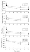
Fig. 4 shows the time course of myoplasmic [Ca2+], MRLC phosphorylation, 1/half-time for the force redevelopment, and active stress in tissues depolarized with 70 mM KCl at both 37℃ and 22℃. Basal MRLC phosphorylation was 12.7±3.3 (37℃) and 18.0±2.8 (22℃). Stimulation with 70 mM KCl at both 37℃ and 22℃ resulted in a transient increase in myoplasmic [Ca2+], MRLC phosphorylation, 1/half-time, and active stress. These values then declined in a time-dependent manner and reached intermediate values. 70 mM KCl-induced increase in MRLC phosphorylation, and active stress were higher and the 1/half-time was lower at 22℃ when compared to 37℃. Myoplasmic [Ca2+], MRLC phosphorylation, 1/half-time, and active stress increased rapidly to a maximal values within 30 seconds at 37℃ and 1 min at 22℃ of KCl stimulation, suggesting that KCl-stimulated responses at 22℃ were more slowly activated than at 37℃ by myosin light chain kinase (MLCK). As shown in Fig. 5, 1 µM CCh-induced increase in active stress was sustained at peak values and did not decreased in spite of the decreases in myoplasmic [Ca2+], MRLC phosphorylation and 1/half-time at 37℃. These results suggest that the latch phenomenon was present in rabbit bladder in response to CCh stimulation at 37℃. It is known that "latch" formation is abolished when temperature was lowered to room temperature. Similar protocols were performed at 22℃. The time course of myoplasmic [Ca2+], MRLC phosphorylation, 1/half-time, and stress were similar to that observed at 37℃. CCh-induced active stress at 22℃ was higher than that of at 37℃, and this is related to the increase in MRLC phosphorylation.
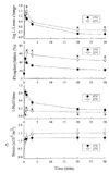
Relationship between 1/ half-time and MRLC phosphorylation
In order to know that latchbridge is temperature or not, we plotted the relationship between 1/half-time and MRLC phosphorylation. As shown in Fig. 6, average shortening velocity as estimated by 1/half-time was also proportional to MRLC phosphorylation over a wide range of phosphorylation values and was independent of stimulus. The relationships between 1/half and MRLC phosphorylation was shifted toward lower-right, suggesting that latchbridge formation is decreased by lowering temperature.
Relationship between active stress and MRLC phosphorylation
When we plotted the relationships between MRLC phosphorylation and steady-state active stress, there was a quasi-hyperbolic dependence of active stress on MRLC phosphorylation at 37℃ (Fig. 7). At 22℃ this relationship was more linear, suggesting that lacthbridge formation was decreased by lowering temperature to room temperature.
DISCUSSION
Ca2+-calmodulin dependent phosphorylation of 20 kD MRLC (crossbridge) by MLCK and dephosphorylation by MLCP may regulate smooth muscle contraction [16,17]. Latch (sustained contraction with a reduction in crossbridge cycling rates) characterizes tonic arterial smooth muscles [1,5]. However, latch is not evident in phasic smooth muscles exhibiting spontaneous rhythmic contractile activity at 37℃. So, we tested the hypothesis that 4-state crossbridge model that predicts latch and other properties of tonic smooth muscles is applicable to phasic smooth muscle and that lower temperatures used to block spontaneous phasic contractions explain many differences in the observed properties of tonic and phasic tissues.
When exposed to CCh, the rabbit bladder preparation exhibited contractile properties observed in tonic smooth muscles including a decline in average crossbridge cycling rates in associated with a fall in Ca2+-dependent phosphorylation to lower steady-state levels, "latch" state. We found that by altering either the temperature or the type of stimulus, stress generation and maintenance were similar but the time course of both crossbridge phosphorylation and shortening velocity (expressed as 1/half-time) were affected. Stimulation with 70 mM KCl or 1 µM CCh at 37℃ resulted in high initial levels of both crossbridge phosphorylation and shortening velocity with a subsequent time-dependent decrease in both parameters to suprabasal levels. Stimulation with 70 mM KCl at 22℃ resulted in a crossbridge transient similar to that at 37℃ but values estimated for shortening were significantly lower; both parameters were transient. Stimulation with 1 µM CCh at 22℃ resulted in high levels of crossbridge phosphorylation that were maintained at higher level throughout activation with lower estimated shortening velocity. Stimulation with high K+ in rabbit bladder smooth muscle induced the phasic contractions at both 37℃ and at 22℃, and these phasic contractions were mediated by transient increases in intracellular Ca2+ and crossbridge phosphorylation. 70 mM KCl-induced contractions at 22℃ is greater than that at 37℃ and this might related to the increases in intracellular Ca2+ and crossbridge phosphorylation. In contrast to this, CCh-induced increases in both intracellular Ca2+ and crossbridge phosphorylation were transient at 37℃, suggesting that CCh-induced tonic response is related to latchbridge formation [1,3,8,9,18]. There was a linear correlation between the level of crossbridge phosphorylation and shortening velocity (expressed as 1/half-time), supports the hypothesis that MRLC phosphorylation in smooth muscle fucntions in regulating crossbridge cycling rates, and similar results were reported previously [10,19]. The average shortening velocity was also proportional to MRLC phosphorylation over a wide range of phosphorylation values and was independent of stimulus. Lowering temperature decreased the slope of the relationship, suggesting that latchbridge formation were diminished by lower temperature as predicted by the 4-state crossbridge hypothesis [1,6,8]. There was a marked suppression of the time-dependent fall in phosphorylation and crossbridge cycling rates characteristic of latch as predicted by the 4-state crossbridge hypothesis.
At 37℃ the Ca2+-sensitivity of myosin light chain phosphorylation was similar in high K+-depolarized and CCh-stimulated bladder tissues. Uncertainties about the aequorin calibration at 22℃ only allow the inference that lower temperature must alter Ca2+-mobilization and the Ca2+-sensitivity for phosphorylation induced by at least one of the two types of activation [20,21]. Lowering the temperature to 22℃ increased crossbridge phosphorylation significantly at all times showing an increase in the MLCK/MLCP activity ratio and the actin-activated myosin ATPase. However, the rates of force generation and MRLC phosphorylation and crossbridge cycling rates are highly temperature dependent, suggesting that the temperature dependence of the regulatory system are likely responsible for the differences. If the in situ temperature dependency of MLCK and MLCP were the same, one would predict that lowering temperature would not increase net MRLC phosphorylation in response to a specific stimulus-induced elevation in myoplasmic Ca2+. It has been reported that MLCK/MLCP activity ratio are higher in phasic smooth muscles than in tonic smooth muscles [17,22]. Blumenthal and Stull [23] reported that Q10 of MLCK equal to 2, whereas Q10 of myosin light chain phosphatase equal to 5.2 [13,24]. Murphy also determined a Q10 of 2.2 for the actomyosin ATPase activity of swine carotid artery [25]. The maximal force generation is only moderately affected by change from 37 to 22℃. Therefore, the increase in crossbridge phosphorylation levels induced by lowering temperature to 22℃ might be due to the changes in MLCK/MLCP activity ratio. The steady-state dependence of stress on crossbridge phosphorylation is very steep at 37℃ in the agonist-stimulated or K+-depolrized phasic bladder. A sigmoidal dependence of force on MRLC phosphorylation were also observed in guinea-pig ileal smooth muscle, frog bladder and rat anococcygeus [9,26,27]. Reduced temperature flattened the dependence of stress on crossbridge phosphorylation compared to 37℃. The latchbridge hypothesis also predicts that a decrease in MLCP activity should alter the dependent steady-state stress on crossbridge phosphorylation [28-30]. When MLCP activity was inhibited by lowering temperature the dependency of steady-state active stress on crossbridge phosphorylation was flattened (linear), not a quasi-hyperbolic relationship between active stress and crossbridge phosphorylation which probably by preventing latchbridge formation upon dephosphorylation as predicted by the inhibition of K2 and K6 in the latchbridge hypothesis [1,9,30].
We concluded that phasic smooth muscle have comparable contractile system function and that mechanisms regulating the myoplasmic [Ca2+] are primarily responsible for functional diversity. The 4-state crossbridge model may adequately predict the behavior of the rabbit bladder although the various rate constants would differ in the relatively fast tissue. Temperature changes have important effects on the response of a muscle with covalent crossbridge regulation. The high temperature dependence of myosin light chain phosphatase (Q10=5.2) is a likely contributing factor.

 XML Download