

 PDF
PDF ePub
ePub Citation
 Print
Print


Epiphora is one of the most common problems encountered by ophthalmologists, and accurate evaluation of the lacrimal drainage system is crucial in making a diagnosis and establishing an effective treatment plan. Although there are various in-office examinations and imaging studies for the lacrimal drainage system, the location and extent of lacrimal obstruction are sometimes elusive. This can be especially true if patients with epiphora symptoms have common canalicular obstruction (CCO) or canalicular obstruction; it is usually very difficult to accurately assess the status of the lacrimal sac using preoperative tests such as lacrimal irrigation, lacrimal probing, or dacryocystography. Therefore, research using these examinations seems to not be adequate to evaluate the obstruction site of the lacrimal drainage system [1]. We observed that the status of the lacrimal sac was variable in cases with CCO or canalicular obstruction during external dacryocystorhinostomy (DCR); some cases had a clear lacrimal sac, while others showed mucosal swelling, erythema, and contracture of the lacrimal sac by intraluminal fibrosis and luminal stricture of the proximal nasolacrimal duct. However, there have been few studies that have conducted a detailed examination of the obstruction characteristics of the lacrimal drainage system in patients with epiphora.
External DCR is the standard management option for most cases with lacrimal drainage obstruction including nasolacrimal duct obstruction (NLDO), CCO, and distal canalicular obstruction [234]. In addition to the excellent surgical outcomes, external DCR has another advantage over endonasal DCR, as the external approach offers direct visualization of the lacrimal sac. During the procedure, the medial wall of the lacrimal sac is incised, and the lumen and internal punctum of the lacrimal sac can be clearly visualized. Thus, the detailed features of the lacrimal drainage obstruction can be assessed more precisely during external DCR.
The purpose of this study was to classify the locational patterns of lacrimal drainage obstruction and the status of the lacrimal sac in CCO or canalicular obstruction cases based on intraoperative observation during external DCR surgery. Recently, we reported the surgical outcomes of 10 years of external DCR and the risk factors associated with functional failure [5]. Anatomical success was achieved in 98.8% of cases (760 / 769) and functional success in 81.9% (630 / 769). When analyzing 760 anatomically successful DCRs, CCO (odds ratio [OR], 1.752; p = 0.014) and canalicular obstruction (OR, 2.058; p = 0.015) were independent risk factors for functional failure. In subgroup analysis of patients with primary NLDO, patients with a small lacrimal sac had a significantly higher risk of functional failure. In that report, the surgical outcomes according to obstruction site were not specifically described, and the lacrimal sac status in CCO or canalicular obstruction patients was not evaluated. In this study, we investigated the surgical outcomes according to obstruction site and effect of lacrimal sac status on surgical outcomes in CCO and canalicular obstruction groups.
Materials and Methods
Institutional review board approval was obtained for this study. A retrospective review of the electronic medical records of all patients who had undergone external DCR at Seoul National University Hospital between 2005 and 2014 was performed. Patients who were presumed to have NLDO, CCO, or distal canalicular obstruction preoperatively were indicated for external DCR. Patients with secondary NLDO who had a history of ocular adnexal tumors, failed DCR, traumatic NLDO, or previous nasal cavity surgery and patients with events or diseases that can affect the lacrimal drainage system such as facial nerve palsy or lower lid ectropion were excluded. Patients who had a history of chemotherapy or radioiodine therapy were included in this study if the lacrimal obstructive feature indicated DCR surgery. Preoperatively, all patients underwent an evaluation of the lacrimal drainage system including the lacrimal syringing test and probing. Dacryocystography or lacrimal scintigraphy was not routinely performed.
All surgeries were performed by a single, experienced oculoplastic surgeon (SIK) under general anesthesia. The detailed surgical procedure was described in our previous reports [678]. A Bowman's probe was inserted through the upper and lower puncta, and the lacrimal sac was opened with a vertical incision along its medial wall. During the procedure, lacrimal obstructive features were carefully inspected and described in detail. The obstruction site was determined by canalicular irrigation and probing and inspection of the internal punctum of the lacrimal sac (common internal ostium) during the surgical procedure. If upper, lower, and common canaliculus were patent and the lacrimal sac was easily tented, primary NLDO was defined. CCO was defined when the canalicular irrigation showed an opposite punctal reflux but canalicular probing showed a soft stop with impossible tenting of the lacrimal sac. When the tip of the probe was visible under the thin membrane at the internal punctum on canalicular probing after opening the medial wall of the lacrimal sac, thin membranous CCO was diagnosed. When the tip of the probe was not visible at the internal punctum on canalicular probing, thick CCO was diagnosed. Also, the status of the inside of the lacrimal sac was inspected whether the lacrimal sac lumen showed a clean, normal appearance or showed intraluminal swelling, erythema, or scarring. In most cases, a silicone bicanalicular tube (solid tube, 0.064-cm diameter, C-line canaliculus intubation set 8590450; Medtronic Ophthalmics, Jacksonville, FL, USA) was inserted. In cases with CCO or canalicular obstruction, double silicone intubation was performed after internal excision of the obstructed portion of the canaliculus.
Postoperatively, all patients were followed up at 1 week, 1 month, and between 4 and 6 months postoperatively and then variably thereafter. The silicone tube was removed 4 to 6 months after surgery, and only patients who were followed up more than 4 months were included in this study. The surgical outcome was assessed anatomically and functionally based on records from the last visit. Anatomical success was defined as good passage without significant reflux on the lacrimal syringing test. Functional success was defined as absence of a tearing symptom as assessed using Munk score. Statistical analyses were performed using IBM SPSS ver. 21.0 (IBM Corp., Armonk, NY, USA). Chi-square test, t-tests, and Fisher exact tests were used to determine statistical significance.
Results
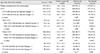
A total of 769 eyes of 603 patients received external DCR for primary lacrimal drainage obstruction during the enrollment period. The mean age of the patients was 57.7 ± 12.2 years (range, 3 to 84 years), and 468 patients (77.6%) were female. The mean duration of tearing symptom was 62.8 ± 92.3 months (range, 1 to 720 months). Table 1 shows the distribution of the detailed lacrimal obstructive features observed during external DCR surgery. Primary NLDO accounted for 56.2% (432 / 769) of primary lacrimal drainage obstruction. CCO was diagnosed in 32.9% (253 / 769); thin membranous CCO accounted for 3.3% (25 / 769), thick CCO accounted for 29.6% (228 / 769), and canalicular obstruction was diagnosed in 10.9% (84 / 769). Significant lacrimal sac changes, such as intraluminal swelling, erythema, or fibrosis, were present in 51.8% (131 of 253 eyes) of the CCO group and 58.3% (49 of 84 eyes) of the canalicular obstruction group. The frequency of lacrimal sac changes was similar between thin CCO (52%, 13 of 25 eyes) and thick CCO (51.8%, 118 of 228 eyes).
The overall anatomical and functional success rate was 98.8% (760 of 769 eyes) and 81.9% (630 of 769 eyes). The anatomical success rate was not significantly different among primary NLDO, CCO, and canalicular obstruction groups (p = 0.484), but the functional success rate was significantly higher in the primary NLDO group than in the CCO or canalicular obstruction group (p = 0.004 and p = 0.001, respectively). The subgroup analyses for success rate according to the status of the lacrimal sac are described in Table 1. When there were lacrimal sac luminal changes, the functional success rate significantly decreased in the CCO group (p = 0.044). Subgroup analysis revealed that the functional success rate was higher in cases without lacrimal sac changes than in those with lacrimal sac changes in the thick CCO group, while the functional success rate was not different regardless of the status of the lacrimal sac in the thin CCO group. In the canalicular obstruction group, the functional success rate was not different according to the status of the lacrimal sac.
Discussion
This study analyzed the distribution of lacrimal drainage system obstruction patterns in patients who required DCR surgeries based on intraoperative findings of the canaliculus and lacrimal sac. In 769 cases with primary lacrimal drainage obstruction, 56.2% had primary NLDO with patent canaliculi; 32.9% had CCO; and 10.9% had canalicular obstruction. Approximately half of the cases of CCO and canalicular obstruction (131 of 253 eyes and 49 of 84 eyes) demonstrated simultaneous lacrimal sac mucosal changes, such as inflammation or fibrosis.
The level of lacrimal drainage obstruction can be roughly estimated preoperatively using several examinations. However, in cases with upper lacrimal drainage system obstruction, the precise status of the lower lacrimal drainage system cannot be accurately assessed using these tests. Consequently, lacrimal drainage obstruction is only able to be broadly classified as NLDO, CCO, or canalicular obstruction. In this study, we describe the detailed characteristics of primary lacrimal drainage obstruction based on intraoperative observations of the canaliculus and lacrimal sac at the step of lacrimal sac incision during the external DCR procedure. When a lacrimal probe could not be inserted into the lacrimal sac, CCO or canalicular obstruction was diagnosed according to the soft stop level. CCO can be subdivided into thin membranous CCO and thick CCO based on the nature of the soft tissue at the internal punctum of the lacrimal sac. In cases with CCO or canalicular obstruction, if the incised lacrimal sac showed significant intraluminal swelling, erythema, or scarring, we assumed that there were concomitant lacrimal sac luminal changes. This allowed us to make a more detailed classification of primary lacrimal drainage obstruction; primary NLDO with patent canaliculus (56.2%), CCO without lacrimal sac luminal change (15.9%), CCO with lacrimal sac luminal change (17.0%), canalicular obstruction without lacrimal sac luminal change (4.5%), and canalicular obstruction with lacrimal sac luminal change (6.4%) (Table 1).
Even though we often observed gross changes of the lacrimal sac mucosa during DCR, these changes have been rarely noted in other studies. It is well known that chronic inflammation and fibrosis are the most common histopathologic changes in lacrimal sacs of patients with NLDO [91011]. We speculated that the cases with CCO or canalicular obstruction, who showed intraluminal lacrimal sac changes, presumed to have simultaneous NLDO. In these patients, CCO or canalicular obstruction might develop secondary to a chronic inflammatory reaction of the lacrimal sac. In a study using dacryoendoscopy, the authors tried canalicular incision and tube intubation in patients with CCO and reported that there was simultaneous NLDO in one-quarter of patients, which makes intubation difficult, supporting our concept [12]. In the present study, approximately half of the cases with CCO or canalicular obstruction (53.4%, 180 / 337 cases) showed intraluminal mucosal swelling, erythema, or fibrosis in the lacrimal sac. Our findings support the assertion that DCR is a more suitable option for management of CCO or canalicular obstruction rather than canalicular trephination or canaliculoplasty, considering the possibility of combined lacrimal sac changes. In terms of success rate of external DCR, the functional success rate was higher in the primary NLDO group than in CCO or canalicular obstruction, in agreement with previous studies [51314]. In cases of thick CCO, the status of the lacrimal sac affected the functional success rate, and this is a novel finding that has not been previously reported. The difference was not significant in the thin CCO group or the canalicular obstruction group. We think that the number of cases in the thin CCO group was too small to allow a statistical conclusion. In canalicular obstruction, canalicular obstruction itself is a strong, unfavorable factor for functional success, so the influence of lacrimal sac status may be limited.
In conclusion, lacrimal sac mucosal changes were confirmed intraoperatively during external DCR in a significant proportion (53.4%) of cases with CCO or canalicular obstruction. In the CCO group, those changes were associated with lower functional success rate.
 XML Download