

 PDF
PDF ePub
ePub Citation
 Print
Print


Anatomically, the lower eyelid can be divided into the anterior (skin and orbicularis oculi muscle), middle (orbital septum and orbital fat), and posterior lamellae (tarsus and conjunctiva) sections. The location of the lower eyelid is maintained by the following factors: horizontal lower eyelid laxity, distensibility of the lower eyelid retractor, length and degree of tension of the lower eyelid, adequacy of the fornix and palpebral conjunctivae, location of the canthal ligament, degree of eyeball protrusion, and degree of orbicularis oculi tension [1]. Pathologic changes in these factors can lead to lower eyelid retraction [12].
Lower eyelid retraction occurs most frequently because of thyroid-associated orbitopathy (TAO) associated with fibrosis of the capsulopalpebral fascia, and can also occur because of extraocular muscle surgery or injury, tissue loss or cicatricial change after lower eyelid blepharoplasty, or weakening of the orbicularis oculi [3]. Exposure of the infracorneal bulbar conjunctiva in lower eyelid retraction can cause various complications such as dry eyes, exposure keratitis, and corneal ulcer. Although conservative treatment, such as the use of artificial tears, can be sufficient when symptoms are mild to moderate, surgery is required for severe symptoms. Furthermore, an increasing number of patients are demanding surgical correction of lower eyelid retraction for aesthetic reasons.
Although many studies have evaluated the advantages and disadvantages of various surgical grafts and the outcomes of surgical procedures conducted with these grafts, there have been few reports on surgical methods and outcomes for cases involving retraction [45]. Therefore, we aimed to investigate the causes of lower eyelid retraction and evaluate the outcomes of various surgical procedures.
Materials and Methods
The institutional review board of Kim's Eye Hospital in Seoul, Korea, approved this study. All methods were in accordance with the Declaration of Helsinki.
Study subjects
We retrospectively analyzed the medical records of patients who were diagnosed with lower eyelid retraction and received surgical treatment by a single surgeon (JWJ) at Kim's Eye Hospital between January 2006 and December 2013. If the sclera was visible above the lower eyelid (inferior sclera show) or if the margin reflex distance 2 (MRD2) exceeded 6 mm, lower eyelid retraction was diagnosed. To investigate the preoperative horizontal laxity of lower eyelids, we used the eyelid distraction test. The eyelid distraction test is performed by grasping the lower eyelid skin over the central aspect of the lower eyelid tarsal plate, pulling the lower eyelid away from the globe, and measuring the distance between the globe and the posterior aspect of the lower eyelid. Horizontal laxity was diagnosed when the measurement exceeded 5 mm.
We excluded patients who had post-surgical follow up times of less than 1 month, had ectropion, had factors that could influence the size of cornea (such as glaucoma and phthisis bulbi), or had artificial eyes. Patients with corneal opacity were also excluded because the condition prevents MRD2 from being measured.
Ocular examinations and surgical procedures
To compare preoperative and postoperative conditions, facial photographs of patients were taken. We measured MRD2 from photographs of patients taken at each visit, in which patients were looking straight into the camera. Because the measured MRD2 values were obtained from magnified images, we divided the MRD2 value by the horizontal corneal diameter and multiplied the resulting value with the mean horizontal corneal diameter of Korean patients (men, 11.4 mm; women, 11.2 mm) to obtain a value closer to the actual value [6]. All photographs were reviewed in a standard manner using ImageJ (National Institutes of Health, Bethesda, MD, USA) by JSB and JWJ. We measured MRD2 (mm) and lower eyelid asymmetry (difference between MRD2 values of both eyes, in mm) before surgery. MRD2 was measured again 1 week and 1 month after the surgery and the final follow-up visit.
Degree of retraction was categorized as mild if lower eyelid asymmetry was less than 2 mm and if the MRD2 was less than 6.5 mm for both lower eyelids. If the results exceeded these values, the degree of retraction was categorized as severe.
In cases of mild lower eyelid retraction, lateral canthal tightening was performed, whereas in most cases of severe lower eyelid retraction, spacer grafts were used. If the lower eyelid exhibited horizontal laxity, the lateral tarsal strip procedure was used for horizontal tightening. Furthermore, when the volume needed to be supplemented because of soft tissue loss, an autogenous dermis fat graft was performed.
Data collection and analysis
We investigated the causes of lower eyelid retraction, past trauma or surgery history, clinical characteristics, and surgical outcomes in these patients.
Surgical outcomes were evaluated during the final follow-up visit. If only one eye exhibited lower eyelid retraction, the surgical outcome was classified as successful when the difference between the affected and unaffected eyes was less than 1 mm, good when the difference was between 1 mm and 2 mm, and poor when the difference exceeded 2 mm. If both eyes exhibited lower eyelid retraction, the surgical outcome was considered successful when the sclera was not visible around the lower margin of the cornea, good when less than 1 mm of sclera was visible, and poor when more than 1 mm of sclera was visible.
Data are presented as the mean ± standard deviation, and paired t-tests were conducted to compare preoperative and postoperative values. The SPSS ver. 19.0 (IBM Corp., Armonk, NY, USA) was used for statistical analysis, and the results were considered statistically significant when the p-value was less than 0.05.
Results
This study included a total of 19 eyelids in 14 patients; 12 eyelids of nine men and seven eyelids of five women. Patient age at the time of surgery ranged from 12 to 57 years (mean, 37.2 ± 15.8 years). Both eyes were affected in five patients (35.7%), while only the right or left eye was affected in seven patients (50.0%) and two patients (14.3%), respectively. The postoperative follow-up period ranged from 1 to 52 months (mean, 15.8 ± 17.6 months).
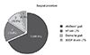
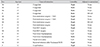
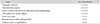
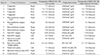
Among the 19 eyelids investigated, the most common cause of lower eyelid retraction was congenital retraction, which occurred in seven eyelids (36.8%); these eyelids required aesthetic correction. Lower eyelid retraction occurred after extraocular muscle surgery in four eyelids (21.0%); among these, two eyelids belonged to a patient who received an inferior rectus recession to correct strabismus associated with TAO. Lower eyelid retraction occurred after lower eyelid blepharoplasty in three eyelids (15.8%), after orbital wall fracture surgery in two eyelids (10.5%), because of bacterial infection after endonasal dacryocystorhinostomy in one eyelid (5.3%), because of facial nerve palsy in one eyelid (5.3%), and because of exophthalmos of a n unknown cause in one eyelid (5.3%) (Tables 1 and 2).
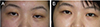
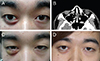
Surgical procedures varied with the degree and cause of retraction; lower eyelid retractor recession or lateral canthal tightening was performed in cases of mild retraction, whereas grafts were used in severe cases. In cases of severe retraction, the acellular dermal allograft (AlloDerm®, LifeCell Corp., Branchburg, NJ, USA) was most frequently used (Figs. 1, 2A, 2B). In the four cases of lower eyelid laxity, the lateral tarsal strip procedure was used for horizontal tightening (21.1%), and a suborbicularis oculi fat (SOOF) lift was conducted in one eyelid of a patient with midface ptosis. In two eyelids that exhibited loss of fat and soft tissue, dermis fat grafts were performed (10.5%) (Figs. 1, 3A, 3B, Table 3).
The mean preoperative MRD2 was 7.49 ± 0.85 mm, whereas the mean MRD2 measured 1 month after surgery was 5.65 ± 0.71 mm. The average MRD2 change after surgery was 1.84 ± 0.63 mm. The difference between preoperative MRD2 and postoperative MRD2 was statistically significant (paired t-test, p < 0.001).
Discussion
The lower eyelid is supported and maintained by the medial canthal tendons, lateral canthal tendons, capsulopalpebral fascia, tarsus, and orbicularis oculi muscle. Lower eyelid retraction is usually caused by contraction of the lower eyelid retractor (capsulopalpebral fascia and inferior tarsal muscle) and the posterior lamella as a result of various pathological changes in the structures supporting the lower eyelid [2]. TAO is reported to be a common cause of shortening of the lower eyelid posterior lamella. Shortening of the posterior lamella can also occur because of injuries and scars, and after lower eyelid blepharoplasty. Additionally, shortening of the posterior lamella because of exophthalmos can cause lower eyelid retraction [12]. In addition, rare cases of idiopathic congenital lower eyelid retraction have also been reported [78910]. The number of patients who want to undergo surgery for aesthetic reasons is increasing because the condition known as three white eyes, in which the sclera is visible above the lower eyelid margin, is not favored in East Asian countries, including Korea. Conservative treatment, such as administration of artificial tears, is used when symptoms are not severe, and surgical correction is required in cases of severe exposure keratitis.
Surgery to correct lower eyelid retraction can be approached with grafting or nongrafting techniques. The decision to use grafts is based on the degree of retraction, and surgical procedures vary based on the causes and degrees of retraction. Mild lower eyelid retraction can be corrected without grafts. Procedures such as lower eyelid retractor recession, which relieves vertical traction, and the lateral tarsal strip procedure (for lateral canthal tightening), which corrects horizontal laxity, are often utilized [311]. When retraction is severe and exceeds 2 mm, spacer grafts are used to push the lower eyelid margin upwards and to support it from below [12]. Autografts, such as hard palate mucosa and ear cartilage, allografts, such as preserved dermis (AlloDerm®, Surederm®) and preserved sclera, and synthetic grafts, such as polytetrafluoroethylene (Gore-Tex) and porous polyethylene (Medpor), can be utilized as spacer materials [451314]. The purposes of spacer graft surgery are to create a recess in the capsulopalpebral fascia and support the tarsus with the graft and consequently push the lower eyelid vertically up to a normal anatomic height and position. Recently, an acellular dermal allograft (AlloDerm®) has been used as a spacer graft in lower eyelid retraction surgery. AlloDerm® is a dermal matrix that is processed to remove cells containing antigenic targets for infection and thereby leaves the dermal matrix immunologically inert. It is made from human skin allografts obtained from cadavers [515]. In this study, AlloDerm® spacer grafts were placed via the subconjunctival approach in 13 eyelids of 10 patients with severe lower eyelid retraction. Graft contraction occurs over time; hence, the size of the AlloDerm® graft is usually 1.5- to 2-fold the size of the deficiency [16]. In this study, we conducted overcorrection procedures such that at the time of surgical completion, the grafts were slightly higher (approximately 1 mm) than lower eyelid margins when patients were measured in the sitting position. All surgical outcomes were satisfactory. None of the included patients complained about symptoms associated with their procedures nor were any complications or malfunctions of the lower eyelid reported.
The most common cause of retraction in this study was congenital lower eyelid retraction. A few cases of primary or idiopathic congenital retraction of the lower eyelid have been described in the literature [78910]. Collin et al. [7] reported four cases with lower eyelid retraction affecting one eye, and Mee and McNab [9] reported two cases of lower eyelid retraction with abnormal ocular movement. In the present study, we report seven cases of congenital lower eyelid retraction. The mean patient age was 25 years; lower eyelid laxity was absent; patients had not received any prior ophthalmological surgery; and no abnormal findings were observed in thyroid function or ocular movement. Congenital lower eyelid retraction occasionally occurs in Asian populations; the number of surgeries conducted for aesthetic purposes has been increasing because retraction is physiognomically unfavorable in these populations. In this study, all patients with idiopathic lower eyelid retraction showed good aesthetic outcomes with the AlloDerm® graft.
To correct strabismus associated with TAO, we grafted an AlloDerm® graft in two eyelids with severe lower eyelid retraction that occurred after inferior rectus recession. In TAO, chronic inflammation causes fibrosis of the orbital fascia and suspensory tissue; this subsequently causes shortening of the posterior lamella and retraction of the lower eyelid. Furthermore, fibrosis of the inferior rectus muscle, hormonal stimulation of the sympathetic nervous system, and exophthalmos can also contribute to retraction. In patients who undergo inferior rectus recession for the correction of strabismus associated with TAO, lower eyelid retraction can occur if the inferior rectus muscle is recessed by more than 4 mm [21718]. To reduce the occurrence of lower eyelid retraction in cases of inferior rectus recession, Jampolsky [19] reported a procedure in which the capsulopalpebral head was reattached to the inferior rectus 15 mm apart from the corneal limbus. In treating lower eyelid retraction associated with TAO, orbital decompression is initially required when exophthalmos is severe. In severe retraction cases without severe exophthalmos, when the degree of retraction exceeds 2 mm, correction with a spacer graft should be performed to supplement and support the posterior lamella.
In this study, some patients exhibited fat and soft tissue atrophy around the lower eyelid. The causes were inflammation and bacterial infection after endonasal dacryocystorhinostomy or orbital wall reconstruction surgery through subciliary incision. In these patients, we conducted autogenous dermis fat grafting and obtained satisfactory outcomes. We also used the lateral tarsal strip procedure to correct retraction that occurred after lower eyelid blepharoplasty (two eyelids) and orbital wall reconstruction surgery (one eyelid). Malpositioning of the lower eyelid is a complication that occurs in 2.7% to 16.6% of procedures, regardless of the type of surgical approach adopted [20]. Lower eyelid retraction occurs in 15% to 20% of lower eyelid blepharoplasty cases [212223]. Various factors can cause lower eyelid retraction after surgery; these include inappropriate skin management, scar contraction (anterior lamella), horizontal laxity of the lower eyelid, and inflammation and scarring of the orbital septum (middle lamella) [202425]. We preoperatively evaluated the state of the anterior, middle, and posterior lamella by pulling the eyelid upward and horizontal eyelid laxity and conducted orbital computed tomography to assess the state of the orbital fat and soft tissue. In lower eyelid retraction that occurs after surgery, scarred tissue is dissected and relaxed, and surgical procedures such as the lateral tarsal strip procedure are required to tighten the lateral canthal angle [262728]. In patients with volume deficiency due to shortening of the posterior lamella and atrophy of tissues surrounding the eyelids, autogenous dermis fat grafting can be performed. Korn et al. [29] reported the results of this procedure, which was conducted in 11 patients with malpositioned lower eyelids. Autogenous dermis fat grafting has recently been used for the correction of lower eyelid retraction caused by atrophy or contraction of periorbital tissues. This is because it can simultaneously supplement both the volume and the posterior lamella and autogenous dermis fat is easily available [30]. Furthermore, because grafted dermis fat tissue can suppress fibrotic effects and scarring in the middle lamella during the cicatricial phase of the recovery process, autogenous dermis fat grafting can be particularly useful for lower eyelid retraction caused by injuries or scarring of the middle lamella after lower eyelid surgery.
We obtained satisfactory surgical outcomes for the facial palsy patient (one eyelid) on whom we simultaneously performed a SOOF lift and a lateral tarsal strip procedure. Long-standing facial palsy often occurs with lower eyelid retraction and midface ptosis because the paralysis of the orbicularis oculi and the progressive weakening of tendinous structures (medial and lateral canthal tendons, etc.) weaken lower eyelid support. These types of cases require surgery, such as the lateral tarsal strip procedure, to correct horizontal laxity and support the lateral canthal angle. Furthermore, a SOOF lift is also required to lift and maintain the midfacial connective tissue and skin supporting the lower eyelids. A SOOF lift simultaneously lifts the anterior lamella, posterior lamella, and tissues surrounding the lower eyelid, and is performed in plastic surgery and oculoplastic procedures to correct cicatricial lower eyelid ectropion or retraction after lower eyelid blepharoplasty. A SOOF lift can also be used to correct midface ptosis caused by aging. The lateral tarsal strip procedure alone may be insufficient to lift the lower eyelid. Furthermore, because palpebral-malar sulcus defects are not corrected by this procedure, it alone is not effective in correcting midface ptosis. Therefore, simultaneously performing a SOOF lift and lateral tarsal strip procedure is required for aesthetic improvements. Olver [31] reported cases in which a SOOF lift and lateral tarsal strip procedure were simultaneously and successfully conducted in facial palsy patients with lower eyelid retraction and midface ptosis.
In our study, the minimal follow-up period was 1 month. Among the included eyes, 16 eyes were observed for longer than the 1 month follow-up period. Additionally, 15 eyes were observed for longer than 6 months and 12 eyes were observed for more than 1 year. The cause of using a short follow-up period seems to easily visit the hospital without an appointment because of good accessibility to the hospital. Patients who were satisfied with the surgery did not visit the hospital for follow-up visits, despite having appointments. We concluded that this period and sample size were sufficient to determine surgical outcomes in our study.
The present study has a few limitations. The medical records were retrospectively analyzed and the sample size was small. Furthermore, in contrast to previous studies that reported lower eyelid retraction associated with TAO as the most common reason for surgery, in the present study, the most common reason for surgery was aesthetic improvement (36.8% of eyelids). This discrepancy between studies may be attributed to the fact that our hospital is a secondary medical facility; therefore, severe TAO patients may have been transferred to tertiary medical facilities. This possibility limits the present study, because severe TAO patients may have been excluded from the analysis. Therefore, future studies should include a greater number of patients and a greater diversity of reasons for surgery in lower eyelid retraction cases. The most common cause of lower eyelid retraction in the present study was congenital retraction, present in four patients (seven eyelids) who received surgery for aesthetic purposes; AlloDerm® grafting resulted in good aesthetic outcomes in all cases. In East Asia, lower eyelid retraction is known as three white eyes, which means that there are three white parts around the cornea, and it is considered a physiognomic disadvantage. Consequently, in East Asia, surgery is frequently conducted for aesthetic correction of congenital eyelid retraction, and this is suggestive of a new indication. Surgeons are required to consider such East Asian cultural and aesthetic perspectives.

 XML Download