Abstract
Purpose
To investigate periorbital lipogranuloma cases that developed after autologous fat injection and to determine various treatment outcomes from these cases.
Methods
This retrospective study involved 27 patients who presented with periocular mass (final diagnosis of lipogranuloma) and had history of facial autologous fat injection. The collected data included information on patient sex, age, clinical presentation, number and site of fat injections, interval between injections, duration from injection to symptom onset, fat harvesting site, use of cryopreservation, and treatment outcome.
Results
The most common presenting symptom was palpable mass (92.6%), followed by blepharoptosis and eyelid edema. The mean time from injection to symptom onset was 13.6 ± 29.2 months (range, 2 to 153 months). Patients were managed by intralesional triamcinolone injection (six patients) and surgical excision (three patients); 18 patients were followed without treatment. Among the six patients who underwent intralesional triamcinolone injection, five showed complete resolution, and one showed partial resolution. Among the 18 patients who were followed without management, three showed spontaneous resolution over a 5-month follow-up period.
Conclusions
Lipogranuloma can develop in the eyelid after autologous fat injection into the face. Both surgical excision and intralesional triamcinolone injection yield relatively good outcomes. Simple observation can be a good option because spontaneous resolution can occur in a subset of patients.
Facial autologous fat injection is a commonly used cosmetic procedure for facial augmentation and recontouring [1]. Autologous fat as a soft tissue filler has many advantages including that it is abundant, inexpensive, easy to harvest, and autogenic and thereby does not result in the severe side effects or potential risks that come with allogenic fillers [2]. Autologous fat injection has therefore been thought to be a safe technique, with relatively minor complications such as chronic edema and calcification [3]. However, several recent studies have reported cases of periorbital lipogranuloma after facial autologous fat injection [45]. Clinicians have also encountered patients with periorbital lipogranuloma after facial autologous fat injection a t o utpatient c linics. However, to the best of o ur knowledge, there have been only case or series reports with a small sample size, and the largest report includes only nine cases. Here, we present findings on 27 patients who developed periorbital lipogranuloma after autologous fat injection for facial augmentation.
Twenty-seven patients presenting with periocular masses (final diagnosis of lipogranuloma) between December 2010 and May 2015 and who underwent facial autologous fat injection were recruited, and their medical records were reviewed for this study. Collected data included information on patient sex, age, clinical presentation, margin reflex distance, exophthalmometric value, number and site of autologous fat injections, interval between injections, duration from injection to symptom onset, fat harvesting site, use of cryopreservation, radiological findings, pathological reports, and treatment outcome. Margin reflex distance was defined as the distance between the center of the pupillary light reflex and the upper eyelid margin with the eye in primary position. Exophthalmometric values were investigated using Hertel exophthalmometry (Carl Zeiss, Jena, Germany). Results were analyzed using the t-test (SPSS ver. 15.0; SPSS Inc., Chicago, IL, USA); a p-value less than 0.05 was considered significant for all statistical tests. The research conducted in this study met the tenets of the Declaration of Helsinki and was approved by the institutional review board of Seoul National University Bundang Hospital before data collection.
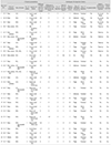
Twenty-seven patients with periorbital lipogranuloma were recruited and enrolled in this study (Table 1). All but one patient (case 2) were female, with a mean age of 40.2 years (range, 23 to 63 years). Presenting symptoms were palpable mass in 25 patients (92.6%), eyelid edema in nine patients (33.3%), and eyelid erythema in three patients (11.1%). Facial autologous fat injection was performed by a plastic surgeon in 23 cases (85.2%) and by a dermatologist in two cases (7.4%). The site of injection was the forehead in 19 patients (69.2%), the whole face including the forehead in six patients (23.1%), and the upper eyelid in two patients (7.7%). The number of injections was two in 23 patients, three in one patient, and one in three patients. All patients who received two or more injections were treated with cryopreserved fat at the second or third injection. The mean interval between first and second autologous fat injections was 2.65 ± 1.07 months (range, 1 to 5 months), and the mean time from the final injection to symptom onset was 13.6 ± 29.2 months (range, 2 to 153 months). The site of fat harvesting was the thigh in 18 cases (66.7%), abdomen in six cases (22.2%), and buttocks in one case (3.7%). Blepharoptosis, defined as a difference in margin reflex distance >1 mm, was observed in 11 cases (40.7%). Exophthalmos, defined as an exophthalmometric value >1 mm, was found in four cases (15.4%). Sixteen patients underwent radiologic imaging (orbital computed tomography [CT] in eight, orbital magnetic resonance imaging [MRI] in eight, both in two). Orbital CT images showed a focal ill-defined soft tissue enhancing lesion. Orbital MRI images showed ill-defined heterogeneous enhanced soft tissue swelling with fat-containing lesion with or without rim enhancement.
Surgical excision was performed in three patients to remove the mass and to identify pathological abnormalities. Histopathological examination revealed variable-sized fat tissue with foreign body reaction and fat necrosis, features characteristic of lipogranuloma. One month after surgery, the masses and eyelid swelling had disappeared. Intralesional triamcinolone injection (Triam, 40 mg/mL, 0.1 mL) was performed in six other patients; five of whom showed complete resolution, and one showed partial resolution. Among the five patients with complete resolution, one experienced recurrence 2 months after resolution. There were no significant side effects except mild bruising. Eighteen patients refused any management; among them, three patients (cases 15, 22, and 24) showed spontaneous resolution over 5 months of follow-up. Cases with spontaneous resolution presented with only one mass and no other symptoms. They also had trends toward relatively shorter intervals between injections (1.33 ± 1.52 vs. 2.65 ± 1.07 months, p = 0.335) and shorter times from injection to symptoms (6.65 ± 4.73 vs. 13.6 ± 29.2 months, p = 0.144), though these differences were not statistically significant.
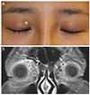
A 29-year-old woman presented with a 3-month history of palpable mass in her right upper eyelid (Fig. 1A). She had received autologous fat injection twice on her forehead at a local plastic surgery clinic 1 year prior. The fat was harvested from her thighs, injected, and the remained was cryopreserved until used for the second injection. The interval between the first and second injections was 2 months. The mass was a 5-mm-sized round fixed mass just below the superior orbital rim margin. Orbit MRI scan showed a fat-containing lesion with peripheral enhancement and adjacent skin enhancement in the right upper eyelid area (Fig. 1B). The mass was excised through eyelid crease incision: after the skin and orbicularis muscle were incised and dissected along the preseptal plane, the 8-mm-sized round mass was exposed superficial to orbital fat. Histological examination revealed foreign body reaction–induced lipogranuloma with fat necrosis (Fig. 1C and 1D). The mass and swelling completely disappeared 1 week after the surgery.
A 31-year-old woman was referred to Seoul National University Bundang Hospital with a 3-week history of swelling and palpable mass in her right upper eyelid (Fig. 2A). Seven months prior to her referral, she had autologous fat harvested from her thigh and injected into her forehead by a plastic surgeon, followed by a second injection 2 months later with cryopreserved fat. On physical examination, her right eye was 2 mm ptotic and also 2 mm proptotic. There was an approximately 5-mm-sized palpable mass in the superomedial periorbital area. Orbital MRI showed multiple bubble-shaped enhancing lesions compatible with lipogranulomas in both upper eyelids (Fig. 2B). The patient received intralesional triamcinolone (4 mg/0.1 mL) injections on her right eye, at the deep medial orbit and central eyelid. At a 1-week follow-up after receiving these injections, the swelling had decreased, and the ptosis had improved to an margin reflex distance difference of 1 mm. Three months after injection, the mass had completed disappeared, and the ptosis was completely resolved.
This study analyzed 27 cases of periorbital lipogranuloma following autologous fat injection into the face. Presenting symptoms were palpable mass, eyelid edema, eyelid erythema, blepharoptosis, and exophthalmos. The mean time from injection to symptom onset was 13.6 ± 29.2 months (range, 2 to 153 months). The diagnosis of periorbital lipogranuloma is based on characteristic clinical manifestations with history of autologous fat injection into the face and radiologic tests including CT and MRI if necessary. Due to its retrospective nature at a tertiary referral center, the study included only patients who had clinically demonstrable lesions; however, there are likely a larger number of patients with subclinical lipogranuloma among those who undergo facial autologous fat injection.
Two different mechanisms have been proposed for the etiology of lipogranuloma. One is an exogenous mechanism through foreign body reactions to lipid or oil-like substances, which result from the inability of the body to metabolize exogenous lipids in the tissue interstitium. The other mechanism is endogenous degeneration of lipids secondary to allergic reactions and/or trauma [6]. In our study, 24 of 27 patients (88.9%) underwent two or more autologous fat injections, and all but two with unverifiable data had used cryopreserved fat in the second injection. The mean duration of fat cryopreservation was 2.65 ± 1.07 months (range, 1 to 5 months). A previous study on fat cryopreservation showed that the viability of adipocytes declines rapidly after frozen storage at both -15℃ and -70℃ for 1 day and decreases gradually over 8 weeks, at which time only approximately 5% of the fat cells are viable [7]. Thus, it is possible that most cryopreserved fat used in secondary or tertiary injections contains non-viable adipocytes and oil-like substances, which could cause lipogranuloma through foreign body reactions. However, in our study, three patients underwent autologous fat injection only once and so were unrelated to cryopreservation. Therefore, the mechanism of lipogranuloma of these cases might not involve foreign body reactions but rather endogenous degeneration of lipids secondary to allergic reactions and/or trauma.
The characteristic predisposition of the upper eyelid to lipogranuloma can be explained by the galea in the superficial musculo-aponeurotic system. Injected fat tissue can migrate through the galea aponeurosis and the retroorbicularis fascia due to gravity, facial muscle movement, and postoperative massage of the injection sites [8]. In this study, orbital CT and MRI scans demonstrated that the most common location of perioribtal lipogranuoloma was the preseptal area (13 cases), followed by the preaponeurotic orbital area (five cases). All patients with preaponeurotic lipogranuoloma had a history of upper eyelid blepharoplasty; considering inevitable septal defect following blepahroplasty, the location of the lipogranuloma might be relevant to whether or not the orbital septum was intact.
Three patients who underwent mass excision reported resolved mass and swelling. Among the six patients who received intralesional triamcinolone injection, five showed complete resolution, and one showed partial resolution. There have been no reports on the effect of triamcinolone injection on lipogranuloma. We managed the lipogranulomas of six patients with intralesional triamcinolone injection, and the results were excellent. These findings suggest that intralesional triamcinolone injection is a good treatment option for patients who do not want to undergo an invasive surgical procedure.
Three patients in this study showed spontaneous resolution, all of whom had only one mass lesion with no other symptoms. These patients had shorter injection intervals (mean, 1.33 months) and shorter symptom onset (mean, 6.65 months) compared to other patients, though these differences were not statistically sigfnificant due to the small number of cases. However, these findings suggest that patients with lipogranuloma presenting with one mass lesion and no other symptoms can be managed by regular follow-up without invasive management, anticipating spontaneous resolution.
The limitations of our study are its retrospective nature and the possible selection bias toward patients more sensitive to symptoms or with good compliance. Pathological diagnosis was performed in only three cases, and systemic corticosteroid therapy was not used as a treatment option for any patients. Therefore, well-controlled prospective studies are warranted to investigate the actual incidence of lipogranuloma, natural history, and treatment outcomes according to type of management.
We present 27 patients who developed periorbital lipogranuloma after autologous fat injection. In conclusion, lipogranuloma can develop in the eyelid following autologous fat injection, and clinicians should be conscious of this possible complication.
Figures and Tables
 | Fig. 1(A) A 29-year-old woman presenting with a 3-month history of palpable mass in the right upper eyelid (asterisk) that developed 10 months after an injection of cryopreserved autologous fat into her face. (B) A T1-weighted axial magnetic resonance image shows a fat-containing lesion with peripheral enhancement (arrow) and adjacent skin enhancement in the right upper eyelid. (C,D) Typical findings of granulomatous foreign body reaction with diffusely infiltrated chronic granulomatous inflammatory cells with fat necrosis and fibrosis (hematoxylin and eosin stain, ×100, ×400). |
 | Fig. 2(A) A 31-year-old woman presenting with a 3-week history of palpable mass in the right upper eyelid (asterisk) that developed 7 months after an injection of cryopreserved autologous fat into her forehead. (B) A T2-weighted coronal image reveals multiple lesions (arrows) of heterogeneous high signal intensity in both upper eyelids. |
References
1. Coleman SR. Facial recontouring with lipostructure. Clin Plast Surg. 1997; 24:347–367.
2. Locke MB, de Chalain TM. Current practice in autologous fat transplantation: suggested clinical guidelines based on a review of recent literature. Ann Plast Surg. 2008; 60:98–102.
3. Hong JW, Kim SM. An analysis of the experiences of 62 patients with moderate complications after full-face fat injection for augmentation. Plast Reconstr Surg. 2013; 131:918e–920e.
4. Paik JS, Cho WK, Park GS, Yang SW. Eyelid-associated complications after autogenous fat injection for cosmetic forehead augmentation. BMC Ophthalmol. 2013; 13:32.
5. Ryeung Park Y, Choi JA, Yoon La T. Periorbital lipogranuloma after cryopreserved autologous fat injection at forehead: unexpected complication of a popular cosmetic procedure. Can J Ophthalmol. 2013; 48:e166–e168.
6. Lemperle G, Morhenn V, Charrier U. Human histology and persistence of various injectable filler substances for soft tissue augmentation. Aesthetic Plast Surg. 2003; 27:354–366.
7. Son D, Oh J, Choi T, et al. Viability of fat cells over time after syringe suction lipectomy: the effects of cryopreservation. Ann Plast Surg. 2010; 65:354–360.
8. Sa HS, Woo KI, Suh YL, Kim YD. Periorbital lipogranuloma: a previously unknown complication of autologous fat injections for facial augmentation. Br J Ophthalmol. 2011; 95:1259–1263.


 PDF
PDF ePub
ePub Citation
 Print
Print


 XML Download