Abstract
Purpose
To investigate the clinical and demographic features of idiopathic macular telangiectasia (MacTel) in Korean patients since the introduction of spectral domain optical coherence tomography (SD-OCT).
Methods
We reviewed medical records of patients who were diagnosed with MacTel from 2009 to 2013. All patients underwent fluorescein angiography and SD-OCT and were classified as type 1 or type 2 according to the classification system proposed by Yannuzzi.
Results
Over a period of 5 years, 4 (18.2%) patients were diagnosed with type 1 MacTel and 18 (81.8%) patients were diagnosed with type 2 MacTel. All patients with type1 MacTel were male, and their mean age was 51 ± 8.6 years. Among patients with type 2 MacTel, 3 (16.7%) were male, 15 (83.3%) were female, and the mean age was 60 ± 13.6 years. Whereas all type 1 MacTel patients had either metamorphopsia or mild scotoma, of the 18 patients with type 2 MacTel, only 4 (22.2%) had those symptoms, 10 (55.6%) complained of only mild visual impairment, and the other 4 (22.2%) had no symptoms. Intraretinal cystoid spaces were observed in 26 (72.2%) of 36 eyes with type 2 MacTel by SD-OCT. These cystoid spaces had irregular boundaries and did not correspond to angiographic leakages.
Idiopathic macular telangiectasia (MacTel) is a rare retinal disease comprised of various vascular diseases affecting the capillaries of the posterior pole [1]. MacTel was formerly called idiopathic juxtafoveolar retinal telangiectasis (IJRT) and was classified into 3 groups by Gass [2,3]. In their 2006 attempt to simplify the Gass classification, Yannuzzi et al. [4] divided IJRT into 2 broad groups based on reconcile optical coherence tomography (OCT) and high speed indocyanine green and fluorescein angiographic findings. According to this classification, groups 1A and 1B IJRT were pooled together into aneurysmal telangiectasia, also known as type1 MacTel, and group 2A IJRT was renamed perifoveal telangiectasia or type 2 MacTel [4]. Types 2B, 3A, and 3B IJRT were eliminated from this classification due to the lack of subjects [1]. Type 2 MacTel is thought to be the most common in Western countries [3,4,5,6]. In contrast, previous studies on Korean patients showed type 1 MacTel to be the most common [7,8].
Recent advances in imaging technology have expanded our knowledge of ocular diseases [9,10]. spectral domain (SD)-OCT has become a valuable tool for diagnosing type 2 MacTel, especially in asymptomatic cases [11,12]. In the present study, we investigated the clinical and demographic features of MacTel in Korean patients since the introduction of SD-OCT.
This study was designed as a retrospective case series. Institutional review board approval was obtained from the Korea University Medical Center, Seoul, Korea and the Dongguk University Ilsan Hospital, Goyang, Korea. All research adhered to the tenets of the Declaration of Helsinki.
We reviewed medical records of consecutive patients who were diagnosed with MacTel from 2009 to 2013. Each patient underwent slit-lamp biomicroscopy, indirect ophthalmoscopy, fundus photography, fluorescein angiography, and SD-OCT. SD-OCT was performed using the 3D OCT (1000 Mark II, software ver. 3.20; Topcon Corp, Tokyo, Japan) or Cirrus HD-OCT (Model 4000, software ver. 6.0; Carl Zeiss Meditec, Dublin, CA, USA). Diagnoses of type 1 or type 2 MacTel were based on biomicroscopic, fluorescein angiographic, and OCT evidence, after excluding age-related macular degeneration, polypoidal choroidal vasculopathy, pathologic myopia, idiopathic choroidal neovascularization, angioid streaks, retinal vein occlusion, radiation retinopathy, and diabetic retinopathy [2,3,4]. All eyes with type 2 MacTel were classified into non-proliferative and proliferative stages [4]. Data, including age, sex, history of diabetes or hypertension, laterality of the eye, presence of cataracts, symptoms, best-corrected Snellen visual acuity (BCVA), and central subfield retina thickness, were assessed. SD-OCT and fluorescein angiographic images were also analyzed.
A total of 22 patients were diagnosed with MacTel during a 5-year period. Of these, 4 (18.2%) patients had type 1 MacTel and 18 (81.8%) patients had type 2 MacTel. Table 1 shows characteristics of each type of MacTel.
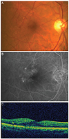
Fig. 1 shows a representative case with type 1 MacTel. Of 4 patients with type 1 MacTel, 2 suffered from metamorphopsia and the other 2 had mild scotoma. In the 1 patient with bilateral type 1 MacTel, the less severe eye was asymptomatic. One of 5 eyes with type 1 MacTel had cataracts. SD-OCT in all eyes with type 1 MacTel showed retinal thickening temporal to the fovea with intraretinal cystoid spaces or hyper-reflective lesions corresponding to fluorescein angiographic leakage.
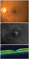
Fig. 2 shows a representative case with type 2 MacTel. Of the 18 patients with type 2 MacTel, 2 suffered from metamorphopsia, 2 from mild scotoma, 10 complained of mild visual impairment, and the other 4 were asymptomatic. All patients with type 2 MacTel had bilateral disease. Metamorphopsia was found in 2 eyes, mild central scotoma in 2 eyes, mild visual impairment in 17 eyes, and no symptoms in 15 eyes. Sixteen of 36 eyes with type 2 MacTel were associated with cataracts. SD-OCT showed no retinal thickening in all eyes with type 2 MacTel, which was in contrast to type 1 MacTel. In 26 (72.2%) eyes, cystoid spaces were observed with SD-OCT. These cystoid spaces had irregular boundaries but did not correspond to angiographic leakages. Disruptions of the line representing the junction of the photoreceptor inner and outer segments (IS/OS line) were observed in 20 (55.6%) eyes with SD-OCT. According to the Yannuzzi classification, 35 (97.2%) eyes were in a non-proliferative stage, and 1 (2.8%) eye was in a proliferative stage.
Type 2 MacTel was most common in the present study. This result coincided with the results of Western studies [3,4,5,6]. In previous Korean studies, Chang et al. [8] and Lee et al. [7] reported that the group 1 subtype of IJRT, i.e., type 1 MacTel, was the most common. This difference could be partially ameliorated by the use of more advanced imaging technology such as SD-OCT, as in the present study [13]. Chang et al. [8] used a time-domain OCT (Stratus OCT Model 3000, Carl Zeiss Meditec) during a study period from 1999 to 2003. Lee et al. [7] investigated patients from 1997 to 2009 during which time SD-OCT was not yet used widely. On the other hand, we used SD-OCT to evaluate macular pathology during a study period from 2009 to 2013. Additionally, increased awareness for the disease through recent promotion activities of the MacTel study group (http://www.mactelresearch.com) possibly affected the higher prevalence of type 2 MacTel in the present study [13].
Even though metamorphopsia and impaired reading ability associated with deep paracentral scotoma are the main symptoms, type 2 MacTel is usually asymptomatic, at least initially [11,14,15,16,17]. BCVA may remain normal or be only slightly affected in many patients until late stages of the disease [13,14,17]. Four (22.2%) of 18 patients had type 2 MacTel in the present study and had no symptoms, and 10 (55.6%) complained of only mild visual impairment. In contrast, all type 1 MacTel patients had either metamorphopsia or scotoma. The macula should be examined in patients with these symptoms. Furthermore, 15 (41.7%) of 36 eyes with type 2 MacTel were asymptomatic. Subtle macular alterations may be missed by standard clinical examination in asymptomatic type 2 MacTel cases, but they may be detected by noninvasive advanced imaging techniques such as SD-OCT [11].
SD-OCT has become a valuable tool for the diagnoses and study of type 2 MacTel [12]. Changes on OCT imaging may include temporal enlargement of the foveal pit, hyporeflective spaces in the inner retina, disruption of the IS/OS line, and with disease progression, atrophy of the outer neurosensory retina [11,12]. There is no angiographic leakage or pooling of fluorescein dye into the intraretinal hyporeflective cystoid spaces in type 2 MacTel, in contrast to other diseases such as diabetic maculopathy [18]. Additionally, the cystoid spaces of eyes with type 2 MacTel have irregular boundaries and low reflectivity [19,20]. Cystoid spaces were observed in 26 (72.2%) eyes and disruption of the IS/OS line was observed in 20 (55.6%) of 36 eyes with type 2 MacTel in the present study.
This retrospective study had several limitations. It was a hospital-based study with a small sample size. During the study period, other advanced imaging technologies such as fundus autofluorescence and confocal blue reflectance imaging previously shown to be more sensitive in detecting early and/or asymptomatic disease stages of type 2 MacTel type were not used [11]. In addition, the prevalence of type 1 MacTel may be underestimated, as it is a monocular condition in most cases [4].
In conclusion, type 2 MacTel was the most common in the present study. The wider availability of SD-OCT may have contributed to the diagnosis of type 2 MacTel. Type 2 MacTel may be more prevalent than type 1 in Koreans, which corresponds to the results of Western countries. Further large-scale studies using more advanced imaging technologies are needed to clarify the demographic features
of type 2 MacTel in the Korean population.
Figures and Tables
 | Fig. 1Type 1 idiopathic macular telangiectasia. (A) Fundus photograph shows multiple microaneurysms and retinal edema with hard exudates. (B) Late phase of fluorescein angiograph shows telangiectasis and multiple microaneurysms. (C) Spectral domain optical coherence tomography image shows retinal thickening and cystoid spaces. |
 | Fig. 2Type 2 idiopathic macular telangiectasia. (A) Fundus photograph shows slight graying of the perifoveolar retina. (B) Late phase of fluorescein angiograph shows hyperfluorescence temporal to the foveola. (C) Spectral domain optical coherence tomography image shows cystoid spaces without retinal thickening. |
Notes
References
1. Engelbert M, Chew EY, Yannuzzi LA. Macular telangiectasia. In : Ryan SJ, editor. Retina. 5th ed. London: Saunders/Elsevier;2013. p. 1050–1057.
2. Gass JD, Oyakawa RT. Idiopathic juxtafoveolar retinal telangiectasis. Arch Ophthalmol. 1982; 100:769–780.
3. Gass JD, Blodi BA. Idiopathic juxtafoveolar retinal telangiectasis: update of classification and follow-up study. Ophthalmology. 1993; 100:1536–1546.
4. Yannuzzi LA, Bardal AM, Freund KB, et al. Idiopathic macular telangiectasia. Arch Ophthalmol. 2006; 124:450–460.
5. Aung KZ, Wickremasinghe SS, Makeyeva G, et al. The prevalence estimates of macular telangiectasia type 2: the Melbourne Collaborative Cohort Study. Retina. 2010; 30:473–478.
6. Klein R, Blodi BA, Meuer SM, et al. The prevalence of macular telangiectasia type 2 in the Beaver Dam eye study. Am J Ophthalmol. 2010; 150:55–62.
7. Lee SW, Kim SM, Kim YT, Kang SW. Clinical features of idiopathic juxtafoveal telangiectasis in Koreans. Korean J Ophthalmol. 2011; 25:225–230.
8. Chang YI, Lee JG, Kim TW, Lee EK. The clinical manifestations and treatments of parafoveal telangiectasis. J Korean Ophthalmol Soc. 2004; 45:576–584.
9. Drexler W, Fujimoto JG. State-of-the-art retinal optical coherence tomography. Prog Retin Eye Res. 2008; 27:45–88.
10. Gabriele ML, Wollstein G, Ishikawa H, et al. Optical coherence tomography: history, current status, and laboratory work. Invest Ophthalmol Vis Sci. 2011; 52:2425–2436.
11. Gillies MC, Zhu M, Chew E, et al. Familial asymptomatic macular telangiectasia type 2. Ophthalmology. 2009; 116:2422–2429.
12. Charbel Issa P, Gillies MC, Chew EY, et al. Macular telangiectasia type 2. Prog Retin Eye Res. 2013; 34:49–77.
13. Heeren TF, Holz FG, Charbel Issa P. First symptoms and their age of onset in macular telangiectasia type 2. Retina. 2014; 34:916–919.
14. Finger RP, Charbel Issa P, Fimmers R, et al. Reading performance is reduced by parafoveal scotomas in patients with macular telangiectasia type 2. Invest Ophthalmol Vis Sci. 2009; 50:1366–1370.
15. Charbel Issa P, Helb HM, Rohrschneider K, et al. Microperimetric assessment of patients with type 2 idiopathic macular telangiectasia. Invest Ophthalmol Vis Sci. 2007; 48:3788–3795.
16. Charbel Issa P, Holz FG, Scholl HP. Metamorphopsia in patients with macular telangiectasia type 2. Doc Ophthalmol. 2009; 119:133–140.
17. Clemons TE, Gillies MC, Chew EY, et al. The National Eye Institute Visual Function Questionnaire in the Macular Telangiectasia (MacTel) Project. Invest Ophthalmol Vis Sci. 2008; 49:4340–4346.
18. Koizumi H, Iida T, Maruko I. Morphologic features of group 2A idiopathic juxtafoveolar retinal telangiectasis in three-dimensional optical coherence tomography. Am J Ophthalmol. 2006; 142:340–343.
19. Oh JH, Oh J, Togloom A, et al. Characteristics of cystoid spaces in type 2 idiopathic macular telangiectasia on spectral domain optical coherence tomography images. Retina. 2014; 34:1123–1131.
20. Barthelmes D, Sutter FK, Gillies MC. Differential optical densities of intraretinal spaces. Invest Ophthalmol Vis Sci. 2008; 49:3529–3534.


 PDF
PDF ePub
ePub Citation
 Print
Print


 XML Download