

 PDF
PDF ePub
ePub Citation
 Print
Print


Bietti crystalline retinal dystrophy is a rare, autosomal, recessively inherited disorder that is more common in patients of East Asian descent. This disease is characterised by the deposition of yellow crystals in the corneal limbus and retina, leading to the development of retinal and choroidal atrophy with resultant visual deterioration [1]. Optical coherence tomography (OCT) may show reflective deposits suggestive of crystalline deposits, and fundus fluorescein angiography (FFA) often demonstrates characteristic retinal pigment epithelium (RPE) atrophy [2]. This condition may also rarely be associated with choroidal neovascularisation (CNV) [3]. The prognosis for this disease is generally poor, with visual decline often progressing to blindness.
Mutations in the CYP4V2 gene, which produces proteins involved in one of the many cytochrome P450 proteins, have been identified as being linked to Bietti dystrophy [4]. Linkage analysis suggests that the affected protein may be involved in fatty acid metabolism [4]. Given that the crystals that are pathognomonic of this disease are thought to be composed of lipids and lipid by-products [1], it is possible that aberrant cytochrome P450 function results in problems with lipid metabolism in retinal tissues, causing crystal deposition. These crystals may then impair cellular function, leading to progressive chorioretinal atrophy.
Case Report
A 32-year-old male of Maltese heritage who was known to have electroretinogram-confirmed Bietti crystalline retinal dystrophy presented with acute bilateral central visual impairment. Upon examination, best-corrected visual acuity (BCVA) was 6 / 60 in the right eye and 6 / 45 in the left eye. At an examination one year previously, BCVA had been 6 / 7.5 in the right eye and 6 / 18 in the left.
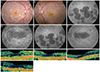
Dilated retinal examination revealed the presence of peripheral corneal and retina crystalline changes characteristic of Bietti dystrophy (Fig. 1A and 1B). Flourescein angiography showed patchy hypof luorescence indicative of chorioretinal atrophy. No evidence of late leakage was observed, excluding the diagnosis of choroidal neovascularisation (Fig. 1C-1F). Time-domain (TD)-OCT was performed and demonstrated bilateral cystic macular lesions (Fig. 1G and 1H) with a central foveal thickness (CFT) of 242 µm in the right eye and 253 µm in the left.
On the basis of the OCT findings, the patient was diagnosed with cystoid macular oedema. Acetazolamide therapy at a dose of 500 mg daily was initiated, and one month later, visual acuity improved to 6 / 18 bilaterally and CFT reduced to 221 µm in the right eye and 225 µm in the left eye. Following one year of acetazolamide therapy, BCVA was 6 / 15 in the right eye and 6 / 24 in the left eye, and CFT had stabilised at 221 µm in the right eye and 219 µm in the left eye (Fig. 1I and 1J).
Discussion
Bietti crystalline retinal dystrophy is an uncommon, inherited, retinal degeneration associated with retinal crystal deposits and progressive visual decline. Cystoid macular oedema is a rare complication of this disease, and may be associated with an acceleration of visual loss [5]. The pathogenesis of the oedema in this disorder is not well understood, and therefore treatment options are limited.
Macular oedema has been documented in association with other inherited retinal dystrophies, notably retinitis pigmentosa [6]. In this setting, it is considered that the oedema may relate to impaired RPE function, resulting in a failure of the RPE pumping mechanism [7]. Inhibition of carbonic anhydrase with acetazolamide has previously been shown to result in increased retinal pump activity [7], and has been used in this regard to treat cystoid macular oedema associated with retinitis pigmentosa [6], X-linked retinoschisis [8] and enhanced S-cone syndrome [9] with some success.
To our knowledge, this is the first case of cystoid macular oedema associated with Bietti dystrophy that was successfully treated with acetazolamide therapy in the literature. Given the reversible nature of this complication, it is important that potential treatments be recognised to preserve optimal vision.
Patients with Bietti dystrophy who develop worsening central vision should be promptly assessed to diagnose cystoid macular oedema. This complication is responsive to therapy with acetazolamide, resulting in improved anatomical outcomes and preservation of visual function.
 XML Download