

 PDF
PDF ePub
ePub Citation
 Print
Print


Vitreous cysts are rare ocular curiosities. They are usually detected during routine ophthalmological examination. Patients can be asymptomatic or may have floaters or transient blurry vision. Vitreous cysts can be congenital or acquired [1].
Case Report
In December 2010, a 50 year-old male was referred to our clinic due to a floating mass in his right eye. The patient complained about floaters in the right eye which he had experienced since childhood. The patient`s uncorrected visual acuity was 10 / 10 in both eyes and he did not have any systemic disorder or history of trauma. His ophthalmological examination revealed an unremarkable anterior segment with no signs of inflammation. The intraocular pressure was 16 mmHg and 15 mmHg in the right and left eyes, respectively.
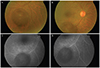
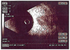
Indirect opthalmoscopy and posterior segment biomicroscopy performed with a 90D lens were unremarkable in the left eye, but a single, oval 6×5 mm diameter cyst was identified in the right eye, floating freely in the vitreous. The cyst was partially masking the underlying retinal vasculature (Fig. 1). Optical coherence tomography was normal in both eyes. A B-scan ultrasound revealed an echo-free, round-shaped cyst that was free from surrounding vitreous strands and retina, and was located in the posterior vitreous (Fig. 2). Fluorescein angiography (FA) ruled out the presence of intra- and overlying vascularization of the cyst. Indeed, FA showed a clear-edged hypofluorescence due to a pre-retinal masking effect (Fig. 1).
The indirect hemagglutinin tests of the patient for ecinococcus and cysticercosis were negative. Eosinophilia was not detected in the preripheral blood smear. Ultrasonography of the liver, spleen and kidney revealed no pathology. Based on these findings the patient was diagnosed with a primary vitreous cyst.
Discussion
Intravitreal cysts are divided into congenital and acquired cysts. Congenital cysts are associated with residues of the hyaloid vascular system and are occasionally present in perfectly normal eyes. These cysts are usually non-pigmented, with a smooth surface, pedunculated or sessile, and located anterior to the optic disc. Some congenital cysts can be limited in movement due to vitreous strands that link them to the optic discs [2]. Aqcuired cysts are more often associated with some type of inflammation due to intraocular infection, such as parasitic vitreitis, toxoplasmosis, and uveitis, or retinitis pigmentosa, choroidal atrophy and retinoschisis [3].
Trauma may play an important role in the generation of the cysts. Awan in 1975, reported a history of trauma in 2.7% of patients with intravitreal cysts. In cases of patients with traumatic cysts, the cyst is mostly transparent, but its wall may be pigmented [4]. Additionally, trauma may dislocate the cyst and move it close to the visual axis. Due to this phenomenon, patients may become symptomatic after traumas. Though the role of the trauma remains unclear. Trauma may trigger cyst formation or dislocate an already formed cyst and cause the patient to become symptomatic [5].
In the presented case, the patient had no history of trauma and the infective causes of the cysts were ruled out by negative serological tests. B-mode ultrasonography did not show any embryonal remnants. FA revealed that the inside and the surface of the cyst were avascular. Upon fundus examination, there was no any accompanying pathology that had been previously reported in the literature, such as retinitis pigmentosa, retinoschisis, etc.
In the literature, patient age has been reported to range from 5 to 68 years, though the majority of the patients are between 20 and 40 years. Vitreous cysts that have been described previously are normally single monolateral, single bilateral, or multiple monolateral. The presented case is of a single monolateral cyst. Bilateral cases are usually observed patients with retinitis pigmentosa [1].
Patients are usually asymptomatic, but because the cysts may be mobile, patients may suffer from floaters. Mioreover, when the cysts approach the visual axis, permanent visual disturbances may occur [6].
There is no treatment indication for asymptomatic or mild cases of vitreous cysts. As reported previously in two cases followed for 17 years, intravitreal cysts do not enlarge in diameter. In previously reported symptomatic cases, argon laser photocystotomy, YAG (Yttrium aluminium garnet) laser cystotomy, and pars plana aspiration have been attempted in order to relieve symptoms [7]. In the presented case, the patient was mildly symptomatic, so we decided to follow up with the patient in order to monitor the cyst, but provide any treatment. In conclusion, intravitreal cysts are rare ophthalmological disorders that have been discussed for a century, but the treatment of these 'ocular curiocities' remain debatable.
 XML Download