Abstract
To report a novel mutation within the CHST6 gene, as well as describe light and electron microscopic features of a case of macular corneal dystrophy. A 59-year old woman with macular corneal dystrophy in both eyes who had decreased visual acuity underwent penetrating keratoplasty. Further studies including light and electron microscopy, as well as DNA analysis were performed. Light microscopy of the cornea revealed glycosaminoglycan deposits in the keratocytes and endothelial cells, as well as extracellularly within the stroma. All samples stained positively with alcian blue, colloidal iron, and periodic acid-Schiff. Electron microscopy showed keratocytes distended by membrane-bound intracytoplasmic vacuoles containing electron-dense fibrillogranular material. These vacuoles were present in the endothelial cells and between stromal lamellae. Some of the vacuoles contained dense osmophilic whorls. A novel homozygous mutation (c.613 C>T [p.Arg205Trp]) was identified within the whole coding region of CHST6. A novel CHST6 mutation was detected in a Korean macular corneal dystrophy patient.
Corneal dystrophies are hereditary diseases involving corneal opacities at different layers of the cornea. They are primary corneal lesions that are not associated with prior inflammation or trauma [1]. Macular corneal dystrophy (MCD) is an autosomal recessive disorder characterized by bilateral progressive stromal clouding and central corneal thinning [2]. It is the least common form of the classic stromal dystrophies in most countries, especially in East Asia [3,4]. MCD occurs in primarily the first decade of life, beginning with a fine superficial stromal haze in the central stroma. Gradually, the opacification extends and involves the entire cornea, resulting in visual impairment, which generally requires penetrating keratoplasty.
Here, we report a patient with MCD and decreased visual acuity, who underwent penetrating keratoplasty. We confirmed the diagnosis via histopathologic studies such as light microscopy, electron microscopy, and DNA analysis. We found a mutation of CHST6 gene mutation (c.613 C>T [p.Arg205Trp]), which has never been reported previously.
A 59 year-old female with a family history of MCD presented to our clinic with progressive loss of vision over a 40-year period. Her visual acuity was finger count/50 cm OU. Slit lamp examination revealed diffusely hazy corneas with bilateral opacities. There were multiple irregular grayish-white, dense, poorly delineated spots in the stroma. Because her parents died at an early age, her family history was unclear. Of note, her only son complained of foggy vision and was 37 years old. His slit lamp examination revealed a similar, but less severe appearing corneal exam compared to that of his mother (Fig. 1).
Penetrating keratoplasty was performed on the right eye. A 7.75 mm donor button was sutured into a 7.5 mm host bed. Histopathologic examinations were conducted on the excised corneal buttons obtained from the procedure. Genomic DNA was extracted from the leukocytes of her peripheral blood via standard procedures. One year post-operatively, that patient had a best-corrected visual acuity of 20 / 100 OD and a clear cornea graft.
Informed consent for both the clinical examinations and DNA analyses was obtained from the patient and her son in accordance with the Declaration of Helsinki. The study was approved by the institutional review board at the Catholic University of Korea, St. Mary's Hospital.
Hematoxylin and eosin (HE), alcian blue, periodic acid-Schiff (PAS), colloidal iron, and Masson's trichrome stains were performed on the specimen using standard techniques. For transmission electron microscopy, the corneal specimen was immersed immediately in a fixative solution containing 3% glutaraldehyde with 0.2 M sodium cacodylate at a pH of 7.4. After overnight fixation, the fixative solution was removed and replaced with a phosphate buffer, followed by 1% osmium tetroxide buffered with sodium cacodylate. After one hour, the osmium was replaced with increasing concentrations of ethanol through propylene oxide, and the tissue was embedded in the epoxy. The embedded tissue was then sectioned with an ultra-microtome into 1 µm-thick sections, and stained with toluidine blue. The area to be observed was placed under a light microscope, ultra-sectioned from 60 to 100 nm in thickness, double stained with uranyl acetate and lead citrate, and examined via transmission electron microscope (JEM 1010; JEOL, Tokyo, Japan).
The unique exon involving the coding region, exon 3, was amplified via polymerase chain reaction (PCR) with the previously described primers [5]. Thermal cycling was conducted via the following protocol. The cycling program began with an initial denaturing step of 5 minutes at 95℃ followed by 33 cycles of 94℃ for 30 seconds, 53℃ to 57℃ for 30 seconds, and 72℃ for 45 seconds with a final extension step at 72℃ for 10 minutes. The PCR products were purified and sequenced directly on both strands using an automatic DNA sequencer (ABIPrism 377XL; Applied Biosystems, Foster City, CA, USA). The nucleotide sequences were compared with the published cDNA sequence of CHST6 (NM_021615) [6].
Light microscopy showed a normal epithelium and Bowman's membrane. HE staining revealed faintly basophilic deposits between the stromal lamellae, and within keratocytes and endothelial cells. These deposits were positive to alcian blue, PAS, and colloidal iron stain, but negative to Masson's trichrome stain (Fig. 2). Electron microscopy revealed keratocytes distended by membrane-bound intracytoplasmic vacuoles containing electron-dense fibrillogranular material. These vacuoles harbored dense osmophilic whorls. Similar vacuoles were also present in the interstromal lamellae and endothelial cells (Fig. 3).
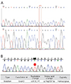
Analysis of the entire CHST6 coding region revealed distinct genetic defects. One missense mutation was identified in a homozygous state (p.Arg205Trp [c.613C>T]), which had not been previously reported. Samples from the patient's son also showed the same missense mutation (p.Arg205Trp [c.613C>T]) (Fig. 4).
In this study, we describe histopathological findings and a novel mutation in the CHST6 gene (p.Arg205Trp [c.613C>T]) in a case of MCD [7]. Light microscopy revealed abnormal deposits of glycosaminoglycans in Bowman's histiocytes and keratocytes, as well as between the stromal lamellae, Descemet's membrane, and endothelium. These glycosaminoglycans stained positively with alcian blue, colloidal iron, and PAS [8]. Electron microscopy revealed that these deposits corresponded to electron-lucent fibrillogranular material visible within membrane-bound intracytoplasmic vacuoles [9]. Such abnormalities have been identified as sequelae of an error in glycosaminoglycans metabolism within the cornea. In particular, errors in proteoglycan keratan sulfate (KS) metabolism have resulted in abnormal intra- and extracellular deposition [10]. The product of the CHST6 gene, corneal N-acetylglucosamine-6-sulfotransferase (C-GlcNac6ST), has been demonstrated to catalyze the sulfation of GlcNAc in KS. In normal corneal tissue, KS is an important glycosaminoglycan, which exists in a highly sulfated form. KS performs a crucial role related to the structure and transparency of the cornea. CHST6 can transfer a sulfuric acid group from the 3'-adenosine 5'-phosphate acid to KS via competition with an endogenous or exogenous substrate [11]. The variations in the coding region of CHST6 may reduce enzyme activity or cause it to be lost, resulting in a low sulfated form or non-sulfated form of KS. Due to the loss of its soluble properties, non-sulfated KS cannot be completely metabolized, inducing the deposition of sediment in the corneal stroma [12].
The mutation identified in this Korean patient is different from those reported previously in Asians (Japanese and Chinese) or whites (American, British, and Icelandic) [5,6,13,14]. Although the p.Arg205Gln mutation has been reported in a Southern Indian patient, the p.Arg205Trp mutation has not been reported previously [15]. The immunophenotype of MCD was not performed in our study since we could not test KS serum levels. Regardless of the type of macular corneal dystrophy, the patient showed indistinguishable clinical characteristics linked to the same responsible gene.
In summary, we identified a novel homozygous missense mutation in CHST6 in a Korean patient with macular corneal dystrophy. This novel gene mutation expands the mutation spectrum of the CHST6 gene and contributes to the study of molecular pathogenesis of corneal dystrophy. Further studies on these mutations would be helpful in further understanding the molecular mechanisms underlying macular corneal dystrophy.
Figures and Tables
Fig. 1
Slit lamp photography of the patient (A,B). (A) Right eye and (B) left eye. Diffusely hazy corneas with bilateral opacities were observed. There were multiple irregular, grayish-white, dense, poorly delineated spots in the stroma. Slit lamp photography of the patient's son (C,D). (C) Right eye and (D) left eye. Haziness of the stroma with diffuse opacities was observed in both eyes. These findings were similar, but less severe in appearance compared to his mother's examination.

Fig. 2
Comparison between light microscopic appearances (×100). The arrows indicate positive findings for glycosaminoglycan. (A) Hematoxylin and eosin stain, (B) alcian blue stain, (C) colloidal iron stain, (D) periodic acid-Schiff stain, and (E) Masson's trichrome stain.

Fig. 3
Transmission electron microscopic findings. (A) Keratocyte distended by membrane-bound intracytoplasmic vacuoles containing electron dense fibrillogranular material (black arrow). Vacuole containing dense fibrillogranular material in the interstromal lamellae (white arrow). Asterisk indicates relatively normal keratocyte (scale bar, 2 µm). (B) Keratocyte with vacuoles containing dense osmophilic whorls (scale bar, 0.5 µm).

Fig. 4
Direct sequencing analysis of the coding region of CHST6. (A) DNA sequence of the patient. (B) DNA sequence of the patient's son. Sequence of the entire coding region of CHST6 revealed a change in the nucleotide at codon 205 (CGG→TGG). The same mutation was identified in the patient's son (p.Arg205Trp [c.613C>T]).

References
1. McTigue JW. The human cornea: a light and electron microscopic study of the normal cornea and its alterations in various dystrophies. Trans Am Ophthalmol Soc. 1967; 65:591–660.
2. Donnenfeld ED, Cohen EJ, Ingraham HJ, et al. Corneal thinning in macular corneal dystrophy. Am J Ophthalmol. 1986; 101:112–113.
3. Jee DH, Lee YD, Kim MS. Epidemiology of corneal dystrophy in Korea. J Korean Ophthalmol Soc. 2003; 44:581–587.
4. Santo RM, Yamaguchi T, Kanai A, et al. Clinical and histopathologic features of corneal dystrophies in Japan. Ophthalmology. 1995; 102:557–567.
5. Akama TO, Nishida K, Nakayama J, et al. Macular corneal dystrophy type I and type II are caused by distinct mutations in a new sulphotransferase gene. Nat Genet. 2000; 26:237–241.
6. Dang X, Zhu Q, Wang L, et al. Macular corneal dystrophy in a Chinese family related with novel mutations of CHST6. Mol Vis. 2009; 15:700–705.
7. Weiss JS, Moller HU, Lisch W, et al. The IC3D classification of the corneal dystrophies. Cornea. 2008; 27:Suppl 2. S1–S83.
8. Snip RC, Kenyon KR, Green WR. Macular corneal dystrophy: ultrastructural pathology of corneal endothelium and Descemet's membrane. Invest Ophthalmol. 1973; 12:88–97.
9. Klintworth GK, Vogel FS. Macular corneal dystrophy: an inherited acid mucopolysaccharide storage disease of the corneal fibroblast. Am J Pathol. 1964; 45:565–586.
10. Hassell JR, Newsome DA, Krachmer JH, Rodrigues MM. Macular corneal dystrophy: failure to synthesize a mature keratan sulfate proteoglycan. Proc Natl Acad Sci U S A. 1980; 77:3705–3709.
11. El-Ashry MF, Abd El-Aziz MM, Wilkins S, et al. Identification of novel mutations in the carbohydrate sulfotransferase gene (CHST6) causing macular corneal dystrophy. Invest Ophthalmol Vis Sci. 2002; 43:377–382.
12. Musselmann K, Hassell JR. Focus on molecules: CHST6 (carbohydrate sulfotransferase 6; corneal N-acetylglucosamine-6-sulfotransferase). Exp Eye Res. 2006; 83:707–708.
13. Aldave AJ, Yellore VS, Thonar EJ, et al. Novel mutations in the carbohydrate sulfotransferase gene (CHST6) in American patients with macular corneal dystrophy. Am J Ophthalmol. 2004; 137:465–473.
14. Liu NP, Dew-Knight S, Rayner M, et al. Mutations in corneal carbohydrate sulfotransferase 6 gene (CHST6) cause macular corneal dystrophy in Iceland. Mol Vis. 2000; 6:261–264.
15. Warren JF, Aldave AJ, Srinivasan M, et al. Novel mutations in the CHST6 gene associated with macular corneal dystrophy in southern India. Arch Ophthalmol. 2003; 121:1608–1612.


 PDF
PDF ePub
ePub Citation
 Print
Print


 XML Download